

Gale Encyclopedia of Genetic Disorder / Gale Encyclopedia of Genetic Disorders, Two Volume Set - Volume 1 - A-L - I
.pdfAmniocentesis
increased incidence of clubfoot and an increased risk of procedure-related pregnancy loss.
Clubfoot, also referred to as talipes equinovarus, occurs in approximately one in 1,000 live births (0.1%) in the general population. It may involve either one foot (unilateral) or both feet (bilateral). Males are affected slightly more often than females. There are several proposed mechanisms by which clubfoot could occur: due to the interaction of several genes during development, as a direct consequence of environmental factors, such as an abnormal position in the uterus, or as a physical component of a single gene disorder. Any such disorder would be expected to also cause other abnormalities.
Overall, the CEMAT study found an incidence of clubfoot in the EA group of 1.3% (29 infants). None of the affected infants had other abnormalities. This is nearly ten times higher than the risk in the general population. The frequency of clubfoot in the MTA group was the same as in the general population (0.1%). Prior studies of mid-trimester amniocentesis did not reveal an increased frequency of infants with clubfoot or other birth defects.
Clubfoot was more common when testing was performed during the eleventh, rather than the twelfth, week of pregnancy. This suggests that there may be a specific window sometime in the eleventh to twelfth weeks during which the fetus may be particularly vulnerable to developing clubfoot. It is possible that EA causes a temporary, but still significant, loss of amniotic fluid. This loss may go unrecognized. However, it could, in turn, affect the flow of blood to the foot or cause direct pressure on the developing limb, either of which could lead to clubfoot. It is difficult to know which potential mechanism could be correct since the number of affected infants born after EA is relatively small.
Of note, a separate, much smaller, study also demonstrated an increased incidence of clubfoot (1.7%) among the set of women who underwent EA. The study consisted of patients randomized between EA and CVS and examined the risk of miscarriage after EA. Enrollment in the study was stopped once the association between EA and clubfoot was identified. There were no birth defects identified after CVS.
An additional concern recognized from CEMAT was a higher rate of miscarriage after EA. A procedurerelated loss was defined as one that occurred either shortly after the testing or before twenty weeks of pregnancy. Fifty-five women (2.5%) experienced a miscarriage after EA. In contrast, miscarriage occurred in seventeen (0.8%) of the MTA patients. An increased rate of loss appeared to more often follow technically challenging procedures. Difficult procedures included those
pregnancies in which bleeding occurred prior to amniocentesis or in which uterine fibroids were present. Tenting of the membranes also made early amniocentesis difficult. Tenting occurs when the amnion and chorion are not yet completely fused, as is true for the majority of first trimester pregnancies. The separation between the membranes makes insertion of the amniocentesis needle more difficult.
In the absence of a difficult EA procedure, a higher rate of loss was also observed among those pregnancies in which the mother experienced obvious leakage of amniotic fluid or vaginal bleeding after testing. The level of physician experience with EA did not influence the rate of loss.
Finally, EA was also linked to an increased number of laboratory culture failures (no growth of cells and no results) compared to MTA. The total waiting time for results was slightly longer in the EA group. This is not entirely a surprise, since a smaller amount of fluid is obtained when EA is performed. Hence, there are fewer cells, and culturing takes longer.
Demographics
According to the National Center for Health Statistics (NCHS), 112,776 amniocentesis procedures were performed in the United States in 1998, the most recent year for which data is available. The annual birth rate that year was approximately 3.9 million infants. Thus, approximately 3% of pregnant women in the United States had this procedure performed. It is likely that this is an underestimate, however. The NCHS obtains information from birth certificates registered in each state and the District of Columbia. Although almost all deliveries are registered in the United States, records are still submitted with incomplete information. It is also not possible to know how many amniocentesis procedures were performed for genetic testing, as compared to other indications, as this information is not requested.
Summary
Amniocentesis is a reliable procedure for prenatal diagnosis in the second trimester of pregnancy. It is primarily offered to pregnant women who are at increased risk, based on their age, family history, or other factor, of having a child with a genetic condition. Amniocentesis provides accurate information about fetal chromosomes or the likelihood of certain physical abnormalities. Additional specialized studies may be performed on an as-needed basis. Despite these benefits, amniocentesis is associated with a slightly increased chance of pregnancy loss. Each woman should discuss the potential risks and
78 |
G A L E E N C Y C L O P E D I A O F G E N E T I C D I S O R D E R S |

benefits of amniocentesis with a doctor or genetic counselor to make a decision about whether or not she has this testing. Early amniocentesis, or procedures performed before the thirteenth week of pregnancy, has been associated with an increased risk of clubfoot and of procedurerelated pregnancy loss.
Resources
BOOKS
“Amniocentesis and chorionic villus sampling (CVS).” In
Medical Tests Sourcebook. 1st ed. Health Reference Series, edited by Joyce Brennfleck Shannon. Detroit: Omniographics Inc., 1999, pp. 517–522.
Elias, Sherman, Joe Leigh Simpson, and Allan T. Bombard. “Amniocentesis and Fetal Blood Sampling.” In Genetic Disorders and the Fetus: Diagnosis, Prevention, and Treatment. 4th ed. Edited by Aubrey Milunsky. Baltimore: The Johns Hopkins University Press, 1998, pp. 53–82.
PERIODICALS
The Canadian Early and Mid-trimester Amniocentesis Trial (CEMAT) Group. “Randomized trial to assess the safety and fetal outcome of early and mid-trimester amniocentesis.” Lancet 351 (January 24, 1998): 242–247.
Farrell, Sandra A., A.M. Summers, Louis Dallaire, Joel Singer, JoAnn M. Johnson, and R. Douglas Wilson, members of CEMAT. “Club foot, an adverse outcome of early amniocentesis: disruption or deformation?” Journal Medical Genetics 36, no. 11 (November 1999): 843–846.
ORGANIZATIONS
American College of Obstetricians and Gynecologists. PO Box 96920, 409 12th St. SW, Washington, DC 20090-6920.http://www.acog.org .
National Center for Health Statistics. Division of Data Services, 6525 Belcrest Rd., Hyattsville, MD 20782-2003.http://www.cdc.gov/nchs .
WEBSITES
“Amniocentesis.” http://www.medicinenet.com/script/main/ Art.asp?li MN1&ArticleKey 268 .
“Amniocentesis.” http://www.modimes.org/HealthLibrary2/ factsheets/Amniocentesis.htm .
“Prenatal diagnosis: Amniocentesis and CVS.” http://www
.familydoctor.org/handouts/144.html .
Terri A. Knutel, MS, CGC
I Amyotrophic lateral sclerosis
Definition
Amyotrophic lateral sclerosis (ALS) is a fatal disease that affects nerve cells in the brain and spinal cord
K E Y T E R M S
Aspiration—Inhalation of food or saliva.
Bulbar muscles—Muscles that control chewing, swallowing, and speaking.
Degeneration—Nerves progressively withering.
Fasciculations—Involuntary twitching of patient’s muscles.
Voluntary muscle—A muscle under conscious control, such as arm and leg muscles.
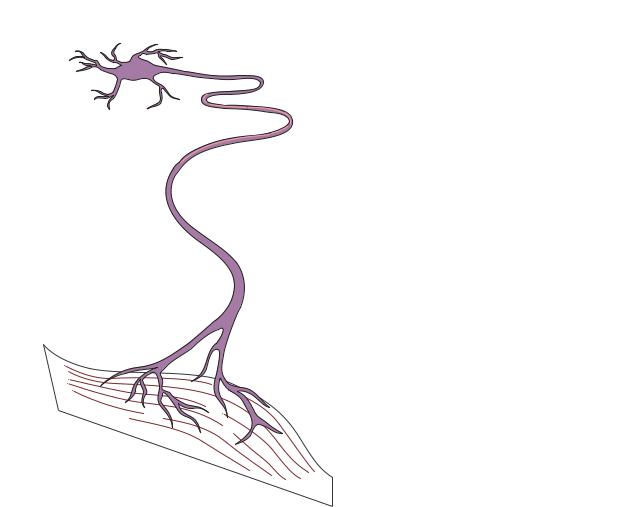
that are responsible for movement. The motor neurons (nerve cells which send an impulse to illicit muscular contraction or movement) in an ALS patient die as a result of rapid degeneration. Voluntary muscles, controlled by motor neurons, lack proper nourishment and will weaken and atrophy (shrink) as a result. Examples of voluntary movement include stepping off of a curb or reaching for the top shelf. These activities rely on the muscles of the arms and legs. Paralysis sets in at the endstages of ALS and leaves the patient unable to function physically, despite remaining mentally intact. There are no known causes or cures for amyotrophic lateral sclerosis, and the disease can afflict anyone. The usual cause of death is paralysis of the respiratory muscles which control breathing.
Description
Amyotrophic lateral sclerosis is a progressive disease of the central nervous system. “A” means “no,” “myo” implies muscle cells, and “trophic” refers to nourishment. The nerve cells that extend from the brain to the spinal cord (upper motor neurons), and from the spinal cord to the peripheral nerves (lower motor neurons), for unexplained reasons, degenerate and die. “Lateral” refers to the areas of the spinal cord that are affected, and “sclerosis” occurs as hard tissue replaces the previously originally healthy nerve.
The parts of the body that are not affected by ALS are those areas not involved in the use of motor neurons. The mind remains very sharp and in control of sight, hearing, smell, touch and taste. Bowel and bladder functions are generally not affected. Amyotrophic lateral sclerosis rarely causes pain, yet leaves patients dependent on the care of others during advanced stages.
At any given time there are about 30,000 people in the United States with amyotrophic lateral sclerosis, and about 5,000 new cases are reported each year. ALS pro-
sclerosis lateral Amyotrophic
G A L E E N C Y C L O P E D I A O F G E N E T I C D I S O R D E R S |
79 |
Amyotrophic lateralsclerosis |
NORMAL SPINAL NEURON |
DISEASED SPINAL NEURON |
|
Normal nerve fiber |
Affected nerve fiber |
||
|
|||
|
Normal skeletal muscle |
Wasted skeletal muscle |
The degeneration and death of motor neurons in the spinal cord and brain results in amyotrophic lateral sclerosis (ALS). These neurons convey electrical messages from the brain to the muscles to stimulate movement in the arms, legs, trunk, neck, and head. As motor neurons degenerate, the muscles are weakened and cannot move as effectively, leading to muscle wasting. (Gale Group)
gresses rapidly and paralyzed patients are usually under the intensive care of nursing facilities or loved ones. This can have a devastating psychological effect on the family members and the patient. In most cases, ALS is fatal within two to five years, although approximately 10% live eight years or more.
Amyotrophic lateral sclerosis is not a rare disease. ALS affects approximately seven people out of every 100,000. Most people with ALS are between 40 and 70 years of age. Approximately 5–10% of cases show a heredity pattern.
ALS, or Lou Gehrig’s disease, is named after the great New York Yankee’s first basemen. Lou Gehrig, known as the “Ironman” of baseball, died two years after he was diagnosed with amyotrophic lateral sclerosis.
Genetic profile
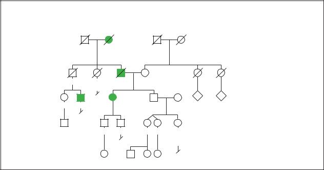
In 1991 a team of ALSA researchers linked familial ALS to chromosome 21. In 1993 it was found that there were structural defects in the SOD1 (superoxide dismutase) gene on chromosome 21. The SOD1 gene is an enzyme that protects the motor neurons from free radical damage. There is a high incidence of ALS on the island of Guam, in the Western New Guinea and on Kii peninsula of Japan leading some theorists to believe that genetic makeup may be susceptible to an environmental cause, such as the high levels of mercury and lead in these areas.
The inheritance pattern is autosomal dominant, which means that children of an affected parent have a 50% chance of inheriting the disorder. The majority of cases are due to a sporadic gene mutation, which means
80 |
G A L E E N C Y C L O P E D I A O F G E N E T I C D I S O R D E R S |
Amyotrophic Lateral Sclerosis
|
|
|
|
Autosomal Dominant |
|
|
|
|
|
|
Representative of Familial Form |
• 90% Sporadic |
|||
|
|
|
|
|
|
|
|
|
|
|
|
|
|
|
• 10% Familial |
|
|
|
|
|
|
|
• Incomplete Penetrance |
|
|
|
|
|
|
|
• Juvenile form (rare) |
|
d.72y |
d.70y |
|
|
|
||
d.81y |
d.60y |
d.74y |
80y |
|
|
||
|
|
Emphysema |
|
Diabetes |
|
|
|
|
|
|
|
|
|
3 |
5 |
65y |
63y |
|
66y |
58y |
58y |
|
|
2 |
|
|
|
|
|
|
|
|
|
|
33y |
33y |
32y 32y |
21y |
|
|
|
|
|
|
Childhood |
|
|
|
|
|
|
|
leukemia at 11y |
|
|
|
|
|
9y |
4y |
1y 3mos |
|
|
(Gale Group)
the mutation ocurrs only in the affected person. It is thought that sporadic mutations result from both biological and environmental causes. In rare cases, a mutation in NFH, the gene encoding for neurofilament (a structure that maintains cell shape) is apparent. Familial amyotropic lateral sclerosis has been linked to other chromosomal locations but the exact genes involved have not been identified. The Institutional Review Board at Thomas Jefferson University in Philadelphia recently approved the ALS gene therapy project. The goal of the project is to inject an adeno-associated virus carrying a normal copy of an EAAT2 gene into an ALS patient’s spinal cord where the motor neurons are dying. The hope is that the cells in that area will not die off.
Demographics
Amyotrophic lateral sclerosis affects anyone and both men and women are at equal risk. ALS may occur at any age, and the odds of developing it increase with age. There have been reported cases of teenagers with ALS. A person only needs to inherit a defective gene from one parent to cause the disease.
Signs and symptoms
The disease starts slowly, affecting just one limb, such as the hands or feet, and steadily progresses to more limbs and muscles. When muscles lack the proper nourishment they require, they begin to thin and deteriorate.
This condition is the hallmark of amyotrophic lateral sclerosis. Muscle wasting is due to the inability of degenerating motor neurons to elicit a signal to the muscles that allow them to function and grow. Common examples of symptoms for ALS are muscle cramps and twitching, weakness in the hands, feet, or ankles, speech slurring, and swallowing difficulties. Other early symptoms include arm and leg stiffness, foot drop, weight loss, fatigue, and difficulty making facial expressions.
One of the earliest symptoms of ALS is weakness in the bulbar muscles. These muscles in the mouth and throat assist in chewing, swallowing, and speaking. Weakness of these muscle groups usually cause problems such as slurred speech, difficulty with conversation and hoarseness of the voice.
Another symptom of ALS that usually occurs after initial symptoms appear is persistent muscle twitching (fasciculation). Fasciculation is almost never the first sign of ALS.
As the disease progresses the respiratory muscles (breathing muscles) weaken, resulting in increased difficulty with breathing, coughing, and possibly inhaling food or saliva. The potential for lung infection increases and can cause death. Many patients find it more comfortable and extend their lives when assisted by ventilators at this stage of the disease. Communication becomes very difficult. One way to accomplish feedback with others is to make use of the eyes. Blinking is one mode that
sclerosis lateral Amyotrophic
G A L E E N C Y C L O P E D I A O F G E N E T I C D I S O R D E R S |
81 |
Amyotrophic lateral sclerosis
patients of amyotrophic lateral sclerosis will be forced to utilize, in order to continue communication.
As the disease progresses, victims gradually lose the use of their feet, hand, leg, and neck muscles, and paralysis results in affected muscle groups. They are able to speak and swallow only with great struggle. Sexual dysfunction is not affected. Breathing will become increasingly difficult and the patients of ALS may decide to prolong life with the use of assisted ventilation, which may decrease the risks of death from infections such as pneumonia.
Diagnosis
ALS is difficult to diagnose. There is no one set way to test for the disease. A series of diagnostic tests will rule out and exclude other possible causes and diseases that resemble ALS. Electro diagnostic tests such as electromyography (EMG) and nerve conduction velocity (NCV) are used to help diagnose ALS. Blood and urine tests, spinal taps, x rays, and muscle and/or nerve biopsy are performed, as well as magnetic resonance imaging (MRI), myelograms of the cervical spine and a complete neurological exam.
A second opinion is frequently recommended if ALS is suspected since it is a fatal neurological disease. After a complete medical exam and family history check has been administered, other tests such as a CT (computed tomography) scan may be done to continue ruling out other causes. Many symptoms mimic ALS such as tumors of the skull base or upper cervical spinal cord, spinal arthritis, thyroid disease, lead poisoning, and severe vitamin deficiency. Other possibilities to rule out are multiple sclerosis, spinal cord neoplasm, polyarteritis, syringomyelia, myasthenia gravis, and muscular dystrophy. Amyotrophic lateral sclerosis is hardly ever misdiagnosed after this intensive series of diagnostic tests.
Treatment and management
Currently, there is no treatment for ALS. Management aims to control the symptoms that patients experience. Emotional, psychological, and physical support are provided to ease the difficulty associated with this disorder.
Moderate activities are recommended in the early stages of the disease. Physical therapy can help muscles stay active and delay the resulting weakness. ALS patients are encouraged to maintain a healthy diet and exercise regularly for as long as possible. Education of ALS is very important in developing an understanding of
the disease, and is vital for family members as well as patients.
Although there are no set treatments for ALS there are still many special considerations that can assist in the quality of lifestyle for the patient. Implementing a physical therapy program, providing a wheelchair or walker, assistance when bathing, and suction machines to help evacuate accumulated secretions all help the ALS patient. Other considerations include providing foods that are soft and easy to swallow, skin maintenance, feeding tubes, ventilation maintenance and emotional support.
Researchers have developed a drug approved by the Food and Drug Administration (FDA) called Rilutek (riluzole). The drug was the first to have a positive effect in that it appears to extend the life of ALS patients by about three months.
Another drug, Myotrophin (somatomedin C), appears to prevent neuron loss and enhance neuron generation in animal studies.
Prognosis
Amyotrophic lateral sclerosis normally progresses rapidly and leads to death from respiratory infection within three to five years. If the person involved is young and the initial symptoms appear in the limbs, the disease tends to develop more slowly. Improved medical care has prolonged the lives of ALS patients and shows promise for more effective treatments in the future.
Resources
BOOKS
Adams, Raymond D., Maurice Victor, and Allan H. Ropper.
Adam’s & Victor’s Principles of Neurology, 6th ed. New York, McGraw Hill, 1997.
Brown, Robert H. “The motor neuron diseases.” In Harrison’s Principles of Internal Medicine, 14th ed., edited by Anthony S. Fauci, et al. New York: McGraw-Hill, 1998, pp. 2368-2372.
Feldman, Eva L. “Motor neuron diseases.” In Cecil Textbook of Medicine, 21st ed., edited by Lee Goldman and J. Claude Bennett. Philadelphia: W.B. Saunders, 2000, pp. 2089-2092.
PERIODICALS
Foubistor, V. “Gene therapy fosters hope.” American Medical News 43 (March 6, 2000).
ORGANIZATIONS
Association of America (ALSA). 27001 Agoura Rd., Suite150, Calabasas Hills, CA 91301-5104. (818) 800-9006. Fax: (818) 880-9006. http://www.alsa.org .
Center for Neurologic Study. 9850 Genesee Ave., Suite 320, Lajolla, CA 92037. (858) 455-5463. Fax: (858) 455-1713. cns@cts.org. http://www.cnsonline.org .
82 |
G A L E E N C Y C L O P E D I A O F G E N E T I C D I S O R D E R S |

Forbes Norris ALS Research Center. Caifornia Pacific Medical Center, 2324 Sacramento St., San Francisco, CA 94115. (415) 923-3604. Fax: (415) 673-5184.
Laith Farid Gulli, MD
Brian Veillette, BS
I Androgen insensitivity syndrome
Definition
Androgen insensitivity syndrome is a genetic condition where affected people have male chromosomes and male gonads (testicles). The external genitals, however, have mild to complete feminization.
Description
Normal sexual development
In normal development, the chromosome sex determines the gonadal sex, which in turns determines the phenotypic sex. The chromosome sex is determined at conception; a male has the sex chromosome pair XY and a female has the chromosome pair XX. During the first 40 days of gestation, a male and female embryo appear the same and have undifferentiated gonads, which have the potential of becoming testes or ovaries. The presence of the Y chromosome in the male directs the undifferentiated gonads to become testicles. If no Y chromosome is present, such as in the female chromosome pair, the undifferentiated gonads become ovaries.
In males, the phenotypic sex, including the internal male structures and the external male genitalia, arises as a result of the hormones secreted from the testicles. The two main hormones secreted by the testicles are testosterone and mullerian duct inhibitor. Testosterone acts directly on the wolffian duct, which give rise to the internal male structures including the epididymides, vasa deferentia, and seminal vesicles. Testosterone is converted into dihydrotestosterone, the hormone responsible for the development of the male urethra and prostate, and the external genitalia of the penis and the scrotum. The mullerian duct inhibitor is the hormone that suppresses the mullerian ducts and prevents the development of fallopian tubes, upper vagina, and uterus in males.
If no testicles are present, as with females, no mullerian duct inhibitor is formed and the mullerian ducts become the fallopian tubes, the upper vagina, and the uterus. The wolffian ducts regress. Due to the lack of
K E Y T E R M S
Androgens—A group of steroid hormones that stumulate the development of male sex organs and male secondary sexual characteristics.
Chromosome—A microscopic thread-like structure found within each cell of the body and consists of a complex of proteins and DNA. Humans have 46 chromosomes arranged into 23 pairs. Changes in either the total number of chromosomes or their shape and size (structure) may lead to physical or mental abnormalities.
Mullerian ducts—Structures in the embryo that develop into the fallopian tubes, the uterus, the cervix and the upper vagina in females.
Wolffian ducts—Structures in the embryo that develop into epididymides, vasa deferentia, and seminal vesicles in males.
dihydrotestosterone, the external genitals are not masculinized and become female. Studies have shown that an ovary is not required for the formation of the internal female structures or the feminization of the genitals. If a testicle is not present, the development of the embryo will default to female development.
In most cases, the chromosomal sex, the gonadal sex, and the phenotypic sex are in agreement. Males have 46,XY chromosomes, testicles, and male internal structures and genitals. Females have 46,XX chromosomes, ovaries, and internal female structures and genitals.
Androgen insensitivity syndrome
Androgen insensitivity syndrome (AIS), also known as testicular feminization, is one of the most common conditions where the chromosome sex and gonadal sex do not agree with the phenotypic sex. Affected people have normal male chromosomes, 46,XY and testicles. The testicles secrete both testosterone and mullerian duct inhibitor as normal and no internal female structures form. However, due to defective androgen receptors, the wolffian ducts and genitals cannot respond to the androgens testosterone and dihydrotestosterone. As a result, no male internal structures are formed from the wolffian ducts and the external genitals are feminized.
The amount of feminization depends on the severity of the androgen receptor defect and is often characterized as complete androgen insensitivity (CAIS), partial androgen insensitivity (PAIS), and mild androgen insensitivity (MAIS). In complete androgen insensitivity, the alteration in the androgen receptor results in complete female
syndrome insensitivity Androgen
G A L E E N C Y C L O P E D I A O F G E N E T I C D I S O R D E R S |
83 |

Androgen insensitivity syndrome
TABLE 1
Classification of AIS Phenotypes
Type |
External genitalia (synonyms) |
Findings |
CAIS |
Female (“testicular feminization”) |
Absent or rudimentary wolffian duct derivatives |
|
|
Inguinal or labial testes; short blind-ending vagina |
|
|
Little or no pubic and/or axillary hair |
CAIS or PAIS |
Predominantly female (incomplete AIS) |
Inguinal or labial testes |
|
|
Labial fusion and enlarged clitoris |
|
|
Distinct urethral and vaginal openings or a urogenital sinus |
PAIS |
Ambiguous |
Microphallus ( 1 cm) with clitoris-like underdeveloped glans; labia majora-like bifid scrotum |
|
|
Descended or undescended testes |
|
|
Perineoscrotal hypospadias or urogenital sinus |
|
|
Excessive development of the male breasts during puberty |
|
Predominantly male |
Simple (glandular or penile) or severe (perineal) “isolated” hypospadias with a normal-sized penis and |
|
|
descended testes or severe hypospadias with micropenis, bifid scrotum, and either descended or |
|
|
undescended testes |
|
|
Excessive development of the male breasts during puberty |
MAIS |
Male (undervirilized male syndrome) |
Impaired sperm development and/or impaired masculinization |
|
|
Overdevelopment of the male breasts during puberty |
external genitals. In partial androgen insensitivity, also called Reifenstein syndrome, partial androgen insensitivity results in female genitalia with some masculinization, ambiguous genitalia, or male genitalia with partial feminization. With mild androgen insensitivity, mild androgen resistance results in normal male genitals or a male with mild feminization.
In both CAIS and PAIS, affected individuals are sterile (can not have a child). In MAIS, the affected male may have fertility problems because of oligospermia, low sperm production, or azoospermia, no sperm production. In all types of AIS, secondary sex characteristics such as body and pubic hair can be abnormal. Mental impairment is not found in any of the types of androgen insensitivity syndromes, though poor visual-spatial ability has been observed. People with AIS can also be rather tall, though bone age is usually normal.
Genetic profile
Androgen insensitivity syndrome is a genetic condition that results from mutations (alterations) of the gene for the androgen receptor. The androgen receptor is located on the long arm of the X chromosome (Xq11q12). As women have two X-chromosomes, they also have two androgen receptor genes. Men have only one X chromosome and a Y chromosome; hence they only have one copy of the androgen receptor gene.
When women have one copy of the androgen receptor altered, they are considered carriers of AIS. In most cases, the second, normal copy of the androgen receptor can compensate for the altered copy. However, in approximately 10% of women who are carriers for the altered androgen receptor gene, clinical signs such as sparse
pubic hair and armpit hair or a delay to the start of their first menstrual period is observed.
46,XY conceptions that have alterations in the androgen receptor gene do not have a second copy to compensate for the altered copy. Hence, these people will have AIS. If the androgen receptor is severely altered, they will have CAIS. If not severely altered, they will have PAIS or MAIS.
All forms of AIS are inherited in an X-linked recessive pattern. This means women who are carriers have a 25% chance of having an affected child. If a carrier woman has a 46,XY conception, there is a 50% chance the child will have AIS. If a carrier woman has a 46,XX conception, there will be a 50% chance the daughter will also be a carrier.
When a person has AIS and has no other family history of the condition, approximately 2/3 of the time the affected person inherited the gene alteration from his or her mother. The other 1/3 of the time, the alteration of the androgen receptor was a new event (new mutation) in the affected person and was not inherited.
Cases of both gonadal mosaicism and somatic mosaicism have been reported with AIS. Gonadal mosaicism occurs when the alteration in the androgen receptor occurred not at conception, but in one of the gamete cells (sperm or egg). The rest of the cells of the body do not have the altered androgen receptor. With AIS, this can occur when one of a woman’s early gamete cell has the new alteration in the androgen receptor but the rest of the cells in her body do not. All the eggs that come from the early gamete cell will also have the alteration. Her risk for having a child with AIS is increased. Somatic mosaicism occurs when the alteration in the
84 |
G A L E E N C Y C L O P E D I A O F G E N E T I C D I S O R D E R S |
androgen receptor occurs after conception but not in a gamete cell. Some of the person’s cells will have the altered androgen receptor and other cells will not. The amount of cells with altered receptors and the location of those cells within the body will determine how severely affected a person will be.
Mutations within the androgen receptor gene are also responsible for the neuromuscular condition spinobulbar muscular atrophy or Kennedy disease. See separate entry for more information.
Demographics
Complete androgen insensitivity syndrome occurs in approximately 1/64,000 46,XY births or 2-5/100,000 births overall. Partial AIS is at least as common as complete AIS. The incident of mild AIS is unknown, but is estimated to account for approximately 40% of male infertility due to severe oligospermia or azoospermia.
Signs and symptoms
Complete androgen insensitivity
Individuals with CAIS are born looking like normal female babies. Often, the condition is discovered in one of two ways. The child can have an inguinal hernia that upon repair is found to contain testicles. The most common presentation is during puberty with primary amenorrhea, or lack of the onset of the menstrual period. Affected individuals have a short, blind ending vagina and no uterus, cervix, fallopian tubes, or ovaries. During puberty, some girls will have absent or decreased sexual hair. Breasts develop normally and can be large in size with pale and immature nipples and areola. People with CAIS are usually raised as females and have normal female sexual orientation. All women with CAIS are sterile. In families with CAIS, all affected members will have complete androgen insensitivity and similar physical features.
Partial androgen insensitivity syndrome
Children with PAIS usually present at birth due to ambiguous genitalia. The genitalia can look like female genitals with some masculinization, completely ambiguous genitals where the sex of the baby cannot be immediately determined, or male genitals with some feminization. The degree of severity is a direct result of the degree of severity of the genetic alteration in the androgen receptor and resulting amount of functional androgen receptor. The internal structures of PAIS are the same as CAIS, with absent fallopian tubes, cervix,
uterus, and ovaries. Testes are present but do not produce sperm. Hence, people with PAIS are also sterile. People with PAIS also have primary amenorrhea, and breast development occurs in puberty. Unlike CAIS, affected individuals in the same family with presumably the same genetic alteration can have varying degrees of masculinization. As a result, some affected people may be raised as females whereas others may be raised as males. Sex assignment is made based upon the structure of the genitals, the surgical correction needed, and the predicted response to androgens during puberty.
Mild androgen insensitivity
Males with mild androgen insensitivity usually have normal male genitals and internal male structures. During puberty, males with MAIS may have breast enlargement, sparse facial and body hair, and small penis. Some affected males may also have impaired sperm production resulting in oligospermia or azoospermia, decreased or absent sperm. As with CAIS, affected men within the same family usually have similar features.
Diagnosis
Diagnosis is usually made based upon clinical features, chromosome analysis, hormone levels, and analysis of androgen receptor function in skin fibroblasts. Clinical features are listed above for CAIS, PAIS, and MAIS. Chromosome analysis reveals normal male chromosomes. Affected individuals can have elevated luteinizing hormone, normal to slightly elevated testosterone, and high estradiol for men. Follicle stimulating hormone may also be normal to elevated. Reduced androgen receptor function in skin fibroblast cells is also used to aid in a diagnosis.
As of 2001, direct genetic testing for molecular defects in the androgen receptor gene is being done on a research basis only.
Treatment and management
Complete androgen insensitivity
Treatment of CAIS requires the removal of the testicles from the pelvis or inguinal canal to decrease risk of testicular malignancy. Because the overall risk of malignancy is approximately 5% and rarely occurs before age 25, the testicles are usually removed after the development of the secondary sex characteristics, as the testes are needed for estrogen formation. After the removal of the testes, estrogen supplementation is started to aid in
syndrome insensitivity Androgen
G A L E E N C Y C L O P E D I A O F G E N E T I C D I S O R D E R S |
85 |

Anemia, sideroblastic X-linked
the development of secondary sex characteristics and to help prevent osteoporosis. Surgery to lengthen the vagina may be necessary.
Partial androgen insensitivity syndrome
For those affected individuals raised as females, treatment is similar to CAIS except the removal of the testicles is done earlier because it may cause enlargement of the clitoris during puberty. Reconstructive surgery of the genitals and lengthening of the vagina may be necessary.
People with PAIS raised as boys may need surgery to improve the appearance of the genitals. Androgen supplementation may be implemented, though long-term affects of androgen therapy are not known. Breast reduction surgery may be necessary after puberty.
Mild androgen insensitivity
Males with MAIS may require no treatment at all or breast reduction surgery after puberty. Males who are infertile may benefit from assisted reproductive technologies.
Prognosis
For CAIS and MAIS, the prognosis is excellent. Generally, gender assignment is not difficult and sexual orientation is female for CAIS and male for MAIS. Treatment usually involves minimal surgery and hormone supplementation. For individuals with PAIS, the prognosis is very dependent upon the severity of the condition. Assignment of gender can be difficult and genital surgery can be more involved. Recently, some individuals with PAIS and other intersex conditions have encouraged the delay of assigning gender until the child is old enough to express a preference. As of 2001, this idea has not been readily embraced in the medical community of the United States.
Resources
BOOKS
Wilson, J. D., and J. E. Griffin. “Disorders of Sexual Differentiation.” In Harrison’s Online, edited by Eugene Braunwald, et al. New York: McGraw-Hill, 2001.
PERIODICALS
Warne, G. L., et al. “Androgen insensitivity syndrome in the era of the molecular genetics and the internet: A point of view.” Journal of Pediatric Endocrinology & Metabolism
11 (1998): 3-9.
ORGANIZATIONS
AIS Support Group (AISSG). PO Box 269, Banbury, Oxon,
OX15 6YT UK http://www.medhelp.org/www/ais .
Intersex Society of North America. PO Box 301, Petaluma, CA 94953-0301. http://www.isna.org .
WEBSITES
Androgen Receptor Gene Mutations Database. http://www
.mcgill.ca/androgendb .
Pinsky, L. P. “Androgen Insensitivity Syndrome.” Gene Clinics: Clinical Information Resource University of Washington, Seattle. http://www.geneclinics.org/profiles/andrgoen/ details.html . February 6, 2001 (Updated March 23, 1999).
Carin Lea Beltz, MS, CGC
I Anemia, sideroblastic
X-linked
Definition
X-linked sideroblastic anemia is a hereditary enzyme disorder in which the body has adequate iron but is unable to incorporate it into hemoglobin.
Description
X-linked sideroblastic anemia is the hereditary form of sideroblastic anemia, also known as iron overload anemia or sideroblastosis. Another, more common type of sideroblastic anemia is called acquired sideroblastic anemia.
In sideroblastic anemia, iron enters a developing red blood cell and is not incorporated properly into the hemoglobin molecule (the cell’s oxygen carrier). This causes iron to accumulate in the mitochondria and sideroblasts. The defective hemoglobin then transports oxygen poorly, resulting in decreased tissue oxygenation.
This build–up of iron gives the cell nucleus its ringed appearance, called ringed sideroblast, which is the primary sign of siderblastic anemia.
Sideroblastic anemia is often mistaken for iron deficiency anemia, but tests usually reveal normal or increased levels of iron.
X-linked sideroblastic anemia
The hereditary form of the disorder is rare. The primary type of inherited sideroblastic anemia was first described in 1945 by Thomas Cooley. He identified cases of X-linked sideroblastic anemia in two brothers from a family with a six-generational history of the inherited disease. The genetic abnormality that causes X-linked sideroblastic anemia was identified almost 40 years later. Identification has aided diagnosis of this disorder.
86 |
G A L E E N C Y C L O P E D I A O F G E N E T I C D I S O R D E R S |

X-linked sideroblastic anemia nearly always manifests in infancy or childhood.
Other inherited forms of sideroblastic anemia
There are other inherited forms of sideroblastic anemia, which are also rare. A rare autosomal recessive form of inherited sideroblastic anemia occurs in both males and females of affected families. Autosomal dominant inheritance has also been reported. The abnormalities that cause these anemias are not yet identified. Also, Pearson’s syndrome, an inherited disorder caused by abnormal mitochondria, is sometimes called sideroblastic anemia with marrow cell vacuolization and exocrine pancreatic dysfunction.
Acquired sideroblastic anemia
Acquired sideroblastic anemia often results from prolonged exposure to toxins (such as alcohol, lead, or drugs), or nutritional imbalances (such as deficiency in folic acid or copper or excess in zinc). Other causes may be inflammatory disease, cancerous conditions, or kidney, endocrine, or metabolic disorders. Acquired sideroblastic anemia sometimes surfaces in the context of a myelodysplastic syndrome.
Removal of the toxin or treatment of the underlying disease will reverse this type of sideroblastic anemia.
Acquired anemia is usually seen in patients over 65, particularly in those cases associated with myelodysplasia. The disorder can appear as early as the mid-fifties.
Genetic profile
Hereditary sideroblastic anemia is most commonly inherited as an X-linked recessive trait.
Typical X-linked genetics
The following concepts are important to understanding the inheritance of an X-linked disorder. All humans have two chromosomes that determine their gender: females have XX, males have XY. X-linked recessive, also called sex-linked, inheritance affects the genes located on the X chromosome. It occurs when an unaffected mother carries a disease-causing gene on at least one of her X chromosomes. Because females have two X chromosomes, they are usually unaffected carriers. The X chromosome that does not have the disease-causing gene compensates for the X chromosome that does. For a woman to show symptoms of the disorder, both X chromosomes would need to have the disease-causing gene. That is why women are less likely to show such symptoms than males.
K E Y T E R M S
Heme—The iron-containing molecule in hemoglobin that serves as the site for oxygen binding.
Hemochromatosis—Accumulation of large amounts of iron in the tissues of the body.
Hemoglobin—Protein-iron compound in the blood that carries oxygen to the cells and carries carbon dioxide away from the cells.
Mitochondria—Organelles within the cell responsible for energy production.
Myelodysplasia—A bone marrow disorder that can develop into aplastic anemia requiring bone marrow or stem cell transplantation.
Nucleus—The central part of a cell that contains most of its genetic material, including chromosomes and DNA.
Red blood cells—Hemoglobin-containing blood cells that transport oxygen from the lungs to tissues. In the tissues, the red blood cells exchange their oxygen for carbon dioxide, which is brought back to the lungs to be exhaled.
If a mother has a female child, the child has a 50% chance of inheriting the disease gene and being a carrier who can pass the disease gene on to her sons. On the other hand, if a mother has a male child who inherits the disease-causing gene, he will be affected and has a 100% chance of passing the disease gene on to his children. Since the gene is defective and in the XY state there is no normal gene, the singular flawed gene is expressed.
Genetics of X-linked sideroblastic anemia
The genetic abnormality that causes X-linked sideroblastic anemia is a mutation in the erythroid (red blood cell) specific form of delta-aminolevulinate synthase (ALAS2). ALAS2 is the first enzyme in the heme biosynthetic pathway and the mutation, when present, results in the inability to transport the heme (iron) into the hemoglobin, making it ineffective.
The ability to test for this genetic disorder has improved diagnosis.
Demographics
X-linked sideroblastic anemia occurs in young men. It may be seen in maternal uncles and male cousins of men with the disorder.
linked-X sideroblastic Anemia,
G A L E E N C Y C L O P E D I A O F G E N E T I C D I S O R D E R S |
87 |