

564 |
J Comp Physiol A (2009) 195:557–569 |
|
|
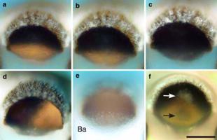
Fig. 7 Optics of the upper lens-eye of Chiropsella bronzie. a–d A point-source casts a distinct shadow onto the retina. The anterior angle of incidence of the point source is 25L in a, 45L in b and 55L in c, the latter angle being just outside the eye’s field of view. In d, the point source is at 25L from the lateral side. e Balloon cells (Ba) are visible as a scattering lump covering the lower part of the aperture of the lens when the eye is viewed 30L from the posterior side of the rhopalium. f A scattering inclusion, or central cataract (white arrow), was regularly observed in lens of the upper lens-eye. Under point source illumination, this inclusion can be seen to cast a faint shadow (black arrow) on the retina. Scale bar 100 lm
present in all jellyfish examined and the occurrence did not seem to be age related.
Unlike the situation in the lower lens-eye, there is no defined vitreous space between the lens and the retina, although cells other than photoreceptors can be found in the area directly underneath the lens (Fig. 6a). The retina of the upper lens-eye consists of only one cell type; photoreceptors, which also contain dark screening pigment confined to a pigment layer. The pigment layer shields the ciliary layer from light entering outside the lens aperture, much in the same way as in the lower lenseye. However, in the medial plane of the rhopalium, at the side closest to the rhopalial stalk, the pigment layer does not extend all the way to the lens, creating a ‘hole’ and consequently exposing part of the retina to direct light (Figs. 1, 6a). Next to this hole, the epithelial cells of the gastric cavity contact the posterior side of the lens (Figs. 1, 6a). The pigment aperture around the lens is fixed, and does not change in response to light as it does in the lower lens-eye. Around the pigment aperture, on the outside of the eye, there are pigment cells containing the same type of white pigment as found in the long pigment cells of the lower lens-eye (Fig. 2a). But in the upper lens-eye, these pigment cells never invade the ciliary layer of the retina, nor do they cover the proximal surface of the retina.
Looking straight into the upper lens-eye, an outward protrusion of the pigment layer can be seen in the centre (Fig. 6b). This protrusion corresponds to the dip seen in the pigment screen at the posterior side of the rhopalium
(Fig. 6a). In the area directly above the upper lens-eye (see Fig. 1a), there is a mass of roughly spherical, large cells with round nuclei (Fig. 6c, d), and in live condition, these cells can be seen to scatter light entering the eye from the posterior side of the rhopalium (Fig. 7e). With light microscopy, these cells stain very lightly, except for the nucleus and abundant large granules (Fig. 6c). TEM reveals that the nucleus of these cells are surrounded by a single layer of densely packed mitochondria, and that the rest of the cytoplasm contains numerous large granules of irregular shape (Fig. 6d). Similar cells have also been found in Carybdea marsupialis (Berger 1898; Conant 1898) and T. cystophora (Skogh et al. 2006). They were referred to as network cells by Conant (1898) and balloon cells by Skogh et al. (2006). We will use the latter nomenclature because it better describes the cells, which have no neural features.
Optics of the upper lens-eye
The rather flat lens, combined with the close proximity of the retina to the lens, suggests that the upper lens-eye does not produce a focused image on the retina. It turned out impossible to isolate the lens of the upper lens-eye in a reasonably undamaged condition, leaving observations of whole intact rhopalia the only direct option for investigating the optical performance of the eye. Luckily, the wide aperture and weak lens, in combination with the short distance between the cornea and retina, allowed us to observe directly how light is distributed on the retina when parallel light is sent into the eye from different directions. When illuminated along the optical axis (from directly above), the entire retina was lit up, indicating that the eye behaves like a lens-less pigment cup, and that the lens is functioning more as a vitreous body than as a focusing element. This is supported by the observation that the damaged lenses we could isolate from upper lens-eyes were soft and appeared to have much lower refractive index than those of the lower lens-eyes.
Shining parallel light into the upper lens-eye from directions of up to 50L from the optical axis created rather sharp shadows of the pigmented aperture on the retina (Fig. 7a–d). As the angle of incidence of the light increased, the percentage of the retina illuminated decreased. At illumination angles exceeding about 50L from the optical axis, the shadow covered the entire retina (Fig. 7c). Extended light sources did not produce obvious images or shadows, and it seems that the upper lens-eye is only capable of informing about the direction to single point sources. Multiple point sources abolished the sharp shadows and would be impossible to distinguish from extended light sources or general ambient light. The sharp shadows from off-axis point-sources were further restricted
123