

Sodium Channels and Neuronal Hyperexcitability
.pdfDENDRITIC Na+ CHANNELS |
63 |
FIG. 1. Dendritic somatofugal glutamate-induced current is increased when soma and proximal dendrite are depolarized. (A) Experimental set-up. (B) Superimposed traces of transmitted current (DI) measured with somatic voltage clamp during one-second dendritic iontophoresis of glutamate. Voltage clamp holding potential at resting potential (776 mV).
(C) Superimposed traces, as in B, at voltage clamp holding potential of 761 mV before (Control) and after application of TTX. (D) Plot of transmitted current (DI) as function of soma voltage clamp holding potential. Arrowheads on abscissa mark resting potential (RP) and ¢ring level (FL). Solid squares are Control and open squares after application of TTX. (E) Current^voltage relationship of baseline soma current before (Control) and after application of TTX. From Schwindt & Crill (1995).
in the glutamate current (Fig. 1D) occurs at the potentials where the persistent TTX-sensitive current is activated. The test solutions contained APV to block voltage-dependent ligand gated current and 2 mM Cs+ to block hyperpolarization activated current (IH). Whereas the glutamate current increased with depolarization (Fig. 1D, dark squares), current evoked by iontophoresis applied to the soma decreased, as expected, when the soma was voltage-clamped to depolarized potentials, closer to the glutamate reversal
64 |
CRILL ET AL |
potential. The somatic voltage-dependent channels cannot contribute to the amplitude because somatic potential is held constant by the voltage clamp.
These results depend upon the soma voltage clamp depolarizing the dendrite between the intracellular electrode at the soma and the iontophoretic site but this region is not isopotential with the soma because the dendrite is an extended cable. Since the transmitted current increases with soma depolarization there must be a voltage-dependent inward current summing with the glutamate current. Otherwise the transmitted current would either decrease with depolarization or show no change at all.
The TTX-sensitive portion of the transmitted current was prolonged indicating a persistent Na+ current in the dendrites which ‘ampli¢es’ the transmitted ligandgated current. The graded increase in the TTX-sensitive transmitted current with dendritic depolarization shown in Fig. 1D indicates that the transmitted current was not caused by a brief TTX-sensitive response evoking a prolonged all-or- nothing Ca2+-mediated action potential. Furthermore, similar results were recorded when MnCl2 replaced Ca2+ in the bathing solution.
Our observations demonstrate that ligand-gated depolarizing currents £owing to the soma are increased by non-inactivating or slowly inactivating dendritic Na+ channels (Alzheimer et al 1993, Stafstrom et al 1985). Note that these voltagedependent dendritic channels need not evoke all-or-nothing dendritic response. All that is required is for the dendritic current^voltage relation to show inward recti¢cation. These biophysical properties of the dendrite e¡ectively shorten the dendritic electrical length of the dendrite increasing the e¡ectiveness of distal dendrites. If the ¢ltering properties of dendrites were changed by synaptic activity the e¡ectiveness of distal synapses would change.
NMDA receptors at the site of iontophoresis contributed to the ampli¢ed transmitted current reaching the soma since part of the voltage-dependent increase in the transmitted current was removed by treatment with APV or MK801.
One can ask whether the ‘ampli¢ed’ glutamate current or synaptic current occurs physiologically since the transmitted current was measured only when the voltage is held constant. During repetitive ¢ring the somatic potential varies and there is good evidence for back-propagated action potentials from the soma into the apical dendrite. This normal physiological activity could a¡ect the current transmitted to the soma. To examine these e¡ects we compared the transmitted current during soma voltage clamp to that measured during repetitive ¢ring.
To estimate e¡ective synaptic current during repetitive ¢ring we used a method developed for spinal motor neurons (Powers et al 1992). Synaptic current and soma-injected current add algebraically. For example, the addition of a steady synaptic current to an injected soma current causes a parallel shift in the frequency^current (f^I) curve. This implies that the faster ¢ring rate caused by
DENDRITIC Na+ CHANNELS |
65 |
synaptic current could not be distinguished from ¢ring evoked by current injected in the soma. Powers et al (1992) have shown that this e¡ective current computed from the f^I curve equals the synaptic current arriving at the soma. This occurs because the spike-generating region is down stream from the soma. Injected current into layer 5 pyramidal neurons causes a parallel shift in the f^I curve (Schwindt & Crill 1996).
In our experiments the f^I relationship of the layer 5 pyramidal cell was measured by injecting constant currents of various strengths into the neuron. We then measured the steady ¢ring rate evoked by dendritic glutamate iontophoresis, which allowed calculation of the e¡ective current depolarizing the soma. The e¡ective glutamate current reaching the soma during repetitive ¢ring was compared with the glutamate current £owing into the soma during voltage clamp (Fig. 1). If the iontophoresed current did not evoke repetitive ¢ring it was added to current injected into the soma to allow calculation of the e¡ective glutamate current by the repetitive ¢ring method.
An example of the results from these experiments in a neocortical layer 5 pyramidal neuron is shown in Fig. 2. Figure 2A shows the response of the neuron to injected soma current alone and Fig. 2B shows the increased ¢ring when added to dendritic glutamate iontophoresis. The instantaneous ¢ring rate is nearly constant (Fig. 2C). The parallel shift of the f^I relationship during iontophoresis is shown in Fig. 2D. In the same cell during soma voltage clamp (Fig. 2E) the e¡ective current during di¡erent levels of soma depolarization (voltage clamp) are plotted (Fig. 2F).
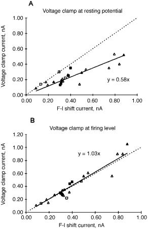
Is the e¡ective glutamate current during repetitive ¢ring similar to the glutamate current measured during voltage clamp of the soma near resting potential when dendritic voltage-dependent conductances are not activated, or is it similar to the transmitted glutamate current near threshold where the attenuation caused by the dendritic cable properties is less? To answer this question the e¡ective glutamate current measured during repetitive ¢ring was compared to the transmitted glutamate current at resting potential and at ¢ring level (Fig. 3). The transmitted glutamate current measured at the ¢ring level corresponds best with the transmitted current measured by repetitive ¢ring.
The repetitive ¢ring experiments show that injected current into the soma causes a parallel shift in the f^I relationship, which supports the concept of a downstream location of the spike-generating site. Secondly these experiments show that the glutamate-injected current evokes repetitive ¢ring in the physiological range. Finally these experiments reveal that the transmitted glutamate current during physiologic repetitive ¢ring is equal to the ‘ampli¢ed’ glutamate current during voltage clamp.
These experiments directly reveal the change in e¡ectiveness of ligand-gated current caused by voltage-dependent dendritic channels. Activation of inward

FIG. 2. Measurement of dendritic glutamate current during repetitive ¢ring and soma voltage clamp. (A) Repetitive ¢ring evoked 0.9 nA current injected into the soma. (B) Superimposed traces dendritic iontophoresis (middle trace) induced depolarization and repetitive ¢ring evoked by same current as in A during iontophoresis. (C) Instantaneous ¢ring during applied soma current (open squares) and during dendritic iontophoresis (closed squares). (D) Iontophoresis of glutamate on the dendrite causes a parallel vertical shift in the frequency (F)^current (I) relationship. (E) Transmitted current (DI in bottom traces) measured at two voltage clamp holding potentials of the soma. Top trace shows glutamate iontophoresis. (F) Transmitted current (DI) as function of soma voltage clamp holding potential. From Schwindt & Crill (1996).
DENDRITIC Na+ CHANNELS |
67 |
FIG. 3. Comparison of transmitted current measured with voltage clamp (ordinate) with current measured by repetitive ¢ring method (abcissa). Each symbol is a di¡erent cell. For perfect agreement the regression line for the points (solid line) would lie on the dashed line. From Schwindt & Crill (1996).
rectifying dendritic current, including a slowly inactivating Na+ current, shortens the electrical length of the dendrite thereby increasing the e¡ectiveness of synaptic input to a particular dendritic site. For example, the uniform excitation of the dendrites by synaptic input will progressively increase the strength of the input and this will occur without the generation of dendritic spikes. Persistent Na+ currents, Ca2+ currents and NMDA synaptic currents all contribute to this increase in synaptic e¡ectiveness. Numerous types of voltage-dependent ion channels have been identi¢ed in the dendrites. At least in the rat neocortical slice
68 |
DISCUSSION |
depolarization causes a net inward recti¢cation rather than voltage-dependent outward currents, which would have the opposite e¡ect. The balance of inward and outward recti¢cation could be modulated by synaptic transmitters and alter the e¡ectiveness of synapses at a speci¢c geometric distance without a change in transmitter release or transmitter receptor properties.
References
Alzheimer C, Schwindt PC, Crill WE 1993 Modal gating of Na+ channels as a mechanism of persistent Na+ currents in pyramidal neurons from rat and cat sensorimotor cortex. J Neurosci 13:660^673
Greene C, Schwindt PC, Crill WE 1994 Properties and ionic mechanisms of metabotropic glutamate receptor-mediated slow afterdepolarization in neocortical neurons. J Neurophysiol 72:693^704
Hu GY, Hvalby O 1992 Glutamate-induced action potentials are preceded by regenerative prepotentials in rat hippocampal pyramidal cells in vitro. Exp Brain Res 88:485^494
Jack JJB, Noble D, Tsien RW 1975 Electric current £ow in excitable cells. Clarendon, Oxford Magee JC, Johnston D 1995 Synaptic activation of voltage-gated channels in the dendrites of
hippocampal pyramidal neurons. Science 268:301^304
Powers RK, Robinson FR, Konodi MA, Binder MD 1992 E¡ective synaptic current can be estimated from measurements of neuronal discharge. J Neurophysiol 68:964^968
Schwindt PC, Crill WE 1995 Ampli¢cation of synaptic current by persistent sodium conductance in apical dendrite of neocortical neurons. J Neurophysiol 74:2220^2224
Schwindt PC, Crill WE 1996 Equivalence of ampli¢ed current £owing from dendrite to soma measured by alteration of repetitive ¢ring and by voltage clamp in layer 5 pyramidal neurons. J Neurophysiol 76:3731^3739
Spruston N, Schiller Y, Stuart G, Sakmann B 1995 Activity-dependent action potential invasion and calcium in£ux into hippocampal CA1 dendrites. Science 268:297^300
Stafstrom CE, Schwindt PC, Chubb MC, Crill WE 1985 Properties of persistent sodium conductance and calcium conductance of layer V neurons from cat sensorimotor cortex in vitro. J Neurophysiol 53:153^170
Stuart GJ, Sakmann B 1994 Active propagation of somatic action potentials into neocortical pyramidal cell dendrites. Nature 367:69^72
DISCUSSION
Spruston: When you do the experiment with the glutamate iontophoresis, the reason that you get additional TTX-sensitive current coming in must be because you have voltage escape in the dendrites. If you had perfect voltage clamp, you wouldn’t see this.
Crill: That’s exactly right.
Spruston: Given that this is the case, how do you know that any small depolarization produced by Na+ channels doesn’t also activate Ca2+ channels? The extra current that you measure could be a mixture of Na+ and other voltageactivated currents that are activated by the subsequent voltage change.
DENDRITIC Na+ CHANNELS |
69 |
Crill: I refer to the response produced by TTX. There is a component of the ampli¢cation that is mediated by NMDA channels, and Ca2+ blockers will knock out part of this. The bulk of the ampli¢cation is TTX-sensitive.
Spruston: How relevant is the depolarization induced by glutamate iontophoresis to those that actually occur with synaptic inputs? How much of the depolarization is occurring in the dendrites and what duration does this have when you do your glutamate iontophoresis, compared to what happens with synaptic stimulation?
Crill: The prolonged plateaus may be relatively unphysiological, although in awake animals there are periods of prolonged ¢ring, which may or may not be due to underlying plateau responses in the dendrites. We have been able to evoke these types of responses by synchronous stimulation of synaptic input to the dendrites. This is still not truly physiological, but it is closer than pouring on glutamate. The reason we picked the plateau is because that was something we can control experimentally and where we could get steady-state ¢ring, so we could make some quantitative measurements. I didn’t point it out, but in the changes in the plateau that you see from evoking at multiple sites, you see similar types of changes occurring in the more transient Ca2+ spikes. We would like to believe that what we say about the plateaus applies also to the more frequently occurring Ca2+ spikes.
Spruston: Going back to my ¢rst question, the main issue I was getting at is that there is still some debate over the question of how common persistent Na+ current is in dendrites of these and other neurons. It is clear that there are Na+ currents. The question is, how much persistent Na+ current in£ux can you get? Given that you showed that there are plateau potentials mediated by Ca2+ currents, it is possible that there is an initial depolarization mediated by Na+ currents which is transient, which subsequently activates a Ca2+ current that is more sustained, producing an unclamped voltage change up in the dendrites. So the TTX-sensitive current may in fact not be a persistent Na+ current, but could be a transient Na+ current giving rise to a depolarization and activating a persistent Ca2+ current. Do you have any evidence that would rule out that scenario?
Crill: We can still see the ampli¢cation in the presence of Ca2+ channel blockers, so that does happen. I would not say it is only Na+ channels, though. I am sure there are some Ca2+ channels: in some imaging studies that I didn’t show, there is signi¢cant Ca2+.
Strichartz: What is the evidence that the passive properties for these very small processes with large surface:volume ratios are changed as a result of Na+ accumulation, a¡ecting for example delayed recti¢er K+ channels or Ca2+-activated K+ channels? In most of the modelling, you assumed that the passive properties were invariant: you just added an active process on top of that.
70 |
DISCUSSION |
Crill: This is Julian Jack’s modelling, not ours. They just assumed that the dendrite has a given current:voltage relationship.
Strichartz: Have you looked for changes in passive properties that would persist after these active properties are over, but during a period when intracellular ions might still be elevated?
Crill: We haven’t done those speci¢c experiments, but it is worth emphasizing that since there is clearly a diversity of channels in the dendrites, there may be signi¢cant variation in the expression and e¡ectiveness of these channels over time. This can markedly shift the electrical closeness of input to the soma and therefore the e¡ectiveness of a given synaptic input. How this happens is a subject for future experiments.
Noebels: I was struck by the resemblance of the linear jump shift transition to a plateau that you saw, and the data that Hodgkin, Stein and others have described for axons (Jack et al 1975). Can intense or persistent depolarization cause not only a spread of the initiation site to properties of proximal dendrites, but also beyond the axon to possibly even the ¢rst node? This would result in spike initiation at the ¢rst node that could back¢re and contribute in some way to the f^I parameters that you see.
Crill: We haven’t asked that question, but I think the evidence against it is that when you inject current into the soma, this isn’t seen at all. We can measure a linear f^I relationship if we inject the current into the soma; it is only when we depolarize the dendrites that we get that saturation of frequency of ¢ring.
Noebels: In some way, perhaps the dendrites are becoming more axon-like, in the sense that many axons have that same f^I behaviour.
Waxman: Some years ago, Mahlon Kriebel, Mike Bennett and I studied oculomotor neurons in ¢sh. They have spike trigger zones in the cell body and in the dendrites, but when they ¢re at high frequency, you can see the spike initiation zone migrating from one site to the other. It is not a ¢xed patch of membrane as you are implying.
Bean: I am curious about the possible involvement of inactivating K+ channels in the jump. Another possible mechanism would be some sort of A-type K+ channels that inactivated with voltage. These could also give you regenerative depolarization. I don’t remember whether A-type K+ channels are present in these dendrites or not.
Spruston: There are two recent papers both showing that these neurons do not have nearly as much IA as hippocampal pyramidal neurons do, for example (Bekkers 2000, Korngreen & Sakmann 2000). They de¢nitely don’t have the strong gradient that is present in CA1 neurons.
Crill: The type of response we get is de¢nitely localized. The Ca2+ goes up wherever you are ionophoresing. Exactly what keeps it localized is unknown. We have done some modelling to try to see whether we could
DENDRITIC Na+ CHANNELS |
71 |
reproduce the behaviour, but this has been unsuccessful. However, it clearly exits experimentally.
Spruston: In CA1 neurons, something analogous happens. If you have a train of spikes that are paired with current injection into the dendrites, you can get Ca2+ spikes. Here, it seems that a crucial role is played by the D-type K+ currents. These D-type K+ currents seem to limit the generation of Ca2+ spikes near the soma, and these currents are activated by the action potentials themselves. So if you block either the D-type K+ currents or the fast Na+ spikes, this dramatically lowers the threshold for Ca2+ spikes in the soma of CA1 pyramidal neurons. It is possible that something similar is happening in the layer 5 neurons (Golding et al 1999). Matthew Larkum has published a very nice paper in which he showed that if you pair back-propagating action potentials with synaptic input in the layer 5 neurons, then you get generation of Ca2+ spikes in the dendrites of the layer 5 cells (Larkum et al 1999). Probably what is also happening with the jump is that when you get high frequency spike trains paired with glutamate ionophoresis, it generates a plateau potential in the dendrite.
Crill: I want to make it clear that although I referred to these as Ca2+ spikes, there are probably other inward currents involved. In every cell that we have examined we could block the plateau of response under Ca2+ spikes with cadmium, but in a few cells we could also block it with NMDA blockers or TTX. There seems to be some variation, but primarily the charge carrier seems to be Ca2+.
References
Bekkers JM 2000 Distribution and activation of voltage-gated potassium channels in cellattached and outside-out patches from large layer 5 cortical pyramidal neurons of the rat. J Physiol 525:611^620
Golding NL, Jung HY, Mickus T, Spruston N 1999 Dendritic calcium spike initiation and repolarization are controlled by distinct potassium channel subtypes in CA1 pyramidal neurons. J Neurosci 19:8789^8798
Jack JJB, Noble D, Tsien RW 1975 Electric current £ow in excitable cells. Clarendon, Oxford Korngreen A, Sakmann B 2000 Voltage gated K+ channels in layer 5 neocortical pyramidal
neurones from young rats: subtypes and gradients. J Physiol 525:621^639
Larkum ME, Zhu JJ, Sakmann B 1999 A new cellular mechanism for coupling inputs arriving at di¡erent cortical layers. Nature 398:338^341
Sodium Channels and Neuronal Hyperexcitability.
Novartis 241
Copyright & 2002 JohnWiley & Sons Ltd
Print ISBN 0-471-48530-6 Online ISBN 0-470-84668-2
Mutations of voltage-gated sodium channels in movement disorders and epilepsy
Miriam H. Meisler, Jennifer A. Kearney, Leslie K. Sprunger, Bryan T. MacDonald, David A. Buchner and Andrew Escayg
Department of Human Genetics, University of Michigan, 4909 Buhl Box 0618, Ann Arbor, MI 48109^0618, USA
Abstract. Spontaneous and induced mutations of neuronal Na + channels in human patients and mutant mice result in a broad range of neurological disease. Epilepsy, a disorder of neuronal hyperexcitability, has been associated with delayed inactivation of SCN2A in mice, and with altered kinetics of SCN1A in human patients. Movement disorders including tremor, ataxia, dystonia and paralysis have been observed in mice with mutations of SCN8A. Electrophysiological recordings from neurons isolated from mice with mutations in individual channels reveal the contributions of each channel to in vivo ¢ring patterns. In addition to monogenic disease, Na+ channel mutations are likely to contribute to polygenic disease susceptibility and to normal variation in neuronal function. Advances in molecular methods coupled with genomic sequences from the Human Genome Project will permit identi¢cation of many new patient mutations and generation of animal models to dissect their physiological and cellular consequences.
2002 Sodium channels and neuronal hyperexcitability. Wiley, Chichester (Novartis Foundation Symposium 241) p 72^86
Other papers in this volume address the biophysical properties of individual voltage-gated Na + channels. In vivo Na + currents in neurons are the result of the combined activity of multiple Na + channels, as well as K + channels and other functionally interacting proteins. The contributions of individual channels in vivo can be inferred by analysis of mice with mutations in a single Na + channel. The neurological abnormalities in the mutant animal provide insight into the role of the mutated channel in genetic disorders, and electrophysiological recordings from brain sections or single cells can detect the e¡ects of the mutations on sodium currents in speci¢c types of neurons. In this chapter we describe neurological and cellular abnormalities caused by mutations of three neuronal Na + channel genes,
SCN1A, SCN2A and SCN8A.
72