

Sodium Channels and Neuronal Hyperexcitability
.pdfNa+ CHANNELS, BLOCKERS, AND NEUROPATHIC PAIN |
203 |
persistent relief of pain that you see with lidocaine and not with mexiletene? Or do you think this re£ects an action on a completely di¡erent target?
Strichartz: Persistent relief means that one week later you still have relief, and all the lidocaine would have cleared from the animal by then.
Bean: I was thinking that there might be di¡erent actions on the Na+ channels while the drug is there.
Strichartz: If you look at the Na + currents in di¡erent tissues with lidocaine and mexiletene, there is nothing obviously di¡erent. The potencies are a little di¡erent, as are the pKas. The octanol:bu¡er partition coe⁄cients, which people usually equate with lipophilicities, are roughly the same. We think the two molecules have a totally di¡erent mechanism.
Bean: Have you any idea what the target is?
Strichartz: No.
Meisler: Are there any good data on the half-lives of the channel proteins in vivo?
Strichartz: I don’t think so. In cultured cells it is a matter of days. If glycosylation is prevented it is hours.
Catterall: We did experiments in cultured primary neurons from rat brain. The Na + channels turn over with a half-life of about 48 h in pulse^chase experiments. But I don’t imagine that a myelinated neuron that runs all the way down to your foot turns over its Na + channels in 48 h.
Strichartz: Although there is this large pool of a subunits that are present inside nodes, are they just sitting there waiting for something unusual to happen? This seems to be a great expenditure of biosynthesis for nothing.
Catterall: We also found in the experiments with the cultured neurons that there is a large intracellular pool of Na + channels that are incompletely assembled with b subunits. Na+ channels must be more stable in mature nerves.
Waxman: When we looked at STX binding in Schwann cells, 50% of the binding sites were not on the membrane but in the intracellular compartment.
Bevan: You have shown the behavioural data from rats and then switched to the frog peripheral nerve, making the connection that the e¡ects you are seeing in vivo are in fact mediated by e¡ects on peripheral nerve. But I think that there is an argument that for some local anaesthetics, some of the anti-nociception e¡ects are centrally mediated.
Strichartz: I don’t think we know whether they are acting on the peripheral nerve or not. We were testing the hypothesis that abnormal activity, which has been reported from peripheral nerve in a number of di¡erent procedures, might be the substrate for local anaesthetics. But you are right: there has been a fair amount of work in spinal cord looking at a number of di¡erent synaptic properties (both peptidergic transmission and glycine/glutamate transmission) showing that low concentrations of local anaesthetic are able to alter transmission.
204 |
DISCUSSION |
Spruston: One thing I noticed is that you didn’t show any examples of bursting, even in the presence of the scorpion toxins. Do you ever see that? I know some people have reported action potential bursting in DRG neurons (White et al 1989).
Strichartz: It could be there. We are looking at the measured discharge from about 103 ¢bres. Although they are in synchrony right after the initial spike, they could go into a burst mode later on. If they were asynchronous at that time, you wouldn’t be able to see it, so you would have to tease out a small ¢bre to look for this. We have looked in peripheral nerve at single units in an anaesthetized rat in vivo, but we have never seen a peripheral sensory ¢bre that showed any burst activity.
Horn: What causes those oscillations that you are seeing?
Strichartz: The a toxin concentrations we use would modify about 10% of the Na + channels in the nerve. We know this from our other voltage clamp studies. So 85^90% of the channels are normal. After a spike, the non-inactivating channels provide su⁄cient current to maintain this plateau. This is at a potential that is su⁄cient to activate the normal channels, which have closed after the spike and will open again. If you use a higher concentration of toxin, as I showed with 30 nM Leiurus toxin, you get a larger plateau, but there are no oscillations there. Our argument is that these large depolarizations have inactivated a substantial fraction of toxin-free channels, now about a third of the total channels.
Horn: This is a compound action potential, so the oscillation frequency is the same in a lot of the ¢bres.
Strichartz: Yes, and we are using a stimulus which selectively activates the largest myelinated ¢bres. We don’t get slower ¢bres whose later pulses might interfere with the signal that we can measure.
Bean: Do you know if you get oscillations and synchronized ¢ring in the allodynia model? Can you take nerves from those animals and see the oscillatory activity?
Strichartz: People have reported spontaneous discharges in Ab ¢bres from rats treated this way. I don’t remember what the frequency of ¢ring is. I think it is between 50 and 100 Hz.
Gold: What Marshall Devor’s group has reported is basically three patterns of activity. One they call tonic ¢rers, another contains bursters with fairly large interburst intervals and the third are irregular ¢rers. The proportions of these di¡erent ¢ring types apparently change throughout the duration of the injury. You start out relatively early on with more of the tonic ¢ring, and then at later dates you move to more phasic and irregular ¢ring.
Strichartz: Aren’t those data from neuromas?
Gold: No, these are recent data from the SNL model.
Bean: Is it known whether this is originating from the cell bodies or the middle of the axon, for example?
Na+ CHANNELS, BLOCKERS, AND NEUROPATHIC PAIN |
205 |
Gold: This is dorsal root recording. The assumption is that it is somewhere around the ganglia. If you cut proximal to the ganglia you eliminate the activity.
Strichartz:Marshall Devor and colleagues studied the discharges in the rat sciatic nerve after neuroma formation, where they sectioned the nerve and then put a cap over the end, so it grows back into some tangled mess (Devor et al 1992). Neuromas have their own pathophysiology, and he gave intravenous bolus injections of lidocaine, but didn’t measure concentrations. He showed that the activity he could record near the ganglion was the most susceptible (i.e. it disappeared ¢rst), activity at the site of injury was next and then activity recorded in branches that were heading centrally was the least susceptible. Whether this was just because of kinetic distribution of the drug is unknown. We are planning to do similar experiments with constant infusions of lidocaine to di¡erent levels.
Gold: Is it known whether mexiletene blocks the Ca2 + ATPase?
Strichartz: I don’t know.
References
Devor M, Wall P, Catalan N 1992 Systemic lidocaine silences ectopic neuroma and DRG discharge without blocking nerve conduction. Pain 48:261^268
White G, Lovinger DM, Weight FF 1989 Transient low-threshold Ca2 + current triggers burst ¢ring through an afterdepolarizing potential in an adult mammalian neuron. Proc Natl Acad Sci USA 86:6802^6806
Sodium Channels and Neuronal Hyperexcitability.
Novartis 241
Copyright & 2002 JohnWiley & Sons Ltd
Print ISBN 0-471-48530-6 Online ISBN 0-470-84668-2
Molecular mechanisms of gating and drug block of sodium channels
William A. Catterall
Department of Pharmacology, University of Washington, Box 357280, Seattle, WA 98195-7280
Abstract. Voltage-gated Na+ channels are composed of an a subunit of 260 kDa associated with b subunits of 33^36 kDa. a subunits have four homologous domains (I to IV) containing six transmembrane a helices (S1^S6). The S4 segments serve as voltage sensors and move outward to initiate activation. The S5 and S6 segments and the short membrane-associated loops between them form the pore. Fast inactivation is mediated by closure of an inactivation gate formed by the intracellular loop between domains III and IV. The 3-D structure of the inactivation gate has been determined by NMR spectroscopy, revealing the conformation of the pore-blocking IFM motif. Peptide scorpion toxins that alter gating of Na+ channels bind to the extracellular ends of the IIS4 and IVS4 segments, trap them in either an activated or non-activated position, and thereby selectively alter channel activation or inactivation. Voltage sensor-trapping may be a general mechanism of toxin action on voltage-gated ion channels. Local anaesthetics block the pore of Na+ channels by binding to a receptor site in segment S6 in domains III and IV. Anticonvulsants and antiarrhythmic drugs also interact with this site. A high- a⁄nity Na+ channel blocker has recently been developed with this site as its target. The emerging knowledge of the molecular mechanisms of Na+ channel gating and drug block may allow development of novel therapeutics for epilepsy, cardiac arrhythmia and persistent pain syndromes.
2002 Sodium channels and neuronal hyperexcitability. Wiley, Chichester (Novartis Foundation Symposium 241) p 206^225
Na+ channels from mammalian brain are complexes of a (260 kDa), b1 (36 kDa) and b2 (33 kDa) subunits (Hartshorne & Catterall 1981, Hartshorne et al 1982). The Na+ channel a subunit was ¢rst cloned from electric eel electroplax (Noda et al 1984), but successful functional expression of the Na+ channel required cloning cDNAs from rat brain. RNA encoding the a subunit proved su⁄cient for functional expression of Na+ currents in Xenopus oocytes (Noda et al 1986a, 1986b, Goldin et al 1986), but the b subunits are required for normal kinetics and voltage dependence of gating (Isom et al 1992, 1995).
Application of molecular modelling to the Na+ channel (Guy & Seetharamulu 1986) predicted six a-helical transmembrane segments (S1^S6) in each of the four
206
MOLECULAR MECHANISMS OF GATING AND DRUG BLOCK |
207 |
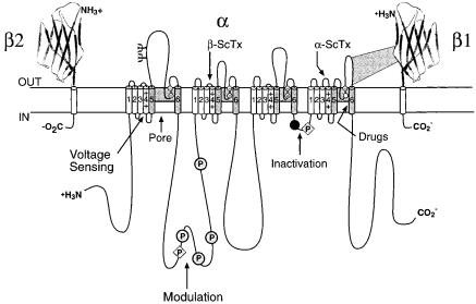
FIG. 1. Subunit structure of the voltage-gated Na+ channels. The primary structures of the subunits of the voltage-gated ion channels are illustrated as transmembrane folding diagrams. Cylinders represent probable a helical segments. Bold lines represent the polypeptide chains of each subunit with length approximately proportional to the number of amino acid residues in the brain Na+ channel subtypes. The extracellular domains of the b1 and b2 subunits are shown as immunoglobulin-like folds. C, sites of probable N-linked glycosylation; P in shaded circles, sites of demonstrated protein phosphorylation by PKA (circles) and PKC (diamonds); shaded S5 and S6, pore-lining segments; clear circles, the outer (EEEE) and inner (DEKA) rings of amino acid residues that form the ion selectivity ¢lter and the tetrodotoxin binding site; shaded, S4 segments, voltage sensors; dark circle, inactivation particle in the inactivation gate loop. Sites of binding of a and b scorpion toxins and a site of interaction between a and b1 subunits are also shown.
homologous domains (I^IV) and a re-entrant loop that dipped into the transmembrane region of the protein between transmembrane segments S5 and S6 and formed the outer pore (Fig. 1). Relatively large extracellular loops were predicted in each homologous domain, connecting either the S5 or S6 transmembrane segments to the membrane-reentrant loop. Even larger intracellular loops were predicted to connect the four homologous domains, and large N-terminal and C-terminal domains were also predicted to be intracellular. Subsequent work on Na+, Ca2+ and K+ channels is consistent with the general features of this early model.
Comparison of the primary structures of the auxiliary b1 and b2 subunits to those of other proteins revealed a clear structural relationship to the family of proteins that contain immunoglobulin-like folds (Isom et al 1995). The extracellular domains of the b1 and b2 subunits are predicted to fold in a similar
208 |
CATTERALL |
manner as myelin protein P0, whose structure is known (Fig. 1; Shapiro et al 1996). The b1 and b2 subunits of the Na+ channel appear to have dual functions . modulation of channel gating and cell^cell interaction. The e¡ect of the b1 subunit on Na+ channel activation and inactivation is mediated by the immunoglobulin-like fold in the extracellular domain (McCormick et al 1998), which is su⁄cient to modulate channel gating when attached to an unrelated transmembrane segment or to a glycophospholipid anchor without the transmembrane and intracellular domains (McCormick et al 1999). Analysis of two di¡erent sets of channel chimeras points to the loop on the extracellular side of transmembrane segment IVS6 as one important point of interaction of the b1 subunit (Fig. 1, Makita et al 1996, Qu et al 1999). Interactions with this extracellular loop serve to modulate channel activation and coupling to fast inactivation via an unknown mechanism.
The structural similarity of the b subunits to cell adhesion molecules suggests that they perform similar functions, and direct experimental support for this idea has come from recent experiments on interaction of the b subunits with extracellular proteins. Na+ channels, and the b2 subunit, bind to the extracellular matrix proteins tenascin C and tenascin R (Srinivasan et al 1998). Transfected cells expressing Na+ channel subunits are repelled by surfaces coated with tenascin R, as though interaction with this extracellular protein is a repellent signal to migrate away from the interacting surface (Xiao et al 1999, Isom 2002, this volume). These interactions may guide the formation of specialized areas of high Na+ channel density such as nodes of Ranvier and axon initial segments and may stabilize the high density of Na+ channels in these locations.
Voltage-dependent activation and voltage-sensor trapping by b scorpion toxins
The voltage dependence of activation of the Na+ channel and other voltage-gated ion channels derives from the outward movement of gating charges in response to changes in the membrane electric ¢eld (Armstrong 1981, Horn 2002, this volume). Approximately 12 electronic charges in the Na+ channel protein move across the membrane electric ¢eld during activation (Hirschberg et al 1995). The novel features of the primary structure of the Na+ channel a subunit led directly to hypotheses for the molecular basis of voltage-dependent gating (Guy & Seetharamulu 1986, Catterall 1986). The S4 transmembrane segments contain repeated motifs of a positively charged amino acid residue followed by two hydrophobic residues, potentially creating a cylindrical a helix with a spiral ribbon of positive charge around it. The negative internal transmembrane electrical ¢eld would exert a strong force on these positive charges arrayed across the plasma membrane, pulling them into the cell in a cocked position.
MOLECULAR MECHANISMS OF GATING AND DRUG BLOCK |
209 |
Depolarization of the membrane is proposed to release the S4 segments to move outward along a spiral path, initiating a conformational change that opens the pore. The proposed outward movement of the S4 segments of Na+ channels has been directly measured using Na+ channel mutants with cysteine residues substituted for positively charged amino acid residues (Yang & Horn 1995, Yang et al 1996, Horn 2002, this volume). Remarkably, these experiments showed that three positively charged amino acid residues in the S4 segment of domain IV become accessible outside the cell during channel gating.
Our recent experiments show that the outward gating movement of the S4 segments during channel activation is also a target for neurotoxin action. b scorpion toxins enhance Na+ channel activation by shifting its voltage dependence to more negative membrane potentials. We analysed toxin binding and action on a family of chimeric Na+ channels in which one of the extracellular loops of the brain Na+ channel was replaced with its counterpart from the toxininsensitive cardiac Na+ channel (Ceste'le et al 1998). The results show that b scorpion toxins bind to a receptor site including the S3^S4 extracellular loop near the extracellular end of the IIS4 transmembrane segment (Ceste'le et al 1998). Toxin binding alone has no e¡ect on activation, but when the channel is activated by depolarization, the bound toxin enhances activation by negatively shifting its voltage dependence (Fig. 2). Once activated, the presence of the toxin greatly slows the deactivation of the channel, creating large tail currents. This e¡ect is thought to be mediated by trapping the activated S4 voltage sensor in its outward, activated position by binding of the S4 segment to the toxin sitting in its receptor site on the extracellular surface of the channel protein (Fig. 2; Ceste'le et al 1998). A prediction of this model is that mutations that allow the IIS4 segment to move outward more easily would enhance toxin action. We found that mutants in which the positive gating charges in the IIS4 segment were neutralized by substitution of glutamine or cysteine for arginine were much more sensitive to enhanced activation by b scorpion toxins. In these mutants toxin binding alone is su⁄cient for voltage sensor trapping without prior activation of the channel. Voltage-sensor trapping appears to be a widespread mechanism through which channel gating is altered by polypeptide neurotoxins, including the e¡ects of a scorpion toxins on Na+ channel inactivation described below (Rogers et al 1996).
Structural basis for Na+ channel inactivation
Na+ channels inactivate within a few milliseconds of opening. Based on its sensitivity to proteases perfused inside the squid giant axon, fast inactivation was thought to be mediated by an intracellular gate that binds to the intracellular mouth of the pore (Armstrong 1981). Site-directed anti-peptide antibodies against the short, highly conserved intracellular loop connecting domains III and
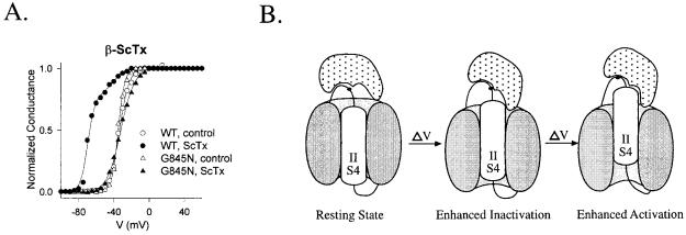
FIG. 2. Voltage sensor trapping by b scorpion toxin. (A) E¡ect of b scorpion toxin on activation of wild-type and mutant Na+ channels. Na+ currents were measured at the indicated test pulse potentials by whole-cell voltage clamp in tsA-201 cells transfected with wild-type Nav1.2 channels or mutant G845N (Ceste'le et al 1998). Where indicated by ScTx, the b scorpion toxin CssIV was present at a saturating concentration (Ceste'le et al 1998). (B) Voltage sensor trapping model for b scorpion toxin action. Domain II and its S4 voltage sensor are illustrated at a negative resting membrane potential (e.g. 7120 mV), a more depolarized resting membrane potential (e.g. 770 mV), and a fully depolarized membrane potential (e.g. +40 mV).
MOLECULAR MECHANISMS OF GATING AND DRUG BLOCK |
211 |
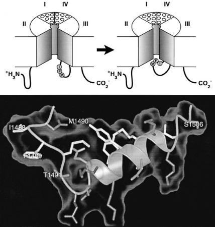
IV of the Na+ channel a subunit (Fig. 1) prevent fast Na+ channel inactivation (Vassilev et al 1989, 1988). Moreover, the accessibility of this site for antibody binding was reduced when the membrane was depolarized to induce inactivation, suggesting that the loop connecting domains III and IV forms an inactivation gate which folds into the channel structure during inactivation (Vassilev et al 1989, 1988). Cutting the loop between domains III and IV by expression of the Na+ channel in two pieces greatly slows inactivation (Stuhmer et al 1989). Mutagenesis studies of this region revealed a hydrophobic triad of isoleucine, phenylalanine and methionine (IFM) that is critical for fast inactivation (Fig. 1, dark circle; West et al 1992), and peptides containing this motif can serve as pore blockers and can restore inactivation to Na+ channels having a mutated inactivation gate (Eaholtz et al 1994). These results support a model in which the IFM motif serves as a tethered pore blocker that binds to a receptor in the intracellular mouth of the pore (Fig. 3A). Inactivation is impaired in proportion to the hydrophilicity of amino acid substitutions for the key phenylalanine residue (F1489), suggesting that it enters into a hydrophobic interaction with an inactivation gate receptor during inactivation (Kellenberger et al 1997). Voltagedependent movement of the inactivation gate has been detected by measuring the accessibility of a cysteine residue substituted for F1489 (Kellenberger et al 1996). This substituted cysteine residue becomes inaccessible to reaction with sulfhydryl reagents as the inactivation gate closes.
The 3-D structure of the central portion of the inactivation gate has been determined by expression as a separate peptide and analysis by multi-dimensional NMR methods (Rohl et al 1999). These experiments reveal a rigid a helix £anked on its N-terminal side by two turns, the second of which contains the IFM motif (Fig. 3B). In this position, F1489 is poised to serve as a tethered ligand that occludes the pore. The nearby threonine (T1491), which is an important residue for inactivation (Kellenberger et al 1997), is also in a position to interact with the inactivation gate receptor in the pore. In contrast, the methionine of the IFM motif (M1490) is buried in the core of the peptide, interacting with two tyrosine residues in the a helix. This hydrophobic interaction stabilizes the fold of the peptide and forces F1489 into its exposed position. The structure of the inactivation gate peptide in solution suggests that the rigid a helix serves as a sca¡old to present the IFM motif and T1491 to a receptor in the mouth of the pore as the gate closes.
Coupling of activation to inactivation
Na+ channel inactivation derives most or all of its voltage dependence from coupling to the activation process driven by transmembrane movements of the S4 voltage sensors (Armstrong 1981). Increasingly strong evidence implicates the S4 segment in domain IV in this process. Mutations of charged amino acid
212 |
CATTERALL |
FIG. 3. Mechanism of inactivation of Na+ channels. (A) The hinged-lid mechanism of Na+ channel inactivation is illustrated. The intracellular loop connecting domains III and IV of the Na+ channel is depicted as forming a hinged lid. The critical residue phenylalanine 1489 (F) is shown occluding the intracellular mouth of the pore in the Na+ channel during the inactivation process. (B) 3D structure of the central segment of the inactivation gate as determined by multidimensional NMR (Rohl et al 1999). Isoleucine 1488, phenylalanine 1489, and methionine 1490 (IFM) are illustrated. Threonine 1491, which is important for inactivation, and serine 1506, which is a site of phosphorylation and modulation by protein kinase C, are also indicated.
residues at the extracellular end of the IVS4 segment have strong and selective e¡ects on inactivation (Chen et al 1996). Toxins that slow coupling of activation to inactivation bind to a receptor site on the extracellular side of the IVS4 segment (Rogers et al 1996). The IIIS4 and IVS4 segments, detected by covalently incorporated £uorescent probes, are speci¢cally immobilized in the outward