

Sodium Channels and Neuronal Hyperexcitability
.pdfNa+ CHANNEL MUTATIONS AND NEUROLOGICAL DISEASE |
83 |
Meisler: Yes, it appears that 50% of the Na + channels are su⁄cient for normal function.
Ptacek: Homozygous would almost certainly be lethal. However, SCN4A, for example, may be a candidate for the cause of a muscle disorder such as congenital ¢bre type disproportion, where there are fewer fast twitch ¢bres. One hypothesis might be that loss of a single allele causing 50% protein loss might lead to a problem of muscle contraction only during maximal e¡ort.
Meisler: It doesn’t seem to be the case in the mice we have looked at: the heterozygotes appear normal.
Ptacek: It would also be interesting to look in the mice at seizure thresholds with proconvulsants. Again, you can imagine a heterozygous loss of function could give a predisposing factor in the human population that you would never see in homozygosity.
Meisler: That would be interesting to look at. My guess would be that you wouldn’t see it, because of the cytoplasmic pool of excess channels. It doesn’t appear that channel production is rate-limiting for these channels. Haploinsu⁄ciency in general only a¡ects fewer than 5% of genes. These are usually structural genes such as collagen. In most cases, 50% is adequate for normal function. However, a loss of function allele of SCN1A was recently found in patients with Severe Myoclonic Epilepsy of Infancy (Claes et al 2001).
Ptacek: Again, when you start moving from Mendelian genetics into susceptbility or predisposition, these would be really attractive alleles to use as candidates.
Bean: Have you looked at pain sensing in these channel mutants? Meisler: No. That would be very interesting.
Segal: Have you looked at the ability to treat the seizures in these mutants with Na + channel acting drugs? This could give us clues about whether the seizures are due to the increase in Na + channels in the present time or whether they are due to some past e¡ect such as kindling that is giving some other type of seizure.
Meisler: That would be one good use for the SCN2A mutant. We haven’t done this yet.
Strichartz: One of the problems with that approach is that you can have a basic pathology whose origins are quite di¡erent than the Na+ channels, but the hyperactivity manifests through the Na+ channels. Suppressing that hyperactivity does not inform you about the basic mechanism of the lesion.
Meisler: What if you are starting with a mouse in which you know the basic lesion is in a Na + channel?
Strichartz: Then I don’t understand the reason for using the pharmacological approach.
Segal: A failure to treat the seizures using Na + channel anticonvulsants would suggest that the seizures were being expressed in a secondary fashion, for example resulting from kindling or neuronal death.
84 |
DISCUSSION |
Noebels: It is curious that the seizures don’t start until the mice are about three months old, but the abnormal currents can be seen much earlier. Is it really because of the delayed current (which you have obviously changed), or does this cause other downstream changes in the nervous system which ultimately make it pathological? We don’t know.
Goldin: The seizures are clearly progressive. Something is getting worse with time, and it is not the channel properties. Cells are dying, and as cells are killed o¡ in the hippocampus, things are getting worse.
Noebels: Is it actually neuronal cell loss that causes seizures in the epileptic brain? Meisler: We can see neuronal cell loss.
Waxman: Why are the cells dying? Are they bursting and burning themselves out, or is there a persistent Na + current letting a Na+ in£ux drive reverse Na + / Ca2 + exchange? Or is it that these animals are becoming transiently hypoxic during seizures and sustaining damage because of that?
Meisler: The damage seems to be localized.
Cummins: It could be hypoxia: the hippocampus is one of the regions that does get hit fairly early by hypoxia.
Strichartz: Is it clear that the cells that express the mutation are the ones that are dying? Are they releasing high concentrations of glutamate locally and killing the cells that are responding?
Meisler: We expect that all of the cells are expressing this construct. It is the NSE promoter for the neural-speci¢c enolase gene. However, we haven’t done histology to demonstrate pan-neuronal expression, because the level of expression is so low it is hard to detect.
Ptacek: It would be interesting to make that mutant under an inducible promoter, because then just by £ipping it on at di¡erent times or in di¡erent locations you could address the question of temporal and spatial e¡ects.
Goldin: We could turn it on at a later time and avoid the issue of developmental changes.
Ptacek: If you were to turn it on at 8 weeks and see seizures immediately, this would suggest that it is not a degenerative disease.
Goldin: It would also allow us to determine how long it takes. NSE is turning on during the ¢rst two or three weeks after birth, and the phenotype is starting after that. It is di⁄cult to know exactly what is going on.
Segal: It would also be helpful if you could target the Na + channel abnormality to certain neuronal populations.
Meisler: We certainly could. Which would you suggest?
Segal: The hippocampus would be of interest because of its sensitivity to epileptiform activity and neuronal death. Perhaps if you lack the Na+ channel abnormality in the hippocampus, then you won’t go on to develop a lot of the
Na+ CHANNEL MUTATIONS AND NEUROLOGICAL DISEASE |
85 |
secondary types of seizures. It would be interesting to turn on or o¡ the Na + channel mutations in CA1 or CA3 pyramidal neurons.
Cummins: I have a general comment about the neuronal disease mutations. Louis Ptacek identi¢ed one of the hyperkalaemic periodic paralysis mutations (T704M) back in the early 1990s and the ¢rst three studies that looked at the T704M mutation found rather subtle defects (Cannon & Strittmatter 1993, Cummins et al 1993, Yang et al 1994). But the T704M mutation actually has a major e¡ect on slow inactivation (Cummins & Sigworth 1996), and none of us caught that in the ¢rst three studies. It could be that the neuronal mutations that Miriam Meisler has described a¡ect some property that hasn’t been looked at yet. There are a lot of di¡erent properties to look at, and therefore it is going to be complex to work out exactly how a given mutation causes a disease.
Goldin: The human Nav1.1 domain 2 mutant that Miriam found in GEFS + doesn’t show any obvious changes. We are sure the changes are there; we just haven’t looked at the right property yet.
Horn: There are a ¢nite number of properties of the Na + channels (at least those that we know about at the moment), which wouldn’t be too hard to put into a screen. You can see activation, fast inactivation, kinetics and steady-state of gating and slow inactivation. Those protocols are not so di⁄cult to do on an individual cell. The slow inactivation ones are tedious to do, however!
Strichartz: I would argue that current clamp, following excitability and action potentials driven by short and long current injections, is a more sensitive assay than voltage clamp. As few as 2% of the channels can be non-inactivating, and give remarkable changes in excitability patterns. It would be very hard to pick this up under voltage clamp.
Horn: But the results would be uninterpretable.
Strichartz: You pick up the abnormality, and then you go hunting for those 2%. What fraction of the Na + channels in the hyperkalaemic mutant are actually non-inactivating? It is just 3^8%. Had you not seen a behavioural mutant, you wouldn’t have picked it up: it would have been written o¡ as biological variation.
Cummins: Even the 3^8% estimate for non-inactivating channels is still considered controversial. Steve Cannon has proposed that mechanism for hyperkalaemic periodic paralysis, but it has not been proven in muscle cells. 10 years following Louis Ptacek’s discovery of the ¢rst hyperkalaemic periodic paralysis mutation, we are still not sure what the underlying mechanism is. I would argue that it is not due to a non-inactivating current, whereas Steve Cannon might argue that it is. We need to get these mutants back into the native cells and do a current clamp, which we can’t do in oocytes or HEK293 cells.
Meisler: This is where the mutant mice will be useful, as a source of neurons for electrophysiology. Also, I think there will be mutations in the loops and in
86 |
DISCUSSION |
the C-terminus and N-terminus which may not a¡ect the kinetics but do change cell biology. If you have a candidate human mutation, the only way to determine the functional consequence may be to introduce the mutation into the mouse.
Noebels: If there are modi¢er genes then you are still never sure.
Meisler: If you get an abnormal phenotype in the mouse you can be sure that the mutation is responsible. If you do not see any abnormal behaviour in the mouse, it would still be possible that the mutation had a negative e¡ect in the human cells.
Ptacek: Are you going to be satis¢ed with the transgenic? To really recapitulate the in vivo situation in humans you would have to use homologous recombination to introduce the mutation into the native gene.
Meisler: Introduction of the mutation into the endogenous gene by targeting in ES cells does produce the best model of a human genetic disease. However, the cost, time and e¡ort involved in ES cell targeting means that only a few mutations can be studied by that method. For mutations with a dominant mode of inheritance in humans, transgenic mice can provide useful and relevant models, much more quickly. It is possible to select transgenic lines in which the level of expression of the transgene is comparable to that of the endogenous gene. The main limitation of transgenes driven by promoters from other genes is that the foreign promoter does not precisely recapitulate the temporal and spatial pattern of expression of the endogenous gene. The use of BAC clones that contain the gene and its regulatory elements to create transgenic lines circumvents this problem. The mutation is introduced into a BAC clone by mutagenesis in bacterial culture, and the BAC DNA is then microinjected into fertilized mouse eggs. Expression of such BAC transgenes is independent of insertion site, and recapitulates the normal expression pattern. This new generation of transgenic mice provides a compromise between the speed of transgenics and the biological relevance of targeted alleles.
References
Cannon SC, Strittmatter SM 1993 Functional expression of sodium channel mutations identi¢ed in families with periodic paralysis. Neuron 10:317^326
Claes L, Del-Favero J, Ceulemans B, Lagae L, Van Broeckhoven C, De Jonghe P 2001 De novo mutations in the sodium-channel gene SCN1A cause severe myoclonic epilepsy of infancy. Am J Hum Genet 68:1327^1332
Cummins TR, Sigworth FJ 1996 Impaired slow inactivation in mutant sodium channels. Biophys J 71:227^236
Cummins TR, Zhou J, Sigworth FJ et al 1993 Functional consequences of a Na+ channel mutation causing hyperkalemic periodic paralysis. Neuron 10:667^678
Yang N, Ji S, Zhou M et al 1994 Sodium channel mutations in paramyotonia congenita exhibit similar biophysical phenotypes in vitro. Proc Natl Acad Sci USA 91:12785^12789
Sodium Channels and Neuronal Hyperexcitability.
Novartis 241
Copyright & 2002 JohnWiley & Sons Ltd
Print ISBN 0-471-48530-6 Online ISBN 0-470-84668-2
Channelopathies: episodic disorders of the nervous system
Louis Ptacek
Howard Hughes Medical Institute, Departments of Neurology and Human Genetics, University of Utah School of Medicine, Eccles Institute of Human Genetics, Building 533, Room 4425, Salt Lake City, UT 84112, USA
Abstract. The ¢eld of channelopathies is a newly recognized group of disorders named after the site of their molecular defects . voltageand ligand-gated ion channels. While voltage-gated ion channel mutants have been recognized for some time in organisms such as Drosophila, the ¢rst channelopathy in humans was reported within the last decade. The recognition of this group of disorders began with the de¢nition of the molecular basis of a group of unusual muscle disorders called the periodic paralysis and non-dystrophic myotonias. Interestingly, this group of muscle disorders share some interesting phenotypic features with a number of seemingly disparate human diseases that involve not only skeletal muscle, but also brain and heart. Some similarities that exist among these di¡erent disorders include their episodic nature, similarities with regard to factors that precipitate attacks, therapeutic agents which can help to treat or prevent attacks, and in some cases, a degenerative component that arises in addition to the episodic attacks. The study of these diseases, along with the recognition of common clinical and pathophysiological themes among these disorders has led to tremendous growth in our understanding of these diseases and the hope of developing better therapies.
2002 Sodium channels and neuronal hyperexcitability. Wiley, Chichester (Novartis Foundation Symposium 241) p 87^108
Ion channel structure
Voltage-gated ion channels are proteins critical for establishment of the resting membrane potential in muscle and the ability of these membranes to generate action potentials. Voltage-gated opening of Na+ channels results in the genesis of ‘all-or-none’ action potentials. These channels close over the course of a few milliseconds. A simultaneous e¡ect of depolarization of the membrane (albeit on a slower time scale) is the opening of voltage-gated K+ channels. Along with the inactivation of the voltage-gated Na+ channels, the movement of positive K+ ions out of the cell through these K+ channels leads to a relatively rapid repolarization of the muscle membranes. Cl7 channels are responsible for a majority of the polarity
87

88 |
PTACEK |
FIG. 1. Voltage-gated K+ channel a subunit. The N-terminus and the C-terminus are intracellular. There are six transmembrane segments. The S4 segments contain positively charged arginine and lysine residues at every third position and are important in voltage sensing. Putative membrane-spanning S5 and S6 segments and the P region linking the two segments line the channel pore. Four subunits come together to form a functional K+ channel.
of resting membranes. The voltage-gated L-type Ca2+ channel in skeletal muscle, also known as the dihydropyridine receptor because dihydropyridines block this channel, allows conductance of Ca2+ into the cell. It is likely that this Ca2+ is important through signalling pathways to e¡ect other downstream changes of the muscle membrane but the Ca2+ conductance itself is not directly important to the depolarization of the muscle membrane. Interestingly, this voltage-gated Ca2+ channel serves as a voltage sensor for excitation^contraction coupling. Depolarization of the muscle membrane leads to changes in the Ca2+ channel protein, which in turn interacts with the ryanodine receptor to open slow release Ca2+ channels in the sarcoplasmic reticulum (SR). It is these slow release channels and the SR Ca2+ stores that lead to elevations in cytosolic Ca2+ that result in the ultimate contraction of muscle.
K+ channels are membrane-bound tetrameric protein complexes. Each subunit is a polypeptide with six putative transmembrane segments (S1^6) (Fig. 1). Four identical subunits may associate to form a homomeric K+ channel. Alternatively, di¡erent subunits may assemble to form a heteromeric K+ channel. Mutagenesis studies have identi¢ed several critical functional domains in K+ channel. The S4 segments with positively charged arginine and lysine residues at every third position appear important in voltage sensing. The P region linking the putative membrane-spanning S5 and S6 contains the K channel signature sequence that is highly conserved and critical for K selectivity of the channel pore.
The voltage dependent Na+ and Ca2+ channels are a large group of homologous genes that are also homologous to the voltage-gated K+ channel genes. Unlike the K+ channel proteins where four subunits must come together to form a homoor hetero-tetrameric functional channel, Na+ and Ca2+ channel a subunits have
CHANNELOPATHIES |
89 |
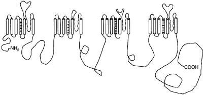
FIG. 2. Diagram of an a subunit of a Na+ channel or a1 subunit of a Ca2+ channel. There are four homologous domains (internally homologous and homologous to the voltage-gated K+ channel), each with six transmembrane segments.
evolved to include in a single transcript four domains (I^IV) in tandem, each with six transmembrane segments (S1^6) homologous to voltage-gated K+ channel genes (Fig. 2). It is thought that this phenomenon resulted from the duplication of a progenitor ‘K+-like’ channel that duplicated and then re-duplicated in the genome. The large, pore-forming a subunit alone is su⁄cient for cation permeability, pharmacologic speci¢city, gating and voltage sensitivity. The auxiliary subunits modulate channel biophysical properties and biosynthesis. Voltage-dependent Na+ channels are composed of a pore-forming, voltagesensing a subunit and a transmembrane b subunit. Voltage-dependent Ca2+ channels are heteromeric complexes composed of a pore-forming a1 subunit, a disul¢de-linked membrane-anchored extracellular a2-d subunit, an intracellular b subunit, and in muscle a g subunit.
Voltage-gated Cl7 channels have been discovered more recently and much less is known about their structure. These proteins have 13 hydrophobic segments but more recent biochemical and immunological data suggest that not all of these traverse the membrane. It appears that voltage-gated Cl7 channels form multimeric functional units but the exact stoichiometry of these channels is not entirely clear. There are no recognized auxiliary subunits of the voltage-gated Cl7 channels at this time.
Periodic paralysis and non-dystrophic myotonia
Clinical manifestations
Periodic paralyses and nondystrophic myotonias include a number of distinct clinical entities as well as some intermediate forms of the various disorders. Myotonia congentia is a group of muscle disorders named for the prominent

90 |
PTACEK |
FIG. 3. Diagram of a subunit of nicotinic acetylcoline receptor channel or inhibitory glycine receptor channel. There are four membrane-spanning segments. These proteins are not homologous to voltage-gated channels.
muscle hyperexcitability or myotonia that is seen in these patients. This myotonia is classical myotonia with the phenomenon of ‘warm up’. These patients experience extreme muscle sti¡ness due to delayed relaxation from repetitive electrical activities in muscle, but this myotonia subsides as their muscles warm up with use. Onset of symptoms is generally in childhood through early adult life. These patients often have hypertrophy of their muscles and a Herculian appearance as a result of their myotonia. Two distinct forms of myotonia congenita are recognized. The ¢rst, named for Julius Thompson who described the disease, is an autosomal dominant form of myotonia congenita (George et al 1994). These patients do not develop degeneration of their muscles even after years of having the disease. An autosomal recessive form of myotonia congenita was described by Becker (1971). These patients have myotonia with ‘warm-up’ phenomenon, may have transient bouts of weakness after periods of disuse, and sometimes develop myopathy as part of their disease.
Hyperkalaemic periodic paralysis is a disorder with myotonia like that seen in the above described disorders. These patients can also have a transition of their muscle membrane hyperexcitability to inexcitability in the form of episodic weakness. This weakness may be so dense as to cause a transient £accid quadraparesis. However, the disorder does not a¡ect the diaphragm and patients are therefore able to continue breathing. First described in 1951 by Tyler (Tyler et al 1951), this disorder is named because of the ability to precipitate attacks in patients by administrating a su⁄cient dose of oral K+. During spontaneous attacks patients may have elevated K+, although this is frequently in the normal range. Attacks of weakness can be precipitated by foods high in K+, rest after vigorous exercise, and with stress and fatigue. The disease is transmitted as an autosomal dominant trait although sporadic cases are sometimes encountered. Percussion and action myotonia are frequently elicited clinically and prominent myotonia can be noted
CHANNELOPATHIES |
91 |
on an electromyographic exam. Patients bene¢t dramatically from treatment with carbonic anhydrase inhibitors.
Paramyotonia congenita is yet another disorder in which myotonia is present. Although the myotonia is somewhat di¡erent as it does not show the classical warm-up phenomenon, but rather is paradoxical. That is, patients frequently have worsening of their myotonia with repeated muscle action. This can be most prominently seen in the obicularis occuli muscles when patients forcefully close their eyes repeatedly and, upon such a manoeuvre, the myotonia of these muscles becomes increasingly severe to the point where patients might have di⁄culty opening their eyes altogether. Of interest, this disorder is a temperature-sensitive mutant of humans. With cooling of their muscles, these patients have worsening of their myotonia and then transition of the hyperexcitability into paralysis. This can be measured quantitatively using electrodiagnostic manoeuvres (Jackson et al 1994). Paramyotonia congenita is transmitted as an autosomal dominant trait, and like hyperkalaemic periodic paralysis, these patients have worsening of their symptoms with stress, fatigue, and rest after vigorous exercise. Patients may be hypo-, normoor hyperkalaemic during attacks. The classical paramyotonia congenita patients are generally hypokalaemic during attacks; those with hyperkalaemia seem to represent a clinical entity somewhere in the spectrum between classical paramyotonia congenita and hyperkalaemic periodic paralysis. Like the hyperkalaemic periodic paralysis patients, these patients bene¢t dramatically from treatment with carbonic anhydrase inhibitors.
K+-activated myotonia is a disorder where patients clinically appear to have myotonia congenita. But their myotonia £uctuates, worsens when K+ is administered, and improves with carbonic anhydrase inhibitors (Trudell et al 1987). Interestingly, these patients do not develop attacks of weakness. This disorder is transmitted as an autosomal dominant trait.
Hypokalaemic periodic paralysis is a disorder of episodic weakness in which a myotonia is not seen. These patients are generally hypokalaemic during an attack and attacks can be precipitated by lowering K+ with administration of glucose and insulin. Furthermore, these attacks can be precipitated by stress, fatigue and rest after vigorous exercise. Dietary precipitants include high carbohydrate meals and salt load. Hyperkalaemic periodic paralysis is transmitted as an autosomal dominant trait although frequent sporadic cases can be seen. Patients K+ levels during hypokalaemia generally remain above 2 mM. K+ levels below 2 mM during an attack of weakness in a sporadic case raises the possibility of thyrotoxic hypokalaemic periodic paralysis, a non-Mendelian form of the disorder which is seen only during periods of thyrotoxicosis (Ptacek 1998). The episodic weakness in patients with hyperkalaemic periodic paralysis, paramyotonia congenita and hypokalaemic periodic paralysis all bene¢t from treatment with the carbonic anhydrase inhibitors diamox and daranide.
92 |
PTACEK |
Genetics
Linkage analysis in large pedigrees with hyperkalaemic periodic paralysis established that the gene for this disorder resided on chromosome 17q (Fontaine et al 1990, Ptacek et al 1991b). Mapping data showed that the paramyotonia congenita and the K+-activated myotonia phenotypes also mapped to the same locus (Ptacek et al 1991c, 1992a). Subsequently, a Na+ channel gene in this region of chromosome 17q was cloned and characterized and shown to be the site of mutations in hyperkalaemic periodic paralysis (Ptacek et al 1991a, Rojas et al 1991), paramyotonia congenita (McClatchey et al 1992, Ptacek et al 1992b), and K+-activated myotonia (Ptacek et al 1994a). The data supporting that this Na+ channel gene, SCN4A was the disease-causing gene included the following:
(1) mutations segregated with the phenotype; (2) they involved highly conserved amino acid residues; (3) these mutations were not found in control individuals; and
(4) some of them occurred as de novo mutations in patients with sporadic disease. A large number of Na+ channel mutations have been identi¢ed in patients with hyperkalaemic periodic paralysis, paramyotonia congenita and K+ aggravated myotonia.
Similar molecular approaches led to mapping of the hypokalaemic periodic paralysis locus to chromosome 1q (Fontaine et al 1994, Ptacek et al 1994b). Subsequently, a voltage-gated Ca2+ channel at this chromosome 1 locus was cloned. This Ca2+ channel gene encodes an L-type Ca2+ channel and three patient-speci¢c mutations have been identi¢ed (Ptacek et al 1994b). The mutations occur at highly conserved residues in the S4 segments of either domain 2 and 4. The domain 4 mutations occur at an analogous position to the most common paramyotonia congenita mutations in the homologous SCN4A Na+ channel (Ptacek et al 1994a).
Genetic linkage analysis in families segregating alleles for both recessive and dominant forms of myotonia congenita showed that both of these phenotypes were linked to a locus on chromosome 7q (Abdalla et al 1992, Koch et al 1992). Subsequent work has identi¢ed a voltage-gated Cl7 channel at this locus, CLCN1, to be the site of defects in myotonia congenita (Abdalla et al 1992, Koch et al 1992). A long list of mutations in this channel gene have since been shown to cause dominant and recessive forms of myotonia congenita (reviewed in Jen & Ptacek 2000). Interestingly, some of these mutations are recognized to cause both dominant and recessive forms (Zhang et al 1996). While it is likely that polymorphisms in the Cl7 channel gene itself may modulate the e¡ect of such mutations, no data is available as yet to substantiate this hypothesis. Many of these Cl7 channel mutants have been expressed in vitro (either in oocytes or by transfection of mammalian cells growing in culture) and the physiologic abnormalities in myotonia congenita are being characterized. Discussion of the