

Sodium Channels and Neuronal Hyperexcitability
.pdfFIG. 1. Autoradiograms of labelled antisense probe hybridized to coronal sections of adult rat brain reveal selective localization of SCNA5A mRNA in limbic regions of rat brain. Strong expression of SCN5A is shown in amygdalar nuclei (left) and piriform cortex (right). (From Hartmann et al 1999.)
114 |
NOEBELS |
result of these defects is to disrupt pacemaking properties in ways that favour sudden ¢brillation of the cardiac syncytium.
SCN5A mutations are likely to contribute to similar instabilities of neuronal rhythmic ¢ring within limbic networks. The presence of veri¢ed epileptic seizures in individuals with LQT mutations (Herman et al 1992, Pacia et al 1994) supports the possibility that a subset of inherited epilepsy may arise from SCN5A channelopathies. Piriform cortex is the most susceptible of all forebrain regions to the induction of seizures, and regulates excitability of the amygdala, a nuclear group with a similar low threshold for epileptogenesis. Persistent depolarization by abnormally prolonged Na+ currents in these neurons would favour epileptiform bursting, synchronous network activation and seizures. Alterations in SCN5A Na + channel inactivation kinetics within the limbic system are also predicted in the previously mentioned human SCN1B mutations with epilepsy. SCN1B subunits interact with SCN5A to markedly increase the amplitude (but not kinetics or voltage dependence) of the Na+ current (Qu et al 1995), and mutations in the SCN5A gene that alter a1^b1 subunit interactions also produce a variant of LQT3. Since the two subunits co-associate, the expression data therefore suggest that SCN5A should be considered along with SCN1B as a candidate Na + channel gene for idiopathic seizure disorders, and that epileptogenic SCN1B mutations may likely reveal a novel a candidate gene for human cardiac arrhythmias, although this phenotype has not yet been described in the GEFS + 1 pedigree.
Ectopic localization of type II channels to hypomyelinated axons
A second major category of ion channelopathy arises from defects in neuronal regulatory pathways that control the expression, location and membrane stabilization of Na + channels. The molecular plasticity of Na + channels that follows injury to the adult axon is well described (see Waxman 2000, for review), but less is understood about inherited defects that trigger Na + channel rearrangements during brain development.
Molecular plasticity of Na + channel gene expression: the dysmyelinating shiverer mouse
One example of Na + channel plasticity in central pathways subsequent to a nonion channel gene deletion is revealed by analysis of the mutant mouse shiverer, which bears a major disruption of the myelin basic protein gene, leading to absence of the glial-speci¢c intracellular membrane protein myelin basic protein (MBP) (Roach et al 1985). Lack of MBP prevents developmental formation of the major dense line essential for the compaction of glial wrappings that form the mature myelin sheath. As a result, large calibre axons in the shiverer brain are
Na+ CHANNELS AND EPILEPSY |
115 |
severely hypomyelinated, with initial apposition of glial^neuronal membranes but a subsequent failure to form compact myelin and white matter in the brain.
As might be predicted from the neurological viability of the phenotype, the profound loss of myelin that should eliminate saltatory conduction is functionally rescued by striking excitability changes in the internodal axon membrane caused by up to 10-fold increases in Na + channel density in these otherwise inexcitable regions of axon membrane. Examination of speci¢c 1:1 binding of [3H]saxitoxin to shiverer brain revealed that the increase of Na + channel density was essentially con¢ned to subcortical pathways containing hypomyelinated sensory (optic nerve), association (corpus callosum and related commissures, the centrum semiovale, and cerebellar corpus medullare), and longer-range projection ¢bres travelling in the internal capsule, hippocampal ¢mbria and fornix, while the density in grey matter regions containing the parent cell perikarya showed little increase (Fig. 2A,B). This pattern indicated that the Na + channel plasticity was targeted ectopically to hypomyelinated regions of central axons.
Additional analysis using subtype-speci¢c antibodies revealed that the ectopic channel expression could be accounted for by increases in type II, but not type I or III Na + channels (Fig. 2B) (Westenbroeck et al 1992).
Interestingly, in the shiverer mutant, the plasticity of Na + channel expression appears to play both salutory and pathogenic roles. While the ectopic expression of internodal channels restores most developmental neurological function (with the exception of a residual cerebellar action tremor), shiverer mutant mice develop behavioural seizures beginning at approximately 3 months of age, culminating in premature death at 4^5 months. The seizure episodes, which occur unprovoked one or more times per day, are stereotyped and consist of sudden arrest of movement and tonic £exor spasms. The animal appears alert during the episodes, which typically last under one minute and terminate abruptly without postictal depression. The mouse promptly regains full locomotor ability, and with exception of the body tremor, appears una¡ected
The mechanism of the seizure discharge in ageing shiverer mice is still unknown. However, it is tempting to speculate that continued accumulation of the ectopic Na + channels lowers the threshold for ephaptic cross-talk between juxtaposed axons within the large hypomyelinated ¢bre tracts, thus creating a basis for aberrant synchronization. However, this seizure activity apparently does not reach the level of the neocortex, since electroencephalogram (EEG) recordings obtained during these episodes display a characteristic electrodecremental pattern without the abnormal synchronous discharges typical of other epileptiform abnormalities. The shiverer lesion thus provides an example of inherited Na + channel-based subcortical excitability changes leading to behavioural seizures.
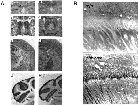
FIG. 2. (A) Selective increase of axonal Na + channels in hypomyelinated brain of the mutant mouse shiverer. Autoradiograms of speci¢c [3H]saxitoxin binding to + / + (a,c,e,g) and shiverer (b,d,f,h) brain sections. Arrows indicate high density regions of Na + ion channel axon expression. (From Noebels et al 1991.) (B) Ectopic axonal Na + channels in shiverer brain are strongly labelled by antibody to Type II channels. Neocortex in + / + (upper) and shiverer (lower). Note dense staining of commissural axon band (seen in cross section at cortical^ subcortical junction) and descending fascicles of hypomyelinated axons in shiverer brain compared to absence of labelling in wild-type nervous system. (From Westenbroek et al 1992.)
Na+ CHANNELS AND EPILEPSY |
117 |
The dysmyelinating human 18q^ syndrome
The channelopathy present in the shiverer mutant may be relevant to understanding epileptogenesis in an inherited human clinical syndrome, 18q^. This mental retardation syndrome is of particular interest since the deleted chromosomal region contains the human orthologue of the myelin basic protein gene, and is accompanied by hypomyelination and characteristic seizures.
White matter changes in this neurological disorder were described only recently (Miller et al 1990, Loevner et al 1996). Both the deleted region (18q21.3^18q22.2 to the q terminus), and the clinical phenotype are highly variable, but epilepsy is present in a substantial fraction of cases, and correlates with the presence and severity of the multifocal dysmyelination (Sturm et al 2000). Video EEG monitoring of a typical seizure in an 18q^ patient with epilepsy and multifocal white matter lesions revealed a brief bilateral asymmetric tonic seizure terminating in 40 seconds. The EEG showed an electrodecremental pattern within the ¢rst second, followed by generalized rhythmical a and y rhythms with no epileptiform spiking. Interestingly, in this case, both the behavioural description and the EEG recorded during these episodes resemble, in important respects, those observed during seizure activity in the shiverer mouse model, since both show tonic contraction seizures with an absence of the high amplitude cortical discharges that typically accompany generalized tonic^clonic convulsive episodes. This pattern is consistent with a subcortical behavioural seizure origin, presumably coinciding with the hyperexcitable axons within the white matter lesions that are likely to express ectopic internodal Na + channels, as seen in the MBP7 shiverer mutant.
Plasticity of Na + channel expression in other CNS demyelinating conditions
It is worth noting that demyelinated axons studied in clinical cases of multiple sclerosis may also display continuous increases of [3H]saxitoxin binding, as seen in shiverer (Moll et al 1991), indicating that similar plasticity of Na + channel expression can occur in some types of this heterogeneous human disorder, where it may contribute to functional neurological recovery following intermittent demyelinating attacks. In addition, multiple sclerosis is often accompanied by tonic seizures that respond to Na + channel blockers such as lidocaine (Sakurai & Kanazawa 1999). Thus dynamic changes in Na + channel biology may be part of the clinical problem, as well as part of the solution at various stages of these diseases, and excessive axonal hyperexcitability with seizures and spasms may represent the price that is paid to restore function to inexcitable ¢bres.
118 |
NOEBELS |
Discussion
While mutation of the Na + channel can probably qualify as one of the most elementary mechanisms of inherited epilepsy, and by that right should be the simplest to understand, signi¢cant complexity underlying the basis for the epileptic phenotype remains to be explored. How does one explain the considerable latency, often measured in years, from birth until seizure onset, and what are the factors that precipitate the recurring synchronous event? The induced excitability changes may also change over time. Seizures themselves up-regulate both a1 and b Na + channel subunit gene expression (Bartholomei et al 1997, Gastaldi et al 1998), and this could provide yet one additional means for changes in synchronization threshold as a seizure disorder progresses. Does the location of mutant gene expression determine where the seizure actually originates, or is local connectivity more important? Do Na + channelopathies, like their Ca2 + channel counterparts (Burgess & Noebels 1999), initiate signi¢cant downstream molecular plasticity in the developing brain? Finally, how many other potential genes for epilepsy manifest their ¢nal hyperexcitability phenotype, or mask it, through downstream alterations in Na + channel gene expression?
Acknowledgements
Supported by NIH 29709 and the Blue Bird Circle Foundation.
References
Alekov A, Rahman MM, Mitrovic N, Lehmann-Horn F, Lerche H 2000 A sodium channel mutation causing epilepsy in man exhibits subtle defects in fast inactivation and activation in vitro. J Physiol 529:533^539
An RH, Wang XL, Kerem B et al 1998 Novel LQT-3 mutation a¡ects Na + channel activity through interactions between a- and b1-subunits. Circ Res 83:141^146
Bartolomei F, Gastaldi M, Massacrier A, Planells R, Nicolas S, Cau P 1997 Changes in the mRNAs encoding subtypes I, II and III sodium channel alpha subunits following kainateinduced seizures in rat brain. J Neurocytol 26:667^678
Black JA, Dib-Hajj S, Cohen S, Hinson AW, Waxman SG 1998 Glial cells have heart: rH1 Na + channel mRNA and protein in spinal cord astrocytes. Glia 23:200^208
Bland BH, Colom LV 1993 Extrinsic and intrinsic properties underlying oscillation and synchrony in limbic cortex. Prog Neurobiol 41:157^208
Burgess DL, Noebels JL 1999 Voltage-dependent calcium channel mutations in neurological disease. Ann N Y Acad Sci 868:199^212
Chen Q, Kirsch GE, Zhang D et al 1998 Genetic basis and molecular mechanism for idiopathic ventricular ¢brillation. Nature 392:293^296
Escayg A, MacDonald BT, Baulac S et al 2000 Mutations of SCN1A, encoding a neuronal sodium channel, in two families with GEFS + 2. Nat Genet 24:343^345
Felts PA, Yokoyama S, Dib-Hajj S, Black JA, Waxman SG 1997 Sodium channel alpha-subunit mRNAs I, II, III, NaG, Na6 and hNE (PN1): di¡erent expression patterns in developing rat nervous system. Brain Res Mol Brain Res 45:71^82
Na+ CHANNELS AND EPILEPSY |
119 |
Gastaldi M, Robaglia-Schlupp A, Massacrier A, Planells R, Cau P 1998 mRNA coding for voltage-gated sodium channel b2 subunit in rat central nervous system: cellular distribution and changes following kainate-induced seizures. Neurosci Lett 249:53^56
Goldin AL 2001 Resurgence of sodium channel research. Annu Rev Physiol 63:871^894 Hartmann HA, Colom LV, Sutherland ML, Noebels JL 1999 Selective localization of cardiac
SCN5A sodium channels in limbic regions of rat brain. Nat Neurosci 2:593^595
Herman LL, Stoshak M, Rittenberry TJ 1992 Long QT syndrome presenting as a seizure. Am J Emerg Med 10:435^458
Isom 2002 b subunits: players in neuronal hyperexcitability? In: Sodium channels and neuronal hyperexcitability. Wiley, Chichester (Novartis Found Symp 241) p 124^143
Kˇhn FJ, Greef NG 1999 Movement of voltage sensor S4 in domain 4 is tightly coupled to sodium channel fast inactivation and gating charge immobilization. J Gen Physiol 114:167^183 Loevner LA, Shapiro RM, Grossman RI, Overhauser J, Kamholz J 1996 White matter changes associated with deletions of the long arm of chromosome 18:a dysmyelinating disorder? Am J
Neuroradiol 17:1843^1848
Loughney K, Kreber R, Ganetzky B 1989 Molecular analysis of the para locus, a sodium channel gene in Drosophila. Cell 58:1143^1154
Miller G, Mowrey PN, Hopper KD, Frankel CA, Ladda RL 1990 Neurologic Manifestations in 18qsyndrome. Am J Med Genet 37:128^132
Moll C, Mourre C, Lazdunski M, Ulrich J 1991 Increase of sodium channels in demyelinated lesions of multiple sclerosis. Brain Res 556:311^316
Noebels JL, Marcom PK, Jalilian-Tehrani MH 1991 Sodium channel density in hypomyelinated brain increased by myelin basic protein gene deletion. Nature 352:431^434
Otoom S, Tian LM, Alkadhi KA 1998 Veratridine-treated brain slices: a cellular model for epileptiform activity. Brain Res 789:150^156
Pacia SV, Devinsky O, Luciano DJ, Vazquez B 1994 The prolonged QT syndrome presenting as epilepsy: a report of two cases and literature review. Neurology 44:1408^1410
Pape HC, Pare¤D, Driesang RB 1998 Two types of intrinsic oscillations in neurons of the lateral and basolateral nuclei of the amygdala. J Neurophysiol 79:205^216
Ptacek LJ, George AL Jr, Griggs RC et al 1991 Identi¢cation of a mutation in the gene causing hyperkalemic periodic paralysis. Cell 67:1021^1027
Qu Y, Isom LL, Westenbroek RE et al 1995 Modulation of cardiac Na + channel expression in
Xenopus oocytes by b1 subunits. J Biol Chem 270:25696^25701
“
Roach A, Takahashi N, Pravtcheva D, Ruddle F, Hood L 1985 Chromosomal mapping of mouse myelin basic protein gene and structure and transcription of the partially deleted gene in shiverer mutant mice. Cell 42:149^155
Sakurai M, Kanazawa I 1999 Positive symptoms in multiple sclerosis: their treatment with sodium channel blockers, lidocaine and mexiletine. J Neurol Sci 162:162^168
Steinlein OK, Noebels JL 2000 Ion channels and epilepsy in man and mouse. Curr Opin Genet Dev 10:286^291
Sturm K, Knake S, Schomburg U et al 2000 Autonomic seizures versus syncope in 18qdeletion syndrome: a case report. Epilepsia 41:1039^1043
Wallace RH, Wang DW, Singh R et al 1998 Febrile seizures and generalized epilepsy associated with a mutation in the Na + -channel b1 subunit gene SCN1B. Nat Genet 19:366^370
Wang Q, Shen J, Splawski I et al 1995 SCN5A mutations associated with an inherited cardiac arrhythmia, long QT syndrome. Cell 80:805^811
Wattanasirichaigoon D, Vesely MR, Duggal P et al 1999 Sodium channel abnormalities are infrequent in patients with long QT syndrome: identi¢cation of two novel SCN5A mutations. Am J Med Genet 86:470^476
120 |
DISCUSSION |
Waxman SG 2000 The neuron as a dynamic electrogenic machine: modulation of sodiumchannel expression as a basis for functional plasticity in neurons. Philos Trans R Soc Lond B Biol Sci 355:199^213
Westenbroek RE, Noebels JL, Catterall WA 1992 Elevated expression of type II Na + channels in hypomyelinated axons of shiverer mouse brain. J Neurosci 12:2259^2267
Whitaker WR, Clare JJ, Powell AJ, Chen YH, Faull RL, Emson PC 2000 Distribution of voltage-gated sodium channel alpha-subunit and beta-subunit mRNAs in human hippocampal formation, cortex, and cerebellum. J Comp Neurol 422:123^139
White JA, Alonso A, Kay AR 1993 A heart-like Na + current in the medial entorhinal cortex. Neuron 11:1037^1047
Yarowsky PJ, Krueger BK, Olson CE, Clevinger EC, Koos RD 1991 Brain and heart sodium channelsubtypemRNA expression inratcerebralcortex.Proc Natl Acad SciUSA 88:9453^9457
DISCUSSION
Ptacek: What do the shiverer mice look like when they are heterozygous? Noebels: They look ¢ne.
Ptacek: That is interesting in light of the 18q^. Presumably, the second allele is not mutated is it? Clearly, in 18q^ there were a lot of other genes, but they are haploinsu⁄cient for myelin.
Noebels: Exactly, and the dysmyelination could be a lot more severe than what we see by magnetic resonance imaging. That is an interesting point. In fact, I don’t know this patient population very well. It is not clear that they all have epilepsy or severe demyelination. It is not a homogeneous syndrome. It will be interesting to ¢nd out what kind of variability there is, and whether it corresponds to heterozygous e¡ectors.
Waxman: When you see the abnormal expression of the type 2 channel along the hypomyelinated axons in shiverer, is the type 2 channel one that has never been along those axons, or are you seeing the failure to suppress a channel that is present earlier in development?
Noebels: Good question. I think it is a failure to suppress, since type 2 is the type that is normally expressed in unmyelinated axons, for example in cerebellar granule cell axons (Westenbroek et al 1992). One question is how do these ¢bres know that they are not myelinated, and what tells the type 2 gene to turn on and insert channels at internodal sites that normally wouldn’t be a recipient of these channels? There was a recent poster at the Society for Neuroscience meeting from Caldwell’s laboratory in which immunocytochemistry was used to look at type 6 channels in shiverer (Caldwell et al 2000): they can barely see nodes, but there is some nodal assembly occurring. They also see strong expression of internodal type 2 channel isoforms. Our initial proposal was that even though there is one immediate wrap of myelin touching the axon in shiverer mice, the failure of subsequent wraps to compact, somehow the axon has to know this. Is it an inhibitory signal or an excitatory signal?
Na+ CHANNELS AND EPILEPSY |
121 |
Waxman: There is currently a lot of research on how myelinated ¢bres build nodes. But there is a converse issue of how the previously excitable internodes are deconstructed. The yet-to-be myelinated axon has enough channels to conduct relatively securely prior to myelination and then it decreases its channel density when it is myelinated. Joel Black and I showed 15 years ago that the suppression of Na + channel expression in the internode depends critically on the formation of compact myelin (Black et al 1986). It doesn’t occur until there is compact myelin, which makes teleological sense, otherwise we would end up with lethality. However, we don’t yet know the molecular nature of that signal. It is highly focal. We also know that where there are ectopic oligodendrocyte processes touching the myelinated axon at the node, under the footprint of that process Na + channels disappear (Black et al 1985).
Meisler: With regard to the mechanism of the expression of SCN2A, do you know whether there is a change in the mRNA level? Or is it just a change in distribution of the proteins?
Noebels: We did one experiment with Bob Maue some years ago to examine the optic nerve, which was where we saw the largest increase in internodal SCN2A expression. I sent him shiverer tissue so that he could look at the retinal ganglion cell mRNA level to see if they were making more mRNA or not. He didn’t see a di¡erence. Before we conclude that this is just channel redistribution (which it could be), it might be that only a small increase in mRNA is necessary to repopulate axonal internodes. There is not necessarily a linear relationship between mRNA copy number and protein.
Meisler: If it were transcriptional, you ought to see a big change in whole brain.
Catterall: You don’t see a big change in mRNA level in whole brain, but that is not a terribly good experiment because most of the neurons in whole brain aren’t synthesizing the Na+ channels that are in these myelinated tracts.
Goldin: In the normal situation, is SCN2A the channel at the nodes, or is it type 6? Noebels: Type 6 is detectable at the nodes, and type 2 is not really at the nodes,
except in a very small percentage of CNS ¢bres.
Goldin: So in those mice is there a lot of type 2 and 6 still present?
Noebels: There were Nav1.6-positive nodes in shiverer, even without this myelin ever compacting. This is a little di¡erent from demyelinating lesions where the oligodendrocyte is dead. In the case of shiverer, there is an oligodendrocyte that is present but didn’t make compact myelin. Hypomyelination is a very interesting lesion, compared to demyelinating diseases where oligodendrocytes die.
Ptacek: But they do have other myelin components. What happens to those? Noebels: Then it is not called myelin. In fact, you can see that there is no white
matter; the brain is all grey.
Ptacek: So the myelin that is present doesn’t insulate the axon at all.
122 |
DISCUSSION |
Noebels: Again, there is no myelin sheath present. Myelin only comes as a condensation of membranes as the oligodendrocyte wraps onto its compact structure. There is abundant glial membrane, but it doesn’t form a myelin cylinder, and therefore it isn’t seen anatomically as white matter.
Ptacek: The issue I am curious about is that part of this sounds like it is a compensation involving the up-regulation of SCN2A. But does that group of myelin proteins that are not really forming myelin still function?
Waxman: One would expect to have capacitative leak.
Noebels: We haven’t yet looked at impulse conduction in these pathways, and we don’t know whether this type 2 protein is even behaving as a normal type 2 channel would. It is possible that the behaviour is di¡erent. It would be nice to know whether this unmyelinated but repopulated axon is in fact behaving like a normal unmyelinated ¢bre or whether it is conducting better than that.
Waxman: Je¡ Kocsis is doing experiments on transplantation of myelin-forming cells to the demyelinated nervous system. Remyelination can occur either by oligodendrocytes or Schwann cells. One of the things that we found is that oligodendrocytes restore the refractory period and conduction velocity to about normal levels. If we put Schwann cells in, they overcompensate and restore refractory period such that it is shorter than normal, and it is not clear whether this is due the passive properties of the Schwann cell myelin or whether there is a di¡erent signal and one is getting a di¡erence in the Na+ channel organization of those ¢bres.
Noebels: I think the latter is correct, because myelin basic protein is actually present in oligodendrocytes centrally and Schwann cells peripherally. But in the shiverer mouse, Schwann cell myelination of peripheral nerves is relatively una¡ected by the same genetic lesion that prevents formation of the central white matter. The protein must either play a di¡erent role in those two cell types, or they signal to axons in di¡erent ways. Whether it is there or not, if you irritate a glial cell, the oligodendrocyte will say one thing to a central axon and the Schwann cells may say something di¡erent to a peripheral axon.
Strichartz: I have a question in a somewhat di¡erent direction. Since in brain, cardiac tissue and skeletal muscle (in T tubules) there is restricted space, K + accumulation is important, and K + hyperaccumulation during repetitive activity or prolonged depolarization may be part of a pathology. Do any of these patients show unusual sensitivities if they are put on cardiac glycosides that would exacerbate problems from extracellular K + accumulation?
Noebels: That is an interesting question, but I don’t know the answer. High doses of these drugs tend to cause seizures by poisoning the Na + pump. It bears investigation. There are a number of drugs that make people with epilepsy worse, and ouabain would be one of them, particularly in these individuals.
Ptacek: I am not aware of that in muscle disease patients, but we haven’t speci¢cally asked that question.