

Sodium Channels and Neuronal Hyperexcitability
.pdfCHAIRS INTRODUCTION |
|
|
|
3 |
||
TABLE 1 Mammalian sodium channel a subunits |
|
|
||||
|
|
|
|
|
|
|
|
|
Genbank number a |
Gene |
Chromosomal |
Splice |
|
Type |
Alias |
symbol |
location |
variants |
Alias |
|
|
|
|
|
|
|
|
Nav1.1 |
rat I |
X03638 (R) |
SCN1A |
Mouse 2 (36) |
Nav1.1a |
rat Ia |
|
HBSCI |
X65362 (H) |
|
Human 2q24 |
|
|
|
GPBI |
AF003372 (GP) |
|
|
|
|
|
SCN1A |
|
|
|
|
|
Nav1.2 |
rat II |
X03639 (R) |
SCN2A |
Mouse 2 (36) |
Nav1.2a |
rat IIA |
|
HBSCII |
X65361 (H) |
|
Human 2q23-24 |
|
|
|
HBA |
M94055 (H) |
|
|
|
|
Nav1.3 |
rat III |
Y00766 (R) |
SCN3A |
Mouse 2 (36) |
Nav1.3a |
rat IIIa |
|
|
|
|
Human 2q24 |
Nav1.3b |
rat IIIb |
Nav1.4 |
SkM1, m1 |
M26643 (R) |
SCN4A |
Mouse 11 (64) |
|
|
|
|
M81758 (H) |
|
Human 17q23-25 |
|
|
Nav1.5 |
SkM2 |
M27902 (R) |
SCN5A |
Mouse 9 (70) |
|
|
|
H1 |
M77235 (H) |
|
Human 3p21 |
|
|
Nav1.6 |
NaCh6 |
L39018 (R) |
SCN8A |
Mouse 15 (64) |
Nav1.6a |
PN4a |
|
PN4 |
AF049239 (R) |
|
Human 12q13 |
|
|
|
Scn8a |
AF049240 (R) |
|
|
|
|
|
CerIII |
U26707 (M) |
|
|
|
|
|
|
AF049617 (M) |
|
|
|
|
|
|
AF050736 (H) |
|
|
|
|
|
|
AF003373 (GP) |
|
|
|
|
Nav1.7 |
NaS |
U35238 (R) |
SCN9A |
Mouse 2 (36) 2 |
|
|
|
hNE-Na |
X82835 (H) |
|
Human 2q24 |
|
|
|
PN1 |
AF000368 (R) |
|
|
|
|
|
|
U79568 (R) |
|
|
|
|
Nav1.8 |
SNS |
X92184 (R) |
SCN10A |
Mouse 9 (67) 26 |
|
|
|
PN3 |
U53833 (R) |
|
Human 3p22-24 |
|
|
|
NaNG |
Y09108 (M) |
|
|
|
|
|
|
U60590 (D) |
|
|
|
|
Nav1.9 |
NaN |
AF059030 (R) |
SCN11A |
Mouse 9 11,33 |
Nav1.9a |
SCN12 |
|
SNS2 |
AJ237852 (R) |
|
Human 3p21-24 |
|
|
|
PN5 |
AF118044 (M) |
|
|
|
|
|
NaT |
AB031389 (M) |
|
|
|
|
|
SCN12A |
AF126739 (H) |
|
|
|
|
|
|
AF109737 (H) |
|
|
|
|
Nax |
Nav2.1 |
M91556 (H) |
SCN7A |
Mouse 2 (41) |
|
|
|
Na-G |
M96578 (R) |
(SCN6A)b |
Human 2q21-23 |
|
|
|
SCL11 |
Y09164 (R) |
|
|
|
|
|
Nav2.3 |
L36179 (M) |
|
|
|
|
|
|
|
|
|
|
|
aThe letter in parentheses after each accession number indicates the species of origin for the sequence, as follows: H, human; R, rat; M, mouse; GP, guinea pig; D, dog.
bThis gene was originally assigned symbols SCN6A and SCN7A, which were mapped in human and mouse, respectively. The two most likely represent the same gene, and the SCN6A symbol will probably be deleted. Reproduced with permission from Goldin et al (2000).
4 |
WAXMAN |
The past decade has given us new drugs and toxins that modulate channels, and we are gaining an increased understanding of their mechanism of action. We are beginning to de¢ne the substrates for the actions of channel modulating drugs and toxins at an atomic level.
I hope that during this meeting we can pursue several overarching questions. One is, how do di¡erent subtypes of Na+ channels contribute to neuronal hyperexcitability? Second, is it possible to target the di¡erent Na+ channel subtypes, in ways that might control or modulate neuronal hyperexcitability? Third, can we exploit, for therapeutic advances, the selective expression patterns of some types of channels? Obviously, we are not going to solve these questions here, but I hope that we can at least think about them and possibly de¢ne some strategies or paradigms.
This meeting is in many ways an outgrowth of the Yale^London Collaboration, and I am indebted both to Yale and to University College London for encouragement and support. It is also a pleasure to acknowledge John Wood, my friend and co-organizer, who played a major role in organizing this meeting.
Again, I’d like to welcome everyone and thank you for coming. I know we will have an interesting week.
Reference
Goldin AL, Barchi RL, Caldwell JH et al 2000 Nomenclature of voltage-gated sodium channels. Neuron 28:365^368
Sodium Channels and Neuronal Hyperexcitability.
Novartis 241
Copyright & 2002 JohnWiley & Sons Ltd
Print ISBN 0-471-48530-6 Online ISBN 0-470-84668-2
Studies of multimodal gating of the sodium channel
Richard D. Keynes
Physiological Laboratory, University of Cambridge, Downing Street, Cambridge CB2 3EG, UK
Abstract. Chandler and Meves found that in squid axons perfused with NaF a small £ow of Na+ ions persisted in the inactivated state, and that the Na+ channel therefore has more than one open state. Studies by Correa and Bezanilla on single patches in squid axons showed that such steady currents arose from reopening of the channel at a relatively low frequency. Currents with comparable properties are generated in mammalian brain cells and elsewhere. The existence of a third mode of gating was established by Patlak and Ortiz when they showed that in frog muscle ¢bres there were occasionally quite large bursts of late openings. Again, similar behaviour has been observed in other types of muscle and in brain cells. It is suggested that the voltage gating of all ionic channels involves a screwhelical mechanism, operating in steps each transferring unit charge. For segment S4 in domain IV of Na+ channels, three charges have to be transferred to reach the initial open state, and a fourth for fast inactivation to take place. The single late openings in the inactivated steady state may be explained by the transfer of a ¢fth charge in IVS4, while the larger bursts of reopening involve a modulation of the mechanism of fast inactivation.
2002 Sodium channels and neuronal hyperexcitability. Wiley, Chichester (Novartis Foundation Symposium 241) p 5^20
As was recalled by Keynes (1994a), it was ¢rst observed by Chandler & Meves (1970) that in squid axons perfused with NaF a small £ow of Na+ ions persisted in the inactivated steady state. Their tentative conclusion based on the Hodgkin^ Huxley model was to suppose that the inactivation parameter h was the sum of two components h1 and h2, where h1 predominated at negative potentials, and h2 predominated at positive ones. This proposition predicted the existence of one type of Na+ conductance that increased transiently with depolarization as in the Hodgkin^Huxley system, and a second type that persisted with depolarization to give a steady low level of conductance. Combined with m3 kinetics the idea ¢tted well with the experimental data.
It was subsequently suggested that the phenomenon might alternatively be explained by the existence in parallel of two types of channel with di¡erent characteristics. But studies by Correa & Bezanilla (1994) in patch-clamped squid
5
6 |
KEYNES |
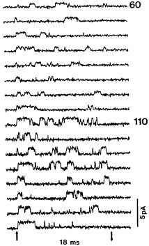
FIG. 1. Reopening of a single channel from a squid axon at potentials of +60 mV (upper block) and +110 mV (lower block), recorded at 5 8C in 270 Na/535 Na. Onset and release of the 18 ms depolarizations are indicated by the arrows at the bottom. Holding potential 100 mV. From Correa & Bezanilla (1994).
axons showed elegantly and conclusively, as may be seen in Fig. 1, that in a patch containing a single channel the steady current arose from the arrival of the channel a short while after its initial opening, followed by its fast inactivation, at a state in which it continued to reopen at a relatively low frequency. It was thus clear that each channel had two di¡erent routes to an open state.
It was suggested mistakenly by Hille (1992) that incomplete inactivation of this type ‘may be unique to squid axons’, but Alzheimer et al (1993a,b) demonstrated the development of a persistent Na+ current in pyramidal neurons from rat and cat sensorimotor cortex, and suggested that a switching between channels exhibiting di¡erent degrees of modal gating might be of importance in early development. Other examples of this type of bimodal gating have since come to light.
Measurements have been reported by Keynes & Meves (1993) of the probability functions both for the initial opening of the Na+ channels in squid axons, and for the reopenings that generate the late current in the inactivated steady state. Plots

MULTIMODAL Na+ CHANNEL GATING |
7 |
FIG. 2. The probability functions PFpeak for initial opening (open circles) and PFss for opening in the inactivated steady state (open squares), plotted against test potential, for a squid axon bathed in 514 mM Na+16 nM TTX and dialysed with 350 mM NaF. Temperature 5 8C. Holding potential 80 mV. The solid symbols show corresponding values of the macroscopic permeability coe⁄cients. From Keynes & Meves (1993).
against potential of the probability functions PFpeak for initial opening, and PFss for
reopening in the steady state, are shown in Fig. 2. It will be seen that whereas PFpeak rises to an early peak as expected, and then declines slightly, PFss rises with a delay
and above 50 mV continues upwards on a straight line. Analysis of the data showed that just as arrival at the initial open state of the channel has been shown by Keynes & Elinder (1999) to involve a series of steps each carrying close to one electronic charge. The late reopenings also require the transfer of slightly less than one charge, though with an equilibrium potential shifted positively by about 95 mV
as compared with that for PFpeak.
The next step in my argument must be to consider the nature of the mechanism responsible for these two di¡erent types of gating. There is now general agreement that the voltage sensors of the system are the positive charges carried by the S4 transmembrane segments in each of the four domains of every channel. The total transfer of charge needed to bring about the normal opening of both K+ and Na+ channels is close to 12 electronic units, and thus three in each S4 segment. A vitally important characteristic of the S4 segments is that the great majority of the positively charged lysine and arginine residues exhibit a one in three spacing with two uncharged residues situated between each of them. This spacing is perfectly conserved across the entire animal kingdom in all voltage-gated ion channels,
8 |
KEYNES |
whether they are selective for Na+, K+ or Ca2+. But the total number of positive charges carried by a single segment varies from four to eight between the several domains. In Na+ channels there are almost always four in domain I, ¢ve in domains
IIand III, and eight in domain IV.
Another less well recognized structural feature of all voltage-gated ion channels,
to which attention was recently drawn by Keynes & Elinder (1999), is the near perfect conservation of the location of negatively charged glutamate and aspartate residues in segments S2 and S3. Thus in S3 there is an aspartate residue located six places from the inner end of the segment, and in S2 invariably a glutamate three places nearer the centre of the segment. The position in every domain of an outermost negative charge, or sometimes of a pair of charges, is conserved with less precision, and they may either be glutamate or aspartate, located either on S2 or S3. It would seem that the prime function of the triplet of ¢xed negative charges is to pair up with three of the mobile positive charges and so stabilize their movements in discrete steps across the membrane. Threedimensional models have shown that if S4 is appropriately tilted relative to S2 and S3 lying parallel to one another, ion pairs can readily be formed. The precision of their location in every voltage gated ion channel in the world, since sodium channels ¢rst evolved around 550 million years ago, con¢rms that their role is of central importance.
The explanation originally put forward by Catterall (1986) and by Guy & Seetharamulu (1986) for the one in three spacing of the positive charges was that their movements take place in a screw-helical fashion, which meant that a 608 twist of the S4 a-helix from a position in which its positive charges were paired up with ¢xed negative ones would move it out 0.45 nm, bringing each positive charge into the position previously occupied by its neighbour, where it would automatically ¢nd a negative charge with which to pair. However, an obvious objection to the screw-helical theory was that the total number of positive charges carried by the four S4 segments was appreciably greater than the number of negative charges carried by the other ¢ve segments. It was also hard to believe that in the open state the outermost arginines of S4 would project as far as 1.45 nm into the external aqueous phase. So for some time the theory lost its initial popularity.
Taking advantage of the naturally occurring mutation R1448C of segment IVS4 in human skeletal muscle Na+ channels in which cysteine replaces the outermost arginine, Yang & Horn (1995) introduced a technique for testing the accessibility of the cysteine residue to hydrophilic methanethiosulfonate (MTS) and methanethiosulfonate-ethyltrimethylammonium (MTSET) reagents, which demonstrated clearly that it was indeed moved into the external aqueous phase by depolarization. This technique was subsequently extended by Yang et al (1997) to carry out more extensive observations on the accessibility of several of the positive charges to either the aqueous external or internal environments when they were
MULTIMODAL Na+ CHANNEL GATING |
9 |
mutated to cysteine. Similar observations were made on Shaker K+ channels by Larsson et al (1996). An important conclusion from this work was that the e¡ective width of the low dielectric constant portion of the membrane across which the electric ¢eld acts to pull the positive charges outwards or inwards must be substantially less than the 3 nm that corresponds to a membrane capacity of 1 mF.cm 2. It is nevertheless wide enough to house the three well conserved negative charges now shown to be located on S2 and S3 in every domain.
Fig. 3 shows a strictly diagrammatic representation of the screw-helical mechanism put forward by Keynes & Elinder (1999) to accommodate the MTSET accessibility data of Larsson et al (1996) and of Yang et al (1997), as applied to segment IVS4 of a human skeletal muscle Na+ channel. In the strongly hyperpolarized closed state, ¢ve of the positive charges project inwards into the aqueous phase, while in the open state depolarized to around 0 mV three of them are exposed to the external aqueous phase, and only two still project inwards. Another positive charge is transferred outwards in order to inactivate the channel, and the last and ¢fth does so for the ¢nal reopening in the inactivated steady state. It should be noted that the data of Keynes (1994a) indicate that hyperpolarization to 180 mV is needed to complete the inward transfer of the gating charge, while depolarization to +100 mV is needed to reopen an appreciable number of channels. The more restricted accessibility of the channels reported by Yang et al (1997) and Horn (2000) refers to a somewhat narrower voltage range.
I should make clear at this point that the relatively straightforward picture of the screw-helical theory shown in Fig. 3, in which a-helix S4 both rotates and moves outwards, con£icts seriously with the views of Bezanilla et al (2001). Bezanilla currently maintains that the four outermost positive charges reside in long crevices with ends too narrow to ¢t MTS reagents, but large enough to allow protons to have access to them. On depolarization, the S4 segment is claimed to rotate with little or no outward translation so that the charges which are initially in an internally exposed crevice move to an externally connected one. It has, however, been pointed out by Horn (2000) that the attachment of large £uorophores to measure such movements of subunits within proteins yields results that are hard to interpret, and there is not yet agreement as to exactly what distances are moved. Most recently, Gandhi et al (2000) have concluded from £uorescence scans of a voltage-gated K+ channel that the rotation of the S4 segment is accompanied by an appreciable outward movement. I have to confess that I am unable to understand where precisely Bezanilla’s crevices are in fact located, the absence of any hard anatomical evidence in support of their existence being for me the major weakness in his arguments. It is also unclear to me how this mechanism is supposed to transfer unit charges in discrete steps, for which there is

FIG. 3. A strictly diagrammatic representation of the screw-helical outward movement of the positive charges carried by segment IVS4 in a human muscle Na+ channel. Each outward step transfers one electronic charge from the interior of the cell to the external solution. The three negative charges shown on the left occupy ¢xed positions on IVS2 and IVS3, and form salt bridges with three of the mobile positive charges. After the version for a Shaker K+ channel of Keynes & Elinder (1999).
MULTIMODAL Na+ CHANNEL GATING |
11 |
incontrovertible evidence in the operation of Na+ channels; nor does it appear to take account of the implications of the conspicuously good conservation of the location of the negative charges on S2 and S3 that I have described.
However, I must also admit that my modi¢ed version of the screw-helical hypothesis is open to serious objection in that the outward movements of more than 2 nm in the IVS4 segment of a Na+ channel shown in Fig. 3 may be hard to reconcile with the shortness of the external links, some of which contain only four residues between the outer ends of segments S3 and S4. I can only suggest as a possible solution to this di⁄culty that the emergence of the ends of the S4 segments into the external aqueous phase may be accompanied by an instantaneous collapse of their a-helical structure.
Although the current generated by reopening of channels in the inactivated steady state may well serve for a ¢ne tuning of the excitability of neurons in the course of development, it could not readily be responsible for the explosive increases in excitability which I take to be the main topic of interest for this meeting. It is therefore necessary to look for a third mode of gating of the Na+ channel. From the extensive literature on the involvement of voltage-gated ion channels in hereditary disease recently summarized by Lehmann-Horn & JurkatRott (1999), the most relevant part of the system to examine would evidently be the mechanism of fast inactivation, for whereas there seem to be few if any hereditary mutations that directly increase excitability, there are many that have been shown to operate indirectly by drastically slowing down fast inactivation. Thus in Fig. 4 you will note that the majority of the symbols are clustered around segment IVS4 itself, and the internal link between domains III and IV where the so-called inactivation particle is located.
A relevant phenomenon that has never been observed in a squid axon, but was ¢rst reported for patches from frog muscle ¢bres by Patlak & Ortiz (1986), and is illustrated in Fig. 5, is the appearance in addition to the single late openings seen in Fig. 1 of occasional bursts of 10 s to 100 s of openings.
This occurrence of multiple gating modes in a single population of channels has been reported in other types of muscle and in brain cells, and is characterized by an alternation between periods of inactivation at two di¡erent rates, one 10 times slower than the other. It has been suggested that the two modes represent two conformations of the a subunit, one of which can be stabilized either by hyperpolarization or, as has been shown by Ma et al (1997), by modulation of the b1-units by G proteins. A third mode of gating of this kind therefore seems to be a good candidate for explaining large increases in excitability.
Nevertheless, while I welcome the recent though belated admission of the truth of the proposition for which I have long fought with very few allies (Keynes 1994b), that fast inactivation of the Na+ channel is indeed a voltage-dependent process, it has to be recognized that a full understanding of the detailed
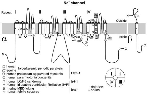
FIG. 4. The transmembrane topology of some mammalian muscle Na+ channels showing the points where single site mutations interfere with fast inactivation and give rise to the hereditary diseases listed. From Lehmann-Horn & Jurkat-Rott (1999).