

Sodium Channels and Neuronal Hyperexcitability
.pdfMOLECULAR MECHANISMS OF GATING AND DRUG BLOCK |
213 |
position by fast inactivation, arguing that their movement is coupled to the inactivation process (Cha et al 1999). Together, these results provide strong evidence that outward movement of the S4 segment in domain IV is the signal to initiate fast inactivation of the Na+ channel by closure of the intracellular inactivation gate
As for the IIS4 segment, the movement of the IVS4 segment is also a molecular target for neurotoxin action. a scorpion toxins and sea anemone toxins uncouple activation from inactivation. They bind to a receptor site including the S3^S4 loop at the extracellular end of the IVS4 segment (Rogers et al 1996, Sheets et al 1999). A single mutation of R1613 in this extracellular loop reduces binding a⁄nity and thus slowing of inactivation by more than 50-fold (Fig. 4). Binding of the toxins in this position is proposed to slow inactivation by preventing the normal outward movement of the IVS4 segment (Fig. 4), evidently trapping it in a position that is permissive for activation but not for fast inactivation. Thus, scorpion venoms contain two di¡erent toxins that act by voltage sensor-trapping . the b scorpion toxins trap the IIS4 segment in an activated position (Fig. 2) and enhance activation while the a scorpion toxins trap the IVS4 segment in an inward, partially activated position that allows activation but not fast inactivation (Fig. 4). The combination of the two e¡ects increases Na+ channel activity inappropriately and causes paralysis. The di¡erential e¡ects of these two toxins also reinforce the evidence for specialization of the S4 segments in di¡erent domains . domain II for activation and domain IV for coupling of activation to inactivation.
The inner pore and local anaesthetic receptor site
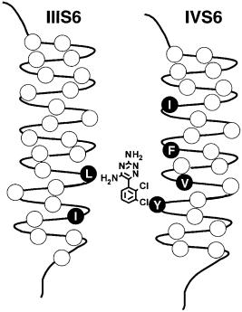
Voltage clamp studies led to the conclusion that local anaesthetics enter from the intracellular side and bind in the inner pore of Na+ channels (Strichartz et al 2002, this volume) and similar work revealed analogous intracellular block of Ca2+ and K+ channels. The ¢rst indication that the S6 segments form the inner pore of the voltage-gated ion channels came from locating a pore-blocker receptor site . the phenylalkylamine receptor site of L-type Ca2+ channels (Striessnig et al 1990). Photoa⁄nity labelling with high-a⁄nity phenylalkylamine pore blockers showed that only the IVS6 segment of the Ca2+ channel b1 subunit was labelled. Subsequently, mutagenesis studies of Na+ channels revealed the local anaesthetic receptor site in an analogous position in the Na+ channel (Ragsdale et al 1994). High a⁄nity binding of local anaesthetics to the inactivated state of Na+ channels requires two critical amino acid residues, phe1764 and tyr1771 in brain type IIA channels, which are located on the same side of the IVS6 transmembrane segment two a-helical turns apart (Fig. 5). It is likely that the tertiary amino group of local anaesthetics interacts with phe1764, which is located more deeply in the pore, and that the aromatic moiety of the local anaesthetics interacts with tyr1771, which is
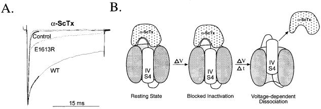
FIG. 4. Voltage sensor trapping by a scorpion toxin. (A) E¡ect of a scorpion toxin on inactivation of wild-type and mutant Na+ channels. Na+ currents were measured at 710 mV by whole-cell voltage clamp in tsA-201 cells transfected with wild-type Nav1.2 channels or mutant E1613R (Rogers et al 1996). Traces with slowed inactivation labelled WT and E1613R were recorded in the presence of 20 nM LqTx, the principal a scorpion toxin of Leiurus quinquestriatus (Rogers et al 1996). (B) Voltage sensor trapping model for a scorpion toxin action. Domain IV and its S4 segment are illustrated at resting membrane potential (e.g. 7100 mV) and at a depolarized membrane potential (e.g. +40 mV) a short time (e.g. 5 ms) and a long time (e.g. 2 s) after depolarization.
MOLECULAR MECHANISMS OF GATING AND DRUG BLOCK |
215 |
FIG. 5. Lamotrigine binding to the local anaesthetic receptor site in transmembrane segments IIIS6 and IVS6 of the rat brain type IIA Na+ channel. Side view of the proposed location of the lamotrigine binding site within the pore.
located nearer to the intracellular end of the pore. Recent work has identi¢ed two amino acid residues in transmembrane segment IIIS6 that also contribute to the local anaesthetic receptor site (Fig. 5).
Like local anaesthetics, certain anticonvulsant and antiarrhythmic drugs are intracellular pore blockers of Na+ channels. We have surveyed a selection of these agents to determine whether they also bind at the local anaesthetic receptor site by testing the e¡ect of the mutations F1764A and Y1771A on the a⁄nity for drug binding to the inactivated state. We have found that a wide range of local anaesthetic, anticonvulsant and antiarrhythmic drugs of di¡erent structure all interact with these two amino acid residues in the local anaesthetic receptor site. The ratio of e¡ects of these two mutations di¡ers for drugs of di¡erent structure, suggesting a speci¢city of interaction based on drug structure. These results lead to a model in which F1764 and Y1771 are common elements of a complex receptor site for Na+ channel blocking drugs, which make additional drug-speci¢c contacts that increase their a⁄nity and alter their pharmacological properties.
216 |
CATTERALL |
The common features of the local anaesthetic receptor site suggest that novel Na+ channel blocking drugs could be developed to ¢t this site that might have higher a⁄nity and speci¢city than presently available compounds. Consistent with this idea, the novel compound BIII 890 CL, a complex ether of benzoyl and modi¢ed benzomorphan moieties, is a potent Na+ channel blocker binding at the local anaesthetic receptor site with a Kd of 50 nM for the inactivated state (Carter et al 2000). It is aimed for neuroprotective therapy in the treatment of stroke and possibly neurodegenerative diseases.
Conclusions
Analysis of the structure and function of Na+ channels has led to many important insights into the molecular basis for channel gating, modi¢cation of gating by polypeptide neurotoxins, and block of Na+ channels by local anaesthetics and related drugs. These studies point to the S4 segments and the S3^S4 loops on their extracellular side as the locus for voltage sensing and for the actions of polypeptide toxins on the gating process. They also point to the S6 segments in domains III and IV as molecular targets for a broad range of Na+ channel blocking drugs, including local anaesthetics, anticonvulsant drugs, and antiarrhythmic drugs. Targeting these regions for future drug design may yield novel agents that can interrupt hyperexcitability in epilepsy, cardiac arrhythmias and persistent pain syndromes.
Acknowledgements
The research from the author’s laboratory was supported by NIH Research Grants NS15751 and NS25704.
References
Armstrong CM 1981 Sodium channels and gating currents. Physiol Rev 61:644^683
Carter AJ, Grauert M, Pschorn U et al 2000 Potent blockade of sodium channels and protection of brain tissue from ischemia by BIII 890 CL. Proc Natl Acad Sci USA 97:4944^4949
Catterall WA 1986 Voltage-dependent gating of sodium channels: correlating structure and function. Trends Neurosci 9:7^10
Ceste'le S, Qu Y, Rogers JC, Rochat H, Scheuer T, Catterall WA 1998 Voltage sensor-trapping: enhanced activation of sodium channels by b-scorpion toxin bound to the S3^S4 loop in domain II. Neuron 21:919^931
Cha A, Ruben PC, George AL Jr, Fujimoto E, Bezanilla F 1999 Voltage sensors in domains III and IV, but not I and II, are immobilized by Na+ channel fast inactivation. Neuron 22:73^87 Chen LQ, Santarelli V, Horn R, Kallen RG 1996 A unique role for the S4 segment of domain 4 in
the inactivation of sodium channels. J Gen Physiol 108:549^556
Eaholtz G, Scheuer T, Catterall WA 1994 Restoration of inactivation and block of open sodium channels by an inactivation gate peptide. Neuron 12:1041^1048
MOLECULAR MECHANISMS OF GATING AND DRUG BLOCK |
217 |
Goldin AL, Snutch T, Lubbert H et al 1986 Messenger RNA coding for only the a subunit of the rat brain Na channel is su⁄cient for expression of functional channels in Xenopusoocytes. Proc Natl Acad Sci USA 83:7503^7507
Guy HR, Seetharamulu P 1986 Molecular model of the action potential sodium channel. Proc Natl Acad Sci USA 83:508^512
Hartshorne RP, Catterall WA 1981 Puri¢cation of the saxitoxin receptor of the sodium channel from rat brain. Proc Natl Acad Sci USA 78:4620^4624
Hartshorne RP, Messner DJ, Coppersmith JC, Catterall WA 1982 The saxitoxin receptor of the sodium channel from rat brain. Evidence for two nonidentical beta subunits. J Biol Chem 257:13888^13891
Hirschberg B, Rovner A, Lieberman M, Patlak J 1995 Transfer of twelve charges is needed to open skeletal muscle Na+ channels. J Gen Physiol 106:1053^1068
Horn R 2002 Molecular basis for function in sodium channels. In: Sodium channels and neuronal hyperexcitability. Wiley, Chichester (Novartis Found Symp 241) p 21^33
Isom LL 2002 b subunits: players in neuronal hyperexcitability? In: Sodium channels and neuronal hyperexcitability. Wiley, Chichester (Novartis Found Symp 241) p 124^143
Isom LL, De Jongh KS, Patton DE et al 1992 Primary structure and functional expression of the b1 subunit of the rat brain sodium channel. Science 256:839^842
Isom LL, Ragsdale DS, De Jongh KS et al 1995 Structure and function of the b2 subunit of brain sodium channels, a transmembrane glycoprotein with a CAM motif. Cell 83:433^442
Kellenberger S, Scheuer T, Catterall WA 1996 Movement of the Na+ channel inactivation gate during inactivation. J Biol Chem 271:30971^30979
Kellenberger S, West JW, Scheuer T, Catterall WA 1997 Molecular analysis of the putative inactivation particle in the inactivation gate of brain type IIA Na+ channels. J Gen Physiol 109:589^605
Makita N, Bennett PB, George AL Jr 1996 Molecular determinants of b1 subunit-induced gating modulation in voltage-dependent Na+ channels. J Neurosci 16:7117^7127
McCormick KA, Isom LL, Ragsdale D, Smith D, Scheuer T, Catterall WA 1998 Molecular determinants of Na+ channel function in the extracellular domain of the b1 subunit. J Biol Chem 273:3954^3962
McCormick KA, Srinivasan J, White K, Scheuer T, Catterall WA 1999 The extracellular domain of the beta1 subunit is both necessary and su⁄cient for beta1-like modulation of sodium channel gating. J Biol Chem 274:32638^32646
Noda M, Shimizu S, Tanabe T et al 1984 Primary structure of Electrophorus electricus sodium channel deduced from cDNA sequence. Nature 312:121^127
Noda M, Ikeda T, Kayano T et al 1986a Existence of distinct sodium channel messenger RNAs in rat brain. Nature 320:188^192
Noda M, Ikeda T, Suzuki H et al 1986b Expression of functional sodium channels from cloned cDNA. Nature 322:826^828
Qu Y, Rogers JC, Chen SF, McCormick KA, Scheuer T, Catterall WA 1999 Functional roles of the extracellular segments of the sodium channel a subunit in voltage-dependent gating and modulation by b1 subunits. J Biol Chem 274:32647^32654
Ragsdale DS, McPhee JC, Scheuer T, Catterall WA 1994 Molecular determinants of statedependent block of Na+ channels by local anesthetics. Science 265:1724^1728
Rogers JC, Qu Y, Tanada TN, Scheuer T, Catterall WA 1996 Molecular determinants of high a⁄nity binding of a-scorpion toxin and sea anemone toxin in the S3^S4 extracellular loop in domain IV of the Na+ channel a subunit. J Biol Chem 271:15950^15962
Rohl CA, Boeckman FA, Baker C, Scheuer T, Catterall WA, Klevit RE 1999 Solution structure of the sodium channel inactivation gate. Biochemistry 38:855^861
218 |
DISCUSSION |
Shapiro L, Doyle JP, Hensley P, Colman DR, Hendrickson WA 1996 Crystal structure of the extracellular domain from Po, the major structural protein of peripheral nerve myelin. Neuron 17:435^449
Sheets MF, Kyle JW, Kallen RG, Hanck DA 1999 The Na channel voltage sensor associated with inactivation is localized to the external charged residues of domain IV, S4. Biophys J 77:747^757
Srinivasan J, Schachner M, Catterall WA 1998 Interaction of voltage-gated sodium channels with the extracellular matrix molecules tenascin-C and tenascin-R. Proc Natl Acad Sci USA 95:15753^15757
Strichartz GR, Zhou Z, Sinnott C, Khodorova A 2002 Therapeutic concentrations of local anaesthetics unveil the potential role of Na+ channels in neuropathic pain. In: Sodium channels and neuronal hyperexcitability. Wiley, Chichester (Novartis Fund Symp 241) p 189^205
Striessnig J, Glossmann H, Catterall WA 1990 Identi¢cation of a phenylalkylamine binding region within the alpha 1 subunit of skeletal muscle Ca2+ channels. Proc Natl Acad Sci USA 87:9108^9112
Stuhmer W, Conti F, Suzuki H et al 1989 Structural parts involved in activation and inactivation of the sodium channel. Nature 339:597^603
Vassilev PM, Scheuer T, Catterall WA 1988 Identi¢cation of an intracellular peptide segment involved in sodium channel inactivation. Science 241:1658^1661
Vassilev P, Scheuer T, Catterall WA 1989 Inhibition of inactivation of single sodium channels by a site-directed antibody. Proc Natl Acad Sci USA 86:8147^8151
West JW, Patton DE, Scheuer T, Wang Y, Goldin AL, Catterall WA 1992 A cluster of hydrophobic amino acid residues required for fast Na+ channel inactivation. Proc Natl Acad Sci USA 89:10910^10914
Xiao ZC, Ragsdale DS, Malhotra JD et al 1999 Tenascin-R is a functional modulator of sodium channel b subunits. J Biol Chem 274:26511^26517
Yang NB, Horn R 1995 Evidence for voltage-dependent S4 movement in sodium channel. Neuron 15:213^218
Yang NB, George AL Jr, Horn R 1996 Molecular basis of charge movement in voltage-gated sodium channels. Neuron 16:113^122
DISCUSSION
Strichartz: One of the interesting things about the high-a⁄nity inactivated state binding is that if we posit that contributions are made from all four S6 helices, then it is odd that when we mutate one amino acid in one of the helices there are huge e¡ects on a⁄nity. You might think that you’d lose a third or a quarter of the binding energy, but in fact it looks like the a⁄nity is near zero.
Catterall: The e¡ects aren’t that large. Binding energy is a logarithmic function, so a 10-fold change in binding constant results from a much smaller change in binding energy. I think it is consistent with the idea that there are multiple sites of attachment. Another correlation which is not strictly quantitative is that the larger the drug, the less impact mutating the two residues in the IV S6 segment has on its binding. Both those mutations have a 300-fold e¡ect on lidocaine, but if you do the same experiment on quinidine or £ecainide, which are much bulkier drugs, they have more like threeor ¢vefold e¡ects. One explanation for this is that
MOLECULAR MECHANISMS OF GATING AND DRUG BLOCK |
219 |
the large drugs touch more places, so when you mutate two residues it has less impact. Our thinking is that the mutations we make are only reducing a portion of the binding energy, and this portion is greatest for the drugs of simplest structure, because they make the fewest contacts with the pore.
Strichartz: An alternative explanation is that these mutations a¡ect the conformation of the channel and indirectly a¡ect the binding site. So when we change them, the channel will still inactivate, but it folds up into a tighter or a looser situation, so this binding site, which has a weak stereoselectivity to begin with, is altered.
Catterall: That is certainly possible. It is almost impossible in a mutagenesis experiment to rule out conformational e¡ects, but it is important to say that in these experiments there is almost no correlation between the e¡ects of mutations on inactivation and their e¡ects on drug block. You can have large e¡ects on inactivation and no e¡ect on drug block, and vice versa. It is not likely that all of the e¡ect here is conformational, simply preventing the inactivated state from forming.
Strichartz: It is not a question of preventing it from forming, but rather of the actual form of the inactivated state.
Catterall: There is no way that we can test that without a crystal structure. For Na+ channels this is still a few years away.
Cummins: I have been told that one of the problems for using these drugs for neuropathic pain is that they are just not speci¢c to peripheral ion channels. There is evidence in the literature, however, that the SNS-type current does have lower a⁄nity to some of these agents, although not all. Does this indicate that we will be able to target this area with subtype-speci¢c drugs?
Catterall: It is a possibility, but the speci¢c residues in the IV S6 segment we have identi¢ed are present in SNS, so it isn’t those residues that are causing the di¡erence. It might be residues in other S6 segments. I haven’t gone back to look at the other residues we have identi¢ed in domain III and domain I to see whether they are present in SNS. There is clearly an a⁄nity di¡erence. It might be that this is due to the slower and weaker inactivation of those channels, and it is not a receptor site phenomenon but instead a conformational change/allosteric-type phenomenon; that is, that the inactivated state is di¡erent for those channels and does not provide such an appropriate binding site for these drugs. This again might argue that you could design a drug that would be speci¢c for these channels and would target this site. I am sure that many drug companies are trying to do this; I think it is a reasonable hypothesis. I think it is encouraging that Boehringer^ Ingleheim was able to make a Na+ channel-blocking drug with a Kd of 50 nM. We don’t have any other Na+ channel-blocking drugs that act at 50 nM concentrations. Their therapeutic target for this is neuroprotection, and they have this drug in stroke trials at present.
220 |
DISCUSSION |
Horn: You talked about a couple of di¡erent classes of compounds that block the pore. One of them was a local anaesthetic and the other was a peptide. Have you ever looked at whether the a⁄nity of this peptide is changed by your S6 mutations?
Catterall: It is not a¡ected by the F1764A or Y1771A mutations, so we think that the binding site for the inactivation gate receptor is di¡erent. I suppose the crystal structure of the Kcsa channel I showed can illustrate the point. We think the drug binds in what I called the cavity of the structure, but the inactivation gate can’t get there. It has to bind more super¢cially to that helix cross near the intracellular end of the S6 segments, where the amino acid residues that we and Al Goldin have found to be essential for inactivation may be located in the Na+ channel structure.
Horn: When Galen Eaholtz was studying that, she looked for a voltage dependence to the block. As I remember, she found some voltage dependence for the peptide reaching its binding site, but it was very super¢cial.
Catterall: Remember, the charges in the KIFMK peptide are not in the natural sequence, and therefore they are probably not binding to the receptor site. We think that the charged peptide is being concentrated in the di¡use double layer by the mechanism that Gary Strichartz suggested, and that you see voltage dependence and a requirement for charge in order to get enough peptide near the mouth of the pore to give block during the millisecond time frame that the Na+ channel is open.
Horn: I don’t see how that is going to give a voltage dependent block. I can understand that the fact that you have lysines on either end of it might concentrate the peptide there, but in order to sense the electric ¢eld it has to get into the electric ¢eld. If it is right on the very mouth of the channel it is not going to feel the electric ¢eld gap.
Catterall: There are two components. You get voltage-dependent block because the channel needs to form the inactivation gate receptor, and then there is voltage dependence to do with the concentration of the drug. In addition, Eaholtz and I found a second form of block by IFM-containing peptides. This second form of block was strongly voltage dependent and slowly reversible, as if the peptide entered more deeply into the pore.
Horn: I had the impression that they found a voltage dependence that looked like open channel block voltage dependence. They had a channel that didn’t inactivate and they could see it as a charged open channel blocker entering the electric ¢eld. It seems to me that there is some inconsistency here: I don’t see how it is entering the electric ¢eld if it is at the very mouth of the channel.
Strichartz: If the ¢eld potential changes when the blocking particle is there, because now no ions can pass through the pore, a voltage dependence will result from the blockade per se. It will show up in the o¡-rate constant.
Horn: You mean that it is there already.
MOLECULAR MECHANISMS OF GATING AND DRUG BLOCK |
221 |
Strichartz: The electric ¢eld across the membrane has to drop over the same potential, from bulk outside to bulk inside. If, when a blocking particle is in place, the pro¢le of ion mobility is altered, then the pro¢le of the ¢eld across the channel is also di¡erent, which can give a voltage dependence that would show up in the o¡-rate constant, but not in the on-rate constant.
Horn: So the ¢eld is actually dropping across the blocker.
Catterall: And the ¢eld drops across that region di¡erently when the channel is open and closed. Again, if the blocker is in that region it is going to feel that change in electrical potential.
Waxman: Earlier Bill Catterall mentioned neuroprotection. In that context, a few years ago Peter Stys, Bruce Ransom and I looked at the issue of Ca2+- mediated injury in white matter, where there are no synapses (Stys et al 1992a). Not surprisingly, glutamate doesn’t trigger Ca2+-mediated injury in white matter. Peter did identify a pathway involving Na+ in£ux via a persistent Na+ conductance which depolarizes axons and also reverses the transmembrane Na+ gradient and drives that Na+/Ca2+ exchanger into a reverse node (Stys et al 1992a). This is the death knell of axons within anoxic white matter. The interesting thing was that one could protect white matter axons from this cascade with both tertiary anaesthetics (procaine and lidocaine), and quaternary anaesthetics (QX314 and QX222). This protection occurred at concentrations that did not impair impulse conduction (Stys et al 1992b). One ¢nal point was that it may have been fortuitous that some of these compounds weren’t clean: they depressed PK as well as PNa, and this may have helped to preserve conduction.
Catterall: I suppose they are acting to preferentially block the long opening Na+ channels and do not have much e¡ect on those open at the peak of the spike.
Bean: What is your view of the origin of the change in a⁄nity between the resting and inactivated states? Do you view that as being a change in the binding site? It couldn’t just be a change in accessibility of the binding site when the channel opens.
Catterall: I imagine it as a conformational change in the binding site. We have one indirect bit of evidence for that. We found that in the III S6 segment, the periodic e¡ect on activation when we make those alanine substitution mutations has the exact periodicity of an a helix. It can be a pretty strong e¡ect (20 mV shifts in activation curve). It is as if one face of the helix makes interactions in the closed state that are di¡erent from those it makes in the open state. We think this helix must be turning during the activation process and exposing a new face to either interactions with solvent or interactions with other transmembrane segments, giving us this periodic pattern of e¡ects on activation. We don’t see the same periodic pattern in the IV S6 segment, but it may be that all of them rotate a little bit. Our model is that these segments rotate and open the pore, sort of like opening a camera lens, and in doing so expose phenylalanine 1764 and tyrosine 1761 to the lumen of the pore, where these drugs can then bind to them. This is why those two
222 |
DISCUSSION |
residues are common determinants of inactivated state binding for all the drugs, because they become accessible to the drug binding site during the channel opening process and inactivation.
Bean: Is it an open possibility that the high a⁄nity binding is a consequence of the activation of the channel . the movements of the S4 regions . and doesn’t necessarily result from the inactivated state? It could be that the movement of the S4s produces a high a⁄nity site for the local anaesthetics, and also promotes formation of the inactivated state.
Catterall: I think that is right, but there is another way to think about this. For most of these drugs, the a⁄nity for the inactivated state is higher. Activation causes rotational movements of helices that reveal the binding site, even in the open state of the channel. The drugs enter and bind. But then when the channel inactivates, the four S6 segments move a little closer, so the drug can bind to all four domains at the same time, or to three of them, rather than just one. So the structure collapses a little bit when you reach the inactivated state. This would be a model that would allow us to understand higher a⁄nity binding to the open state and even higher a⁄nity binding to the inactivated state.
Bean: Can you see a di¡erence in binding if you use the inactivation gate mutants to prevent inactivation?
Catterall: There are some di¡erent results in the literature, but we haven’t looked at this systematically. If we make just the phenylalanine 1489 mutation, which is su⁄cient to substantially block inactivation we still observe pretty good usedependent block by local anaesthetics. Other groups have mutated all three residues of the IFM motif and saw a much bigger loss of local anaesthetic binding, so they concluded that the gate was critical to the receptor site. We don’t ¢nd that when we just use a single mutation. Perhaps the triple mutation has conformational e¡ects beyond just preventing the closure of the inactivation gate.
Strichartz: Moving to peptide toxins for a moment, years ago we did experiments looking at the e¡ect of combining a toxins and b toxins on nodal Na+ currents, and saw that there was a marked synergy, particularly in terms of enhancing and prolonging the slowly declining current induced by b toxins (Wang & Strichartz 1982). Looking at your model, this would suggest that there is some coupling between S4s in domain II and domain IV. Either that, or the two toxins at the surface somehow interact.
Catterall: Aren’t the scorpions clever! They have one peptide toxin pulling out one S4 segment, and the other pushing the other S4 segment in and keeping the channel in an open state.
Strichartz: And then they add K+ channel blockers to make it even worse! Isom: A while ago, we had the idea that the binding site for a scorpion toxin was
the site of interaction between b1 and a. Do you still think that is true?