


Posterior Pituitary |
3 |
HORMONES OF THE POSTERIOR PITUITARY
•Made up of distal neuron terminals
•Secreted hormones;argininevasopressin (ADH), oxytocin (see chapter 1 1 ) - both are peptide hormones.
•Cell bodies located in the supraoptic nucleus and paraventricular nucleus ofthe hypothalamus.
•ADH is a major controller of water excretion and ECF volume. ADH also controls osmolarity.
•The osmoreceptor neurons in the hypothalamus are extremely sensitive and are able to maintain ECF osmolarity within a very narrow range.
•There is a resetting of the osmostat downward in pregnancy, the men strual cycle, and with volume depletion. In the latter case osmoregula tion is secondary to volume regulation; a return of circulating volume occurs even though osmolarity decreases.
•Volume receptors are less sensitive than osmoreceptors and a change of 10-15% in volume is required to produce a measurable change in ADH.
•Cortisol and thyroid hormone restrain the release ofADH.
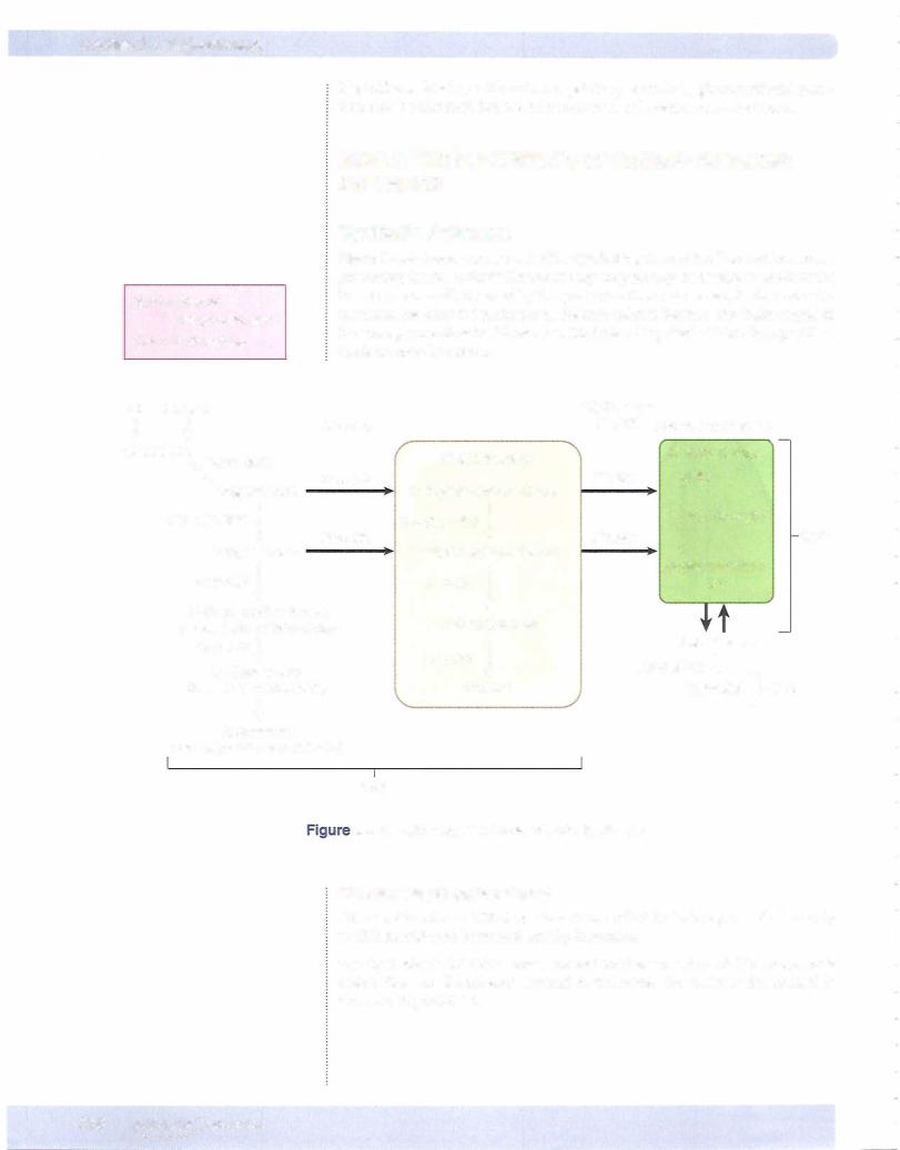
Figure X-3-1 illustrates the neural control mechanisms that regulate secretion of ADH bythe posteriorpituitary. The principal inputs are inhibition by barorecep tor and volume receptor unit and stimulation by osmoreceptors.
MEDICAL 269


Chapter 3 • Posterior Pituitary
Synthesis and Release ofADH
•ADH is synthesized in the supraoptic (SO) and paraventricular (PVN) nuclei ofthe hypothalamus; it is stored and released from the posterior pituitary.
•Osmoreceptors are neurons that respond to increased plasma osmolar ity, principally plasma sodium concentration. They synapse with neu rons ofthe SO and PVN and stimulate them to secrete ADH from the posterior pituitary. They also stimulate consumption ofwater through hypothalamic centers that regulate thirst.
•The SO and PVN also receive input from cardiopulmonary mechano receptors, as well as arterial baroreceptors. High blood volume or blood pressure tends to inhibit secretion ofADH.
•Secretion of ADH is most sensitive to plasma osmolarity (1%); how ever, ifblood volume decreases by 10% (such as hemorrhage) or cardiac output falls, high levels ofADH are secreted even if it causes abnormal plasma osmolarity.
Action ofADH
•The main target tissue is the renal collecting duct (V2 receptors).
•ADH increases the permeability ofthe duct to water by placing water channels in the luminal membrane.
•Water is reabsorbed passively, drawn across the membranes bythe higher osmolarity ofthe interstitium.
•Urea can pass with the water, but electrolytes cannot. ADH stimulates the urea transporter, increasing urea reabsorption.
•In severe hemorrhage, high levels ofADH via Vl receptors on vascular smooth muscle cause a vasoconstriction.
•We see a 10% change in osmolarity, 10% change in volume
REGULATION OF ECFVOLUME AND OSMOLARITY
Volume Regulation
•Stimuli arising from stretch receptors act to chronically inhibit ADH secretion.
•Decreases in blood volume cause venous and arterial stretch receptors to send fewer signals to the CNS, decreasing chronic inhibition ofADH secretion.
•This mechanism is especially important for restoring ECF volume following a hemorrhage.
Osmoregulation
•An increase ofonly 1% in the osmolality ofthe ECF bathing the hypo
thalamic osmoreceptors will evoke an increased rate ofADH secretion.
•A similarly sized decrease in osmolality will decrease ADH secretion.
•In this manner, ECF osmolality is kept very close to 285 mOsm/Kg.
MEDICAL 271
Section X • Endocrinology
Effect ofAlcohol and Weightlessness on ADH Secretion
Ingesting ethyl alcohol or being in a weightless environment suppresses ADH se cretion. In weightlessness, there is a net shift ofblood from the limbs to the abdo men and chest. This results in greater stretch of the volume receptors in the large veins and atria, thus suppressing ADH secretion.
Clinical Correlate
Circulating levels of brain natriuretic peptide (BNP) correlate well with the degree of heart failure. Although very little BNP is synthesized and released in the normal heart, there is a marked elevation as the ventricle dilates, hence the correlation. The functional or pathologic significance is still unknown, but there is a strong correlation.
Atrial Natriuretic Peptide (ANP)
ANP is the hormone secreted by the heart. ANP is found throughout the heart but mainly in the right atrium. The stimuli that release ANP (two peptides are released) are:
•Stretch, an action independent of nervous involvement
•Increased salt intake
•CHF and all fluid overload states
ANP increases sodium loss (natriuresis) and water loss by the kidney because of, in part, an increase in glomerular filtration rate due to:
•ANP-mediated dilation of the afferent arteriole
•ANP-mediated constriction of the efferent arteriole
ANP also increases sodium loss (natriuresis) and water loss (diuresis) by the kid ney because it inhibits aldosterone release as well as the reabsorption of sodium and water in the collecting duct.
The physiologic importance ofANP is not known because it has not been possible to identify or produce a specific deficiency state in humans. However, ANP secre tion increases in weightlessness (submersion to the neck in water), while renin, aldosterone, and ADH secretion decrease. It mayplay a role in normal regulation ofthe ECF osmolality and volume.
ANP tends to antagonize the effects of angiotensin II and ADH.
A normal ANP level is used to exclude CHF as a cause of dyspnea.
PATHOPHYSIOLOGIC CHANGES IN ADH SECRETION
Diabetes lnsipidus
The consequences can be explained on the basis ofthe lack ofan effect ofADH on the renal collecting ducts.
Central diabetes insipidus (CD/)
• Sufficient ADH is not available to affect the renal collecting ducts.
•Causes include familial, tumors (craniopharyngioma), autoimmune, trauma
•Pituitary trauma - transient diabetes insipidus
•Sectioning ofpituitary stalk - triphasic response: diabetes insipidus, fol lowed by SIADH, followed by a return of diabetes insipidus
272 MEDICAL


Chapter 3 • Posterior Pituitary
Subgroups
Hypervolemia
Caused by marked reduction in water excretion and/or increased rate of water ingestion. Would include congestive heart failure and cirrhosis
Hypovolemia
Indicates solute depletion. Would include mineralocorticoid deficiency, diuretic abuse, renal disease, diarrhea, and hemorrhage
Clinical euvolemia
Would include SIADH. A clinically equivalent presentation may occur in gluco corticoid deficiency.
Chapter Summary
•ADH is synthesized in the hypothalamus but is stored and released from the posterior pituitary.
•The major action of ADH is the passive reabsorption of water and urea, but not electrolytes, in the renal collecting duct.
•Osmoreceptors are very sensitive and normally maintain osmolarity in a very narrow range.
•Reduced input from the low-pressure stretch receptors is a strong stimulus for the release ofADH.
•ANP, found mainly in the tissue ofthe right atrium, is released in response to stretch. The major action ofANP is diuresis and natriuresis.
•In diabetes insipidus, central form has low plasma ADH, nephrogenic form has high plasma ADH.
•Easily separated by measuring plasma ADH or injection of desmopressin
•Differential diagnosis followingwater deprivation
•SIADH: Inappropriate elevated secretion ofADH. Characterized by euvolemia but hyponatremia.
•Acute hyponatremia is life threatening if severe. Treated aggressively.
•Chronic hyponatremia is usually well-tolerated. Aggressive treatment is associated with central pontine myelinosis.
MEDICAL 275


Chapter It • Adrenal Cortex
(lipid active
-soluble |
|
Glucuronide group |
|
Cortisol --"'- -.....Glucuronide-conjugated metabolites of cortisol |
|||
hormone) |
Liver |
(inactive water-soluble metabolites) |
|
|
|||
|
|
||
Figure X-4-3. Metabolism of Cortisol
Urinary 17 OH steroids have in the past been measured as an index of cortisol secretion. This has been replaced by the measurement of the 24-hour urine-free cortisol.
C19 steroids (19 carbon atoms)
Adrenal Androgens
•Have a keto group at position 17 and are therefore called 17-ketosteroids.
•Are conjugated with sulfate in the adrenal cortex, making them water soluble. As water-soluble metabolites, they circulate in the bloodstream, are filtered by the kidney, and are excreted in the urine. The sulfated form is not produced in the gonads and is thus considered an index of androgen production by the adrenals.
•The major secreted form is dehydroepiandrosterone (DHEA).
•DHEA, DHEA sulfate, and androstenedione have very low androgenic activity. They function primarily as precursors for the peripheral con version to the more potent testosterone and dihydrotestosterone (men and women).
•In adult males, excessive production of adrenal androgens has no clinical consequences. In prepubertal males it causes premature penile enlarge ment and early development of secondary sexual characteristics. In women excessive adrenal androgens cause hirsutism and virilization.
Testosterone
•Produced mainly by the Leydig cells of testes
•The active hormone is lipid-soluble and not a 17-ketosteroid.
•When metabolized, it is converted to a 17-ketosteroid and conjugated to become water soluble. In this form, it is filtered and excreted by the kidney.
Urinary Excretion
•Urinary 17-ketosteroids are an index of allandrogens, adrenal and testicular.
•In females and prepubertal males, urinary 17-ketosteroids are an index of adrenal androgen secretion.
•In adult males (postpuberty), urinary 17-ketosteroids are 2/3 adrenal and 1/3 testicular, and thus mainly an index of adrenal secretion.
MEDICAL 279

Section X • Endocrinology
C18steroids-estrogens (e.g., estradiol)
• Aromatase converts androgen into estrogen.
Regional Synthesis
Conversion ofcholesterol to pregnenolone
The starting point in the synthesis ofall steroid hormones is the transport ofcho lesterol into the mitochondria by steroidogenic acute regulatory protein (StAR). This is the rate-limiting step.
The enzyme catalyzing the conversion of cholesterol to pregnenolone is side chain cleavage enzyme (SCC).
Synthesis in the zona glomerulosa
Figure X-4-4represents the pathways present inthe zonaglomerulosa. Angiotensin II is the main stimulus to the zona glomerulosa, which produces aldosterone, the major mineralocorticoid.
LDL Acetate |
|
cond |
'-.s ec |
||
Cholesterol |
(m:senger |
|
Pregnenolonei
3 B-OH HSD
21B-OH !
1 1 -Deoxycorticosteronei
1 1 B-OHProgesterone
Corticosterone
......Aldosterone,...
Aldosterone, the major mineralocorticoid
Figure X-4-4. Pathway to Aldosterone Synthesis
280 MEDICAL

Section X • Endocrinology
PHYSIOLOGIC ACTIONS OF GLUCOCORTICOIDS
Stress
Stress includes states such as trauma, exposure to cold, illness, starvation, and exercise. The capacity to withstand stress is dependent on adequate secretion of the glucocorticoids.
Stress hormones usually act to mobilize energy stores. The stress hormones are:
•Growth hormone: mobilizes fatty acids by increasing lipolysis in adi- pose tissue
•Glucagon: mobilizes glucose by increasingliver glycogenolysis
•Cortisol: mobilizes fat, protein, carbohydrate (see below)
•Epinephrine, in some forms ofstress such as exercise: mobilizes glucose via glycogenolysis and fat via lipolysis
Allstress hormones raise plasma glucose. Severe hypoglycemia is a crisis and causes a rapid increase in all stress hormones. By definition, because these hor mones raise plasma glucose, they are referred to as counterregulatory hormones (opposite to insulin).
A deficiency in a stress hormone may cause hypoglycemia.
Metabolic Actions ofCortisol
Cortisol promotes the mobilization ofenergy stores, specifically:
•Protein: Cortisol promotes degradation and increased delivery ofhepatic gluconeogenic precursors.
•Lipids: Cortisolpromoteslipolysis andincreased delivery offree fatty acids and glycerol.
•Carbohydrate: Cortisol raises blood glucose, making more glucose avail able for nervous tissue. Two mechanisms are involved:
-Cortisol counteracts insulin's action in mosttissues (muscle, lymphoid, and fat).
-Cortisol increases hepatic output ofglucose via gluconeogenesis from amino acids in particular (not from liver glycogenolysis).
Permissive Actions ofCortisol
Cortisol enhancesthe capacity ofglucagon andcatecholamines, hence theadjec tivepermissive aptly describes many ofthe actions ofcortisol.
Glucagon
Promotes glycogenolysis in the liver (some lipolysis from adipocytes as well). Without cortisol, fasting hypoglycemia rapidly develops. Cortisol permits gluca gon to break down glycogen and generate glucose from gluconeogenesis.
282 MEDICAL


Chapter If • Adrenal Cortex
SpecificActions ofAldosterone
Aldosterone promotes the activityofNa/K-ATPase-dependent pump that moves Na+ into the renal ECF in exchange for K+. The net effect is to increase Na+ reab sorption, which in turn increases water reabsorption. Aldosterone regulates Na+ to regulate extracellular volume.
The reabsorption of Na+ creates a negative luminal potential promoting K+ excretion.
Aldosterone stimulates H+ secretion by intercalated cells. Thus, excess aldosterone causes alkalosis, while insufficient aldosterone causes acidosis (type IV RTA).
Table X-4-1. Actions ofAldosterone
Na+ |
reabsorption |
i total body Na• |
|
||
K+ |
secretion |
J, plasma [K•] |
H+ |
secretion |
promotes metabolic alkalosis |
HC03- |
production |
promotes metabolic alkalosis |
H20 |
reabsorption |
volume expansion |
CONTROL OF ALDOSTERONE SECRETION
Controlling Factors
Acutely, ACTH increases aldosterone secretion. However, the primary regulators ofaldosterone secretion are circulating levels ofAng II and K+.
Sensory Input-the JuxtaglomerularApparatus
The main sensory cells are the juxtaglomerular cells. They are modified smooth muscle cells which surround and directly monitor the pressure in the afferent arteriole. This signal in many cases is in response to a reduction in circulating fluid volume.
These cells are also innervated and stimulated by sympathetic neuronsvia norepi nephrine and beta receptors. Thus the release ofrenin induced by hypovolemia is enhanced by increased sympathetic neural activity.
Additional sensory input is from the macula densa cells ofthe distal tubule. They perceive sodium delivery to the distal nephron and communicate with the juxta glomerular cells.
The juxtaglomerular apparatus is represented in Figure X-4-8.
MEDICAL 285


Section X • Endocrinology
Any of the 3 stimuli listed at the top of the figure will produce an increase in the secretion ofrenin and circulating angiotensin ILAngiotensin II raises blood pressure by 2 independent actions:
•The direct vasoconstrictive effects ofangiotensin II increase total peripheral resistance.
•It stimulates the adrenal cortex to secrete aldosterone, resulting in increased reabsorption ofNa+.
As Na+ reabsorptionis increased, so is water. This increases thevolumeofthe ECF, the plasma, and the blood, thus raisingcardiac output and bloodpressure.
An increase in blood pressure willsuppress the renin-angiotensin-aldosterone system. This decrease in angiotensin II willdecrease total peripheral resistance.
Reduced activity of aldosterone willcause a urinary loss of sodium and water, lowering cardiac output.
In addition to its effects to serve as a direct vasoconstrictor and increase aldoste rone secretion, angiotensin II also:
•Increases ADH release from posterior pituitary
•Increases thirst
•Increases sodium reabsorption in proximal tubule
Potassium Effect
In addition to the preceding system, elevated plasma K+ (hyperkalemia) increases the secretion ofaldosterone bydirectlystimulating the zona glomerulosa. A small increase in the plasma potassium level can cause a several-fold increase in aldo sterone secretion.
Physiologic Changes in Aldosterone Secretion
Increased aldosteronesecretion
Increased aldosterone secretion is any condition that decreases pressure in the re nal artery (e.g., hemorrhage,prolongedsweating) activates the renin-angiotensin system, increases aldosterone secretion, and increases sympatheticstimulationto return blood pressure toward normal.
Decreased aldosterone secretion
Decreased aldosterone secretion is any condition that increases blood pressure in the renal artery. This includes weightlessness, because blood no longer pools in the extremities when the individual is standing or sitting. A large portion ofthe redistributed blood ends up in the atria and largeveins ofthe chest and abdomen. The increased distention ofthese vessels stimulates baroreceptors located there. Signals from these baroreceptors reach the vasomotor center, where they inhibit sympathetic output, including sympathetic signals that normally promote renin secretion by the juxtaglomerular cells. As a result, less renin, angiotensin II, and aldosterone are secreted, causing individuals to lose Na+ and ECF volume.
288 M EDICAL
Chapter If • Adrenal Cortex
GLUCOCORTICOID DISORDERS
Definitions
Cushing syndrome: hypercortisolism regardless of origin, including chronic glucocorticoid therapy
Cushing disease: hypercortisolism due to an adenoma of the anterior pituitary (microadenoma)
The first step in the evaluation ofpossible hypercortisolism is to establish that the cortisol level is truly elevated. Once this is done, the ACTH level and high-dose dexamethasone suppression tests are used to determine the source or etiology of the hypercortisolism.
Establishingthe Presence of Hypercortisolism
Firstdo a 24-hour urine-free-cortisol or 1 mg overnight dexamethasone suppres sion test. A single random cortisol level is always the wrong answer.
24-hour urine-free-cortisol is harder to do but it has fewer falsely positive tests.
1-mg overnight dexamethasone suppression test
•For the presence of Cushing syndrome regardless of the cause
•Normal; cortisol decreases
•Hypercortisolism; cortisol not suppressed
•False-positives from depression or alcoholism
High-dose dexamethasone
•To differentiate pituitary adenoma from ectopic ACTH secretion and adrenal tumors
•Pituitary source; cortisol decreases
•Ectopic ACTH, adrenal tumor; cortisol not suppressed
ACTHlevel
•Used after 24 hour urine cortisol establishes presence of hypercorti- solism
•ACTH levels establish etiology ofhypercortsolism
•Low ACTH = Adrenal source of cortisol overproduction
•High ACTH = Pituitary or Ectopic source
•High dose dexamethasone distinguishes Pituitary vs Ectopic source
Stimulation Tests
ACTH stimulation test diagnoses adrenal insufficiency.
•To diagnose atrophied adrenal nonresponsive
•Normal; cortisol increases after ACTH
•Adrenal insufficiency: no change in cortisol level
MEDICAL 289

Section X • Endocrinology
Clinical Correlate
Metyrapone testing is always the wrong answer:
•Metyrapone is no longer manufactured
•Will be a wrong answer
•Simulates 11 beta-hydroxylase deficiency
-Normal = ACTH goes up
-Pituitary insufficiency = ACTH fails to rise
Hypercortisolism
Primary hypercortisolism (adrenalsource)
•ACTH independent
•Cortisol elevated,ACTH depressed
•Most are benign adrenocorticol adenomas
•Adrenal adenoma usually unilateral and secretes only cortisol; decreased adrenal androgen and deoxycorticosterone (hirsutism absent)
•Presence of androgen or mineralocorticoid excess suggests a carcinoma.
Secondary hypercortisolism (pituitary vs. ectopic source)
•ACTH dependent
•Hypersecretion ofACTH results in bilateral hyperplasia ofthe adrenal zona fasciculata and reticularis
•Elevated ACTH, cortisol, adrenal androgen, deoxycorticosterone
•Two main subcategories:
-Pituitaryadenoma, usually a microadenoma (< 1 cm dia.) o This is Cushing disease
o Increased ACTH not sufficient to cause hyperpigmentation o Dexamethasone suppressible
-EctopicACTH syndrome:
o Most frequently in patients with small cell carcinoma of the lung
oGreater secretion ofACTH than in Cushing disease and hyperpig mentation often present
oEctopic site nonsupressible with dexamethasone
Differential diagnosis
•Hypercortisolism established by lack of cortisol suppression to 1 mg over night dexamethasone and/or elevated 24-hour urine free cortisol
•Decreased plasma ACTH in adrenal source
•High-dose dexamethasone
-ACTH suppressed = Cushing disease (pituitary source)
-ACTH not suppressed = ectopic ACTH syndrome
Characteristics ofCushing Syndrome
•Obesitybecause of hyperphagia, classically central affecting mainly the face, neck, trunk, and abdomen: "moon facies" and "buffalo hump"
•Protein depletion as a result of excessive protein catabolism
•Inhibition of inflammatoryresponse and poor wound healing
•Hyperglycemia leads to hyperinsulinemia and insulin resistance.
•Hyperlipidemia
•Bone breakdown and osteoporosis
•Thinning ofthe skin with wide purple striae located around abdomen and hips
290 MEDICAL

Chapter 4 • Adrenal Cortex
•Increased adrenal androgens, when present in women, can result in acne, mild hirsutism, and amenorrhea. In men, decreased libido and impotence
•Mineralocorticoid effects of the high level of glucocorticoid and deoxy corticosteroid lead to salt and water retention (hypertension), potassium depletion, and a hypokalemic alkalosis.
•Increased thirst and polyuria
•Anxiety, depression, and other emotional disorders may be present.
Hypocortisolism
Primary Hypocortisotism (in primaryadrenal insufficiency, Addison's disease)
Cortisol deficiencyleads to weakness, fatigue, anorexia, weight loss, hypotension, hyponatremia,hypoglycemia. Increases in ACTH result in hyperpigmentation of skin and mucous membranes.
Aldosterone deficiency leads to sodium wasting and hyponatremia, potassium retention and hyperkalemia, dehydration, hypotension, and acidosis
•Autoimmune origin with slow onset in about 80% ofcases
•Loss of 90% of both adrenals required before obvious clinical mani festations
•With gradual adrenal destruction, basal secretion is normal but secre tion does not respond to stress, which may initiate an adrenal crisis.
•Bilateral hemorrhage as the origin results in an adrenal crisis. Hyperpigmentation, hyponatremia, and hyperkalemia usually absent
•Orthostatic intolerance due to diminished alpha-receptor function.
•Abnormalities in GI function
•Loss ofaxillary and pubic hair in women due to loss of androgens, amenorrhea
•Insufficient glucocorticoids lead to hypoglycemia and an inability ofthe kidney to excrete a water load
•Severe hypoglycemia in children but rare in adults
MEDICAL 291

Chapter 4 • Adrenal Cortex
MINERALOCORTICOID DISORDERS
Hyperaldosteronism with Hypertension
Primary hyperaldosteronlsm (Conn's syndrome)
•Most common cause is a smaU unilateral adenoma, on either side
•Remainder mostly bilateral adrenal hyperplasia (idiopathic hyperaldo- steronism)
•Rarely due to adrenal carcinoma
•Increased whole body sodium, fluid, and circulatingblood volume
•Hypernatrernia is infrequent.
•Increased peripheral vasoconstriction and TPR
•Blood pressure from borderline to severe hypertension
•Edema rare (sodium escape*)
•Modest left ventricular hypertrophy
*A major increase in sodium and water retention is prevented by "sodium escape" in primary hyperaldosteronism. Although the mechanism is not well understood, evidence exists that atrial natriuretic factor plays a role.
•Potassium depletion and hypokalernia create symptoms ofweakness and fatigue.
•Detection ofhypertension with hypokalernia is often the initial clue for Conn's syndrome
•Increased hydrogen ion excretion and new bicarbonate create metabolic alkalosis.
•A positive Chvostek or Trousseau's sign is suggestive ofalkalosis leading to low potassium levels.
•Cortisol is normal.
•Suppression of renin a major feature
Secondary hyperaldosteronism refers to a state in which there is an appropri ate increase in aldosterone in response to activation ofthe renin-angiotensin system.
Secondary hyperaldosteronism with hypertension
•In most cases a primary over-secretion of renin secondary to a decrease in renal blood flow and/or pressure
•Renal arterial stenosis, narrowing via atherosclerosis, fibromuscular hyperplasia.
•Renin-secreting tumor rare
•Modest to highly elevated renin
•Modest to highly elevated aldosterone
•Hypokalernia and metabolic alkalosis
MEDICAL 293

Section X • Endocrinology
Differential diagnosis
•Hypokalemia in a hypertensive patient not taking diuretics
•Hyposecretion of renin with elevated aldosterone that fails to respond to a volume contraction - Conn's syndrome
•Hypersecretion of renin with elevated aldosterone - renal vascular
Hyperaldosteronism with Hypotension
Secondary hyperaldosteronism with hypotension
Sequestration of blood on the venous side of the systemic circulation is a com mon cause of secondary hyperaldosteronism. This results in decreased cardiac output and thus decreased blood flow and pressure in the renal artery. The following conditions produce secondary hyperaldosteronism through this mechanism:
•Congestive heart failure
•Constriction of the vena cava
•Hepatic cirrhosis
Table X-4-3. Summary ofSecondary Hyperaldosteronism
The cause in all cases is a decrease in blood pressure.
1. Plasma renin and angiotensin II activity:
The increased angiotensin II activity will drive the secondary hyperaldosteronism.
2. Total body sodium:
3. ECF volume:
4. Plasma volume:
5. Edema*:
t
t t t
yes
*Na• escape prevents peripheral edema in primary but not secondary hyperaldosteronism. Also note that the increased ECF volume remains mainly on the venous side ofthe circulation, accentuating the venous congestion and preventing a return ofcirculating blood volume to normal.
ENZVME DEFICIENCIES
Single enzyme defects can occur as congenital "inborn errors ofmetabolism:' Con genital defects in any of the enzymes lead to deficient cortisol secretion and the syndrome called congenitaladrenalhyperplasia. Hyperplasia is caused by the exces sive secretion of ACTH that results from the loss of the negative feedback action of cortisol. In allof the following examples, assume the deficiency is significant to the extent that it affects normal hormonal production but not a complete blockade.
A useful summary of enzyme deficiency conditions is that a horizontal cut of the pathway causes decreased production of allsubstances below the cut and in creased secretion of substances above the cut. A vertical cut causes decrease of substances to the right ofthe cut and increase of substances to the left ofthe cut.
294 M EDICAL

Chapter It • Adrenal Cortex
21 -Hydroxylase Deficiency
Tissues affected: zona glomerulosa, zona fasciculata, zona reticularis
21 P-hydroxylase deficiency is the most common ofthe congenital enzyme defi ciencies.
Effect in thezona glomerulosa
Blockade Point
Figure X-4-10 illustrates the blockade point in the zonaglomerulosa.
|
|
|
|
|
|
LDL |
Acetate |
r-:: |
|||
t |
t |
|
' |
cond |
|
|
senger |
||||
ChoIesteroI |
Desmolase |
||||
|
|
|
|||
|
|
|
|
Pregnenolone |
|
|
|
|
|
HSD |
|
|
|
3 -OH |
Progeslerone |
||
|
|
|
21 -OH t |
||
11-Deoxycorticosterone1
1 1 -OH
Second
Ang II
Aldosterone, the major |
Aldosterone J, |
mineralocorticoid |
|
|
|
Figure X-4-1 0. 21 -P Enzyme Deficiency in the Zona Glomerulosa
Consequence: Result is a decreased production ofaldosterone, the main miner alocorticoid.
Cholesterol can be made "de novo" from acetate ifthere is nutritional deficiency.
MEDICAL 295


Section X • Endocrinology
Chapter Summary
•Loss of mineralocorticoid function causes severe hypotension and can be fatal. Lackof glucocorticoids is not life-threatening under normal conditions, but stressful situations can cause severe problems.
•Loss of pituitary function causes loss of glucocorticoids but not mineralocorticoids.
•Cortisol, aldosterone and adrenal androgens can be easily measured in plasma and urine.
• Sulfated androgen is specificto synthesis in the adrenals.
•Zona glomerulosa is stimulated by angiotensin II and K+, and secretion is aldosterone.
•Zona fasciculata and reticularis main stimulus is ACTH and main secretions are cortisol and androgens. Weak mineralocorticoid and glucocorticoid normally unimportant.
•Cortisol is a stress hormone that mobilizes carbohydrate, protein and lipid. Other stress hormones include growth hormone, glucagon, and epinephrine.
•Stress hormones are counter-regulatory because they raise plasma glucose.
•Aldosterone's main action is to increase sodium reabsorption in the kidney. Because the water follows the sodium, aldosterone does not affect sodium concentration.
• Aldosterone also increases potassium and hydrogen loss bythe kidney.
•The renin-angiotensin-aldosterone system represents the long-term regulation of blood pressure. Activation is a decrease in blood pressure inside the kidney.
•Cushing syndrome is hypercortisolism.
•Primary hypercortisolism is usually an adrenal adenoma secreting cortisol. ACTH, adrenal androgens decrease.
•Secondary hypercortisolism is due to an increase in ACTH. Ifthe source is the anterior pituitary it is Cushing disease. Ectopic ACTH hypersecretion is most often due to small cell carcinoma ofthe lung.
•Primary hypocortisolism is usually associated with Addison's disease and a concurrent loss of mineralocorticoid, increased ACTH, renin and hyperpigmentation.
•Secondary hypocortisolism is due to withdrawal of glucocorticoid therapy or an anterior pituitary mass with the loss ofACTH.
•Primary hyperaldosteronism (Conn's) due to an adenoma or hyperplasia of the zona glomerulosa. Hypertension, hypokalemia, alkalosis, low renin, and no edema (sodium escape).
(Continued)
302 MEDICAL

Chapter If • Adrenal Cortex
Chapter Summary (Cont'd)
•Secondary hyperaldosteronism with hypertension usually has a renal vascular origin. Hypertension, hypokalemia, and high renin.
•Secondary hyperaldosteronism with hypotension is usually due to a low cardiac output. Low blood pressure, hyponatremia, and edema (no sodium escape).
•In congenital adrenal hyperplasia, decreased cortisol synthesis causes increased ACTH.
•In 21 -hydroxylase deficiency, there is mineralocorticoid deficiency, salt wasting and hypotension, androgen excess and female virilization.
•In 11 -hydroxylase deficiency, there is mineralocorticoid excess, salt retention and hypertension, androgen excess as in the preceding.
•In 17 a-hydroxylase deficiency, there is mineralocorticoid excess, adrenal androgen deficiency, and hypertension but gonadal steroid deficiency and hypogonadism.
MEDICAL 303
