

Dictionary of DNA and Genome Technology
.pdftarget amplification
adjacent on the oligonucleotide – a quencher of fluorescence. In this (intact) state the probe does not exhibit fluorescence (see also FRET).
TaqMan® probes are added to the reaction mixture in large numbers prior to cycling.
At the primer-binding (annealing) stage, these probes bind to their complementary sequence at a (non-terminal) site on the amplicons. Subsequently, during primer extension, the DNA polymerase exerts 5′-to-3′ exonuclease activity – which degrades the bound probe; as a consequence, the fluorophore (i.e. reporter dye) and quencher are separated, allowing the reporter to exhibit fluorescence. The intensity of fluorescence increases as the number of free (i.e. unquenched) reporter molecules increases during ongoing cycling.
In this technology, the DNA polymerase must be able to exert 5′-to-3′ exonuclease activity. A number of commercial enzymes have this ability; the Stoffel fragment does not.
target amplification An approach used for the detection and/or quantitation of a specific DNA or RNA target sequence in a sample; it involves repeated amplification (i.e. copying) of the target sequence, producing millions of copies, and the detection and/or quantition of these copies.
In practice, the production of large numbers of copies of the target sequence can be problematic: it may be difficult to prevent them from contaminating subsequent assays; such contamination destroys the value of an assay. This problem is
avoided in SIGNAL AMPLIfiCATION.
Methods involving target amplification include
PCR and SDA.
targeted gene methylation See TAGM.
targeted trapping A modification of GENE TRAPPING in which the gene-trap vector is flanked by sequences homologous to genomic sequences; this enables insertion of the vector to be directed to a precise location in the genome by homologous recombination.
[Targeted trapping and gene trapping: Proc Natl Acad Sci USA (2005) 102(37):13001–13002.]
Tat protein (of HIV-1) See RETROVIRUSES.
Tat protein export system In certain bacteria: a system used for exporting folded proteins across the cell membrane.
(See also SIGNAL SEQUENCE.) TATA box See CORE PROMOTER.
Tay–Sachs disease An autosomal recessive neurodegenerative condition which is caused by mutation in the gene encoding the α-subunit of hexosaminidase A – see table in the entry GENETIC DISEASE for further details.
Tay–Sachs disease and related GM2 gangliosidoses (such as SANDHOFF DISEASE) are currently incurable. A study on mice with Sandhoff disease, in which adeno-associated virus vectors (see AAVS), encoding genes for the α and β subunits of hexosaminidase, were introduced by intracranial inoculation, found that the animals survived for an extended period of time (>1 year versus <20 weeks), suggesting the possibility of appropriate GENE THERAPY for treating human Tay–Sachs and related GM2 gangliosidoses [Proc Natl Acad Sci USA
(2006) 103(27):10373–10378].
TDM Tissue-specific, differentially methylated (referring to a particular region of DNA).
TE TRANSPOSABLE ELEMENT.
telomerase In eukaryotes: a composite enzyme, consisting of reverse transcriptase and an RNA moiety, whose function is to prevent reduction in the length of the telomere at each round of replication; telomerase extends the G-rich 3′ overhang of the telomere, using the RNA as template. A primer can then bind to the extended 3′ strand and begin synthesis of the complementary (C-rich) strand.
Elimination of telomerase in the protozoan Trypanosoma brucei was found to cause a progressive shortening of the telomere by 3–6 bp per generation [Nucleic Acids Res (2005) 33(14):4536–4543].
telomere (1) In a eukaryote: a terminal section of a chromosome – involved e.g. in maintaining chromosomal length (see
TELOMERASE).
(See also QUADRUPLEX DNA.)
[Techniques in plant telomere biology (review): BioTechniques (2005) 38(2):233–243.]
(See also VSG.)
(2) In certain prokaryotes with a linear genome (e.g. Borrelia burgdorferi), and in some bacteriophages (e.g. N15, ϕKO2), telomere refers to the closed (hairpin) terminal regions of the genome.
In e.g. BORRELIA BURGDORFERI (q.v. for further details), replication of the genome involves production of a circular intermediate form consisting of a chromosome dimer. Within the dimer, the junction regions between the chromosomes undergo a process called telomere resolution which generates the hairpin terminal regions present in each of the daughter chromosomes; this process is mediated by an enzyme called
PROTELOMERASE.
telomere resolution See TELOMERE (sense 2). telomeric repeat amplification protocol See TRAP. temperate phage See BACTERIOPHAGE.
template-independent polymerase activity See EXTENDASE
ACTIVITY.
template strand (of a gene) See CODING STRAND. template-switching (in cDNA synthesis) See CAPfiNDER. temporal temperature-gradient gel electrophoresis
tenofovir See NUCLEOSIDE REVERSE TRANSCRIPTASE INHIB- ITORS.
terminal deoxynucleotidyl transferase See TAILING. terminal redundancy In a linear molecule of nucleic acid: the
presence of the same sequence of nucleotides at both ends. terminal transferase-dependent PCR A method, involving
PCR, which is used to examine a given sequence of dsDNA (e.g. a gene) for the presence of any obstruction or barrier which may stop the activity of DNA polymerase during DNA replication. [Original description of method: Nucleic Acids Res (1998) 26(7):1807–1811.]
Essentially, a gene-specific primer is initially extended, repeatedly, to form single-stranded products; these products are
258

thermal melting profile
subjected to 3′ homopolymer TAILING with ribonucleotides, using the enzyme terminal deoxynucleotidyl transferase. The (tailed) ssDNA products are then ligated to a dsDNA linker which has a complementary 3′ overhang; the overhang serves to prime the complementary strand.
During the initial primer-extension phase, any barrier that arrests the DNA polymerase will give rise to prematurely terminated ssDNA products. These short products are amplified by PCR and are then displayed by gel electrophoresis; the results are compared with the corresponding normal (i.e. wild-type) sequence which gives rise to full-length products.
Obstructions which can arrest the DNA polymerase during replication include various types of PYRIMIDINE DIMER, including cyclobutyl dimers and (6–4) photoproducts.
Terminal transferase-dependent PCR has been used e.g. for detecting photodimers and strand breaks in the p53 gene that result from exposure to ultraviolet radiation [Proc Natl Acad Sci USA (2005) 102(29):10058–10063].
termination codon See NONSENSE CODON.
Terrific Broth A (liquid) medium that contains peptone, yeast extract, dipotassium hydrogen phosphate and potassium dihydrogen phosphate. It is used e.g. to promote high yields of plasmid DNA in transformed cells of Escherichia coli.
Tetrahymena thermophila A single-celled, free-living ciliate protozoan which has been suggested as a model organism for studies on the molecular and cellular biology of unicellular eukaryotes.
An important (ciliate) feature is the functional and physical separation of the germline and somatic aspects into a micronucleus and a macronucleus. The DNA in the macronucleus (~104 Mb) consists of ~225 chromosomes containing an estimated 27000 protein-encoding genes – of which 15000 are homologous to genes in other organisms; DNA in the macronucleus has been sequenced [see PLoS Biology (2006) 4(9): e286].
tetraplex DNA Syn. QUADRUPLEX DNA.
TEV Tobacco etch virus: a member of the potato virus Y group (the potyviruses).
The TEV protease is used in recombinant DNA technology; it cuts the sequence Glu-Asp-Leu-Tyr-Phe-Gln-Ser between the Gln and Ser residues.
The nucleotide sequence encoding the TEV protease recognition site is included in some types of engineered plasmid in order that this cleavage site be present in an expressed fusion product; this enables e.g. a particular tag to be removed from the protein of interest by cleaving the tag with TEV protease.
Sequences encoding TEV protease cleavage sites have been inserted, in vivo, into various positions within the putA gene of Salmonella in order to investigate predictions regarding domain organization in the PutA protein. Insertion of these sequences was carried out in a two-stage process involving initial insertion of a chloramphenicol-resistance gene followed by replacement of the gene with the TEV sequences. In the recombinant cells, TEV protease activity was induced (with IPTG) from a plasmid (pGEX-2T).
Three types of mutant were found to have resulted from cleavage of the PutA protein at the various TEV protease cleavage sites:
•Mutants in which no functional PutA product was formed. In these mutants, TEV protease cleavage sites were located in positions apparently important for enzymic function, and the insertion of cleavage sites in these positions may prevent normal folding of the protein.
•Mutants in which PutA was produced but was not cleaved by the TEV protease – apparently owing to a lack of accessibility of the protease cleavage sites (which were presumed not to be at the protein’s surface).
•Mutants in which PutA was cleaved by the TEV protease. In the presence of proline, one of these mutants exhibited a deficiency in proline dehydrogenase activity, possibly as a result of decreased interaction between the PutA protein and the membrane (such interaction apparently promoting proline dehydrogenase activity); the location of the relevant inserted TEV protease cleavage site, residue 1224, was close to the region (residues 1204 to 1220) which may be important in membrane binding. In a different mutant (with insertion at residue 48), increased activity of proline dehydrogenase was exhibited in the absence of proline, suggesting that this lesion decreased the ability of PutA to bind to DNA (as PutA–DNA binding represses transcription of the put operon).
[TEV protease cleavage site insertions for probing protein structure in vivo: BioTechniques (2006) 41(6):721–724.]
TFIIB recognition element |
See CORE PROMOTER. |
TFO Triplex-forming oligonucleotide: see TRIPLEX DNA. |
|
TGS Transcriptional gene silencing: see SIRNA. |
|
thalassemia (Cooley’s anemia) |
Any of several types of inherit- |
ed disorder, involving the α-chain or β-chain of hemoglobin, which usually result in anemia and may involve enlargement of the spleen.
(See also SICKLE-CELL ANEMIA.)
THAM Syn. TRIS.
theophylline 1,3-Dimethylxanthine: a competitive inhibitor of cyclic adenosine monophosphate (cAMP) phosphodiesterase, an enzyme that cleaves cAMP to adenosine monophosphate.
Theophylline has also been used e.g. as an effector molecule in an engineered system for controlling gene expression in mammalian cells [RNA (2006) 12:710–716].
thermal cycler Syn. THERMOCYCLER.
thermal melting profile (of dsDNA) Absorption of ultraviolet radiation (260 nm) plotted against temperature for a given sample of dsDNA; absorption increases with the progressive MELTING of dsDNA molecules and the corresponding rise in the proportion of single-stranded DNA molecules present in the sample.
The temperature at which 50% of the dsDNA molecules are dissociated is designated Tm. The Tm depends e.g. on the proportion of GC pairs in a given sample: GC pairs are more stable than AT pairs and they tend to raise the Tm.
Some reagents – such as formamide – destabilize hydrogen bonding and reduce the Tm.
259

thermocycler
thermocycler (thermal cycler) An instrument used for making the cyclical changes in temperature that are needed e.g. for certain nucleic-acid-amplification techniques (such as PCR).
In general, these instruments are designed so that any of a range of time/temperature protocols can be selected.
A number of reaction mixtures are processed simultaneously.
Heating and cooling of the reaction mixtures is achieved in either of two main ways. In one approach (the Peltier system) each reaction mixture is contained within a thin-walled tube that fits snugly inside a hole in a metal block. The block is heated and cooled according to the required protocol. In this system the permitted cycling protocol depends on the rate at which the block can undergo heating and cooling (changes in temperature of several degrees per second). Loss of sample through evaporation may be minimized e.g. with an oil overlay; another solution involves the use of a heated-lid cycler in which the temperature of the cover is maintained up to e.g. 120°C.
In an alternative arrangement for temperature cycling, the sample is sealed into a capillary glass tube that is heated and cooled by a forced-air current; in this procedure, the temperature of the air surrounding the capillary tube can be changed much more rapidly than is achieved with the Peltier system.
In a distinct approach to temperature cycling, heating of the reaction mixture (and monitoring of the temperature) exploits the electrolytic resistance of the mixture itself. In this method heating is accomplished by passing an alternating current of electricity through the reaction mixture, and cooling involves a forced-air flow around the capillary tube containing the mixture; the temperature of the mixture can be monitored by measuring its resistance (exploiting the correlation between resistance and temperature).
thermonuclease See DNASE. thermophilic SDA See SDA.
thi-1 See ESCHERICHIA COLI (table).
thiofusion See entry OVEREXPRESSION (Secreted/intracellular proteins.)
thioredoxin A ubiquitous, heat-stable protein (~110 amino acid residues) with various roles, including e.g. hydrogen donor in the reduction of ribonucleotides (in the formation of deoxyribonucleotides).
Thioredoxin from Escherichia coli (encoded by the trxA gene) promotes processivity in the DNA polymerase from bacteriophage T7.
Thioredoxin is also required for assembly of filamentous phages such as PHAGE M13.
The thioredoxin gene is used e.g. as a fusion partner in the production of recombinant proteins in Escherichia coli: see entry OVEREXPRESSION (Secreted/intracellular proteins).
threshold cycle (Ct) (in real-time PCR) See REAL-TIME PCR. thrombin (DNA technol.) A serine proteinase; in at least some cases it is reported to cleave proteins or peptides between arginine and glycine residues. The enzyme has been used e.g.
for cleaving components of fusion proteins.
(cf. ENTEROKINASE; see also AFfiNITY PROTEIN EXPRESS- ION AND PURIfiCATION and SITE-SPECIfiC CLEAVAGE.)
thymidine See NUCLEOSIDE.
thymidine kinase The enzyme ATP:thymidine 5′-phospho- transferase (EC 2.7.1.21); thymidine kinase is found in both prokaryotic and eukaryotic cells and is also encoded by some DNA viruses. It catalyzes ATP-dependent phosphorylation of thymidine to thymidine 5′-monophosphate.
A virus-encoded thymidine kinase is responsible for in vivo activation of the antiviral agent ACYCLOVIR.
thymine dimer A type of PYRIMIDINE DIMER which is formed by covalent cross-linking between adjacent thymine residues in the same DNA strand; thymine dimers can be produced by
ULTRAVIOLET RADIATION.
A thymine dimer can occur in several isomeric forms, a major form being the cyclobutyl dimer (involving covalent linkage between the 5,6-positions of adjacent residues) which contains a 4-carbon cyclobutane ring (CH2)4.
A thymine dimer, which can block DNA replication, may be lethal in the absence of a suitable repair mechanism.
(See also PHOTOLYASE.)
Ti plasmid See CROWN GALL.
TIGR The Institute of Genomic Research.
TIGR microbial database (microbial genomes) A source of information on whole-genome sequences of a wide range of species which is available at: www.tigr.org/tdb/mdb/mdb.html
TILLING An acronym for: ‘targeting induced local lesions in genomes’: a method devised for the detection of chemically induced point mutations. The method was subsequently used for detecting naturally occurring polymorphisms and was redesignated ECOTILLING (q.v. for principle).
time-resolved emission spectra (TRES) An approach used to detect the fluorescence from one specific source in an experimental system containing more than one source of fluorescence.
The method depends on the difference in the rate of decay of fluorescence from the different sources following a brief (<1 nanosecond) pulse of excitation. That fluorescence which has a longer lifetime is measured only after the shorter-term fluorescence has decayed so that only the longer-term fluorescence is monitored.
[Example of use: Nucleic Acids Res (2006) 34(10):3161– 3168.]
tirandamycin An antibiotic apparently similar in activity, and
structure, to STREPTOLYDIGIN. TK (1) THYMIDINE KINASE.
(2) TYROSINE KINASE.
Tm See THERMAL MELTING PROfiLE.
Tm-shift genotyping (of SNPs) See SNP GENOTYPING.
Tm-shift primers Primers whose (extension) products can be identified (and differentiated from the extension products of other primers) by their melting characteristics – see e.g. SNP
GENOTYPING.
TMA Transcription-mediated amplification: a method used for
260

Tn5-type transposition
the isothermal amplification of target sequences in RNA; the principle of TMA is similar to that of NASBA (q.v.). The commercial applications of TMA and NASBA differ e.g. in the way in which amplified products are detected/quantitated.
Both TMA and NASBA reflect an earlier system for the in vitro amplification of nucleic acids (‘self-sustaining sequence replication’), described by Guatelli et al [Proc Natl Acad Sci USA (1990) 87:1874–1878], which was based on the retroviral mode of replication.
TMA is the basis of certain diagnostic tests that are used e.g. for detecting the pathogens Mycobacterium tuberculosis and Chlamydia trachomatis in clinical specimens. These tests involve amplification of conserved sequences in rRNA and use of a probe-based system (hybridization protection assay: see entry ACCUPROBE) for detecting the products.
TMA-based assays include AMTDT (for M. tuberculosis) and AMP CT (for C. trachomatis).
Compared with (for example) the basic PCR methodology, TMA-based assays have several advantages which include:
•theoretically, an improved capacity to detect small numbers of target organisms owing to the use of rRNA targets (which are abundant in cellular pathogens);
•isothermal amplification, avoiding the need for thermocycling equipment.
Tn Abbreviation for TRANSPOSON.
Tn1 A TN3-like TRANSPOSON that encodes a β-lactamase. Tn2 A TN3-like TRANSPOSON that encodes a β-lactamase. Tn3 A ~5-kb class II TRANSPOSON which includes a gene (bla)
encoding a TEM-type β-lactamase (which inactivates certain β-LACTAM ANTIBIOTICS). The transposon has identical 38-bp terminal inverted repeats.
Insertion of Tn3 appears to occur preferentially in AT-rich regions and it gives rise to a 5-bp target-site duplication.
Transposition of Tn3, involving a replicative mechanism (see figure in TRANSPOSABLE ELEMENT), requires two trans- poson-encoded genes: tnpA and tnpR, which are associated, respectively, with transposase and resolvase activities. These two genes (in Tn3) are transcribed in opposite directions.
Resolution of the cointegrate involves site-specific recombination between two copies of a so-called internal resolution site (IRS) present in the duplicated sequences of Tn3 in the cointegrate.
The resolvase is apparently specific for two IRS sequences in the same replicon and in the same orientation; this makes the reaction irreversible.
A replicon which contains Tn3 exhibits TRANSPOSITION
IMMUNITY.
A number of the transposable elements found in bacteria are related to Tn3. All of these elements have, for example, similar inverted repeats of 35–40 bp (and they also create a 5- bp target-site duplication on insertion). All transpose replicatively, with the formation of a cointegrate, and many exhibit transposition immunity. The Tn3-like elements include Tn1, Tn2, Tn4, Tn21, Tn501, Tn551, Tn1721 and Tn2603.
Collectively, Tn3 and the Tn3-like elements are sometimes
referred to as TnA.
Tn4 A TN3-like TRANSPOSON encoding resistance to ampicillin and e.g. streptomycin.
Tn5 A 5818-bp class I TRANSPOSON that includes a gene which encodes aminoglycoside 5′-phosphotransferase (an enzyme that confers resistance to aminoglycoside antibiotics) as well as other antibiotic-resistance genes. These (contiguous) anti- biotic-resistance genes are flanked by the insertion sequences IS50L and IS50R, which differ slightly in sequence. Each of these two insertion sequences is flanked by a pair of 19-bp sequences called the outer end (OE) and the inner end (IE); the two OE sequences form the two ends of the transposon.
IS50R encodes the transposase, Tnp, and also an inhibitor of transposase, Inh. The frequency of transposition of Tn5 (which occurs by a cut-and-paste mechanism) is regulated by the relative levels of Tnp and Inh. Another regulatory factor is the cell’s Dam methylation system which tends to inhibit transcription from the promoter of the Tnp gene; accordingly, like Tn10, transposition of Tn5 is linked to DNA replication – see the entry TRANSPOSABLE ELEMENT for a rationale.
IS50L encodes two analogous, but non-functional, proteins. The transposase Tnp can mediate transposition of Tn5 as a whole, or it can mediate transposition of IS50; transposition of Tn5 involves both OE sequences, while transposition of
IS50 involves one OE sequence and one IE sequence.
In DNA technology a system for in vivo and in vitro transposition is based on Tn5: see TN5-TYPE TRANSPOSITION.
Tn5-type transposition TRANSPOSITION that can be carried out wholly in vitro with a DNA fragment flanked, on both sides, by the 19-bp outer end (OE) sequences of transposon Tn5 and the enzyme transposase [J Biol Chem (1998) 273:7367– 7374] – or in vivo, using the Transposome™ approach (see below). Insertion into target DNA occurs at random sites.
The system has a number of uses. For example, as insertion occurs at random sites in a population of plasmids, transposition can generate a range of templates (the insert providing known primer-binding sites) that can be used for sequencing the target molecule.
The system can also be used e.g. to introduce marker genes into vector molecules.
Commercially available transposon–transposase complexes (Transposome™; Epicentre Technologies, Madison WI) can be inserted into cells e.g. by ELECTROPORATION – following which the transposons insert randomly. This system has been used for making mutations in Rickettsia prowazekii [Appl Environ Microbiol (2004) 70:2816–2822] and Francisella tularensis [Appl Environ Microbiol (2004) 70:6901–6904], identifying a gene encoding resistance to capreomycin in the bacterial pathogen Mycobacterium tuberculosis [Antimicrob Agents Chemother (2005) 49:571–577], and for studying the production of outer membrane vesicles by Escherichia coli [J Bacteriol (2006) 188(15):5385–5392].
(See also GENETIC FOOTPRINTING.)
Note. The term ‘transposome’ has also been used e.g. to refer
261

Tn7
(apparently) to the sum total of transposable elements/targets in Drosophila populations [PLoS Genet (2006) 2(10):e165].
Tn7 A TRANSPOSON which is able to insert into the chromosome of Escherichia coli with a high degree of specificity at a target site designated attTn7; insertion depends on several transposon-encoded proteins which jointly contribute transposase activity.
(See also BACMID.)
In another, distinct mode of transposition, Tn7 inserts into certain plasmids and into the E. coli chromosome at locations near double-stranded breaks and at the site of termination of DNA replication.
(See also INTEGRON.)
Tn10 A 9.3-kb class I TRANSPOSON found e.g. in plasmid R100 and encoding resistance to tetracycline. The two ends of the transposon consist of 1.4-kb IS10 elements designated IS10R and IS10L; these two elements are similar, but not identical. A transposase, essential for transposition of Tn10, is encoded by IS10R.
The insertion of Tn10 can occur at various sites, although there are apparently certain preferred sites. Insertion results in a 9-bp target-site duplication.
As in Tn5, transposition of Tn10 is influenced by the cell’s Dam methylation – see entry TRANSPOSABLE ELEMENT for a rationale. It is also influenced by copy number: see MULTI-
COPY INHIBITION.
Tn21 A ~19-kb TN3-like TRANSPOSON, found e.g. in Shigella flexneri plasmid R100, which encodes resistance to mercury ions, streptomycin and sulfonamides.
Transposition of Tn21 is regulated by a putative modulator protein encoded by the tnpM gene; in this transposon, tnpA, tnpR and the tnpM sequence are all transcribed in the same direction. The modulator protein is believed to influence the activities of both the transposase and resolvase – enhancing the former and inhibiting the latter.
(See also TN2610.)
Tn501 An ~8-kb TN3-like TRANSPOSON, found e.g. in Pseudomonas plasmid pUS1, which encodes resistance to mercury ions; the presence of low levels of mercury is reported to enhance the frequency of transposition.
Tn551 A ~5-kb TN3-like TRANSPOSON that encodes resistance to erythromycin; it is found e.g. in a Staphylococcus aureus plasmid.
Tn916 See CONJUGATIVE TRANSPOSITION.
Tn1681 A TRANSPOSON containing the gene for a heat-stable enterotoxin (STa) that is produced by enterotoxigenic strains of Escherichia coli (ETEC).
Tn1721 An ~11-kb TN3-like TRANSPOSON encoding resistance to tetracycline.
(See also TN2610.)
Tn2410 An 18.5-kb TRANSPOSON, present e.g. in a Salmonella typhimurium plasmid, encoding a β-lactamase and resistance to streptomycin and mercury.
Tn2603 A ~22-kb TN3-like TRANSPOSON which encodes a β- lactamase and resistance to mercury, streptomycin and sulf-
onamides.
Tn2610 A 28.8-kb TRANSPOSON, originally isolated from an Escherichia coli conjugative plasmid, which appears to have developed from transposons Tn1721 and Tn21 following extensive recombination [Antimicrob Agents Chemother (2006) 50(4):1143–1147].
Tn4655 A class II TRANSPOSON, found e.g. in Pseudomonas plasmid NAH7, which includes genes encoding catabolism of naphthalene. The tyrosine recombinase, TnpI, apparently catalyzes both intraand intermolecular recombination of attI sites, suggesting that this recombination system may promote mobility of these catalytic genes [J Bacteriol (2006) 188(11): 4057–4067].
(See also TOL PLASMID.)
TnA A designation sometimes used to refer – collectively – to transposon Tn3 and the Tn3-like elements.
TNP Transdominant negative protein: see GENE THERAPY. tnpA gene See TN3.
TnphoA See TRANSPOSON MUTAGENESIS.
TnpI See TN4655. tnpM gene See TN21. tnpR gene See TN3.
TnYLB-1 transposon See MARINER FAMILY.
tobacco etch virus See TEV.
tobramycin See AMINOGLYCOSIDE ANTIBIOTICS.
toeprinting A procedure used for investigating the series of events that occur during translation (i.e. ribosomal synthesis of a polypeptide from an mRNA template). The elongation stage is halted, as required, by adding CYCLOHEXIMIDE; this stops the movement of ribosomes, leaving them at various sites along the length of the (polysomal) transcript. Reverse transcriptase is then used to copy mRNA into cDNA, using a (labeled) mRNA-specific primer; the synthesis of cDNA is blocked by the first ribosome encountered by the transcriptase (‘primer-extension inhibition’), resulting in a prematurely terminated (‘toeprint’) fragment. The resulting products can be examined e.g. by gel electrophoresis.
In toeprinting experiments the primer is often labeled with 32P. However, a rapid and convenient procedure has been reported with fluorescent primers [BioTechniques (2005) 38 (3):397–400].
Toeprinting has been used e.g. for studying pseudoknots in mRNA [Nucleic Acids Res (2005) 33(6):1825–1833].
TOL plasmid A large (~117 kb) plasmid, found in some strains of the Gram-negative bacterium Pseudomonas, that confers the ability to metabolize certain hydrocarbons.
The plasmid encodes enzymes for a two-stage catabolic pathway for the mineralization of toluene and xylene. Both of these hydrocarbons are initially oxidized to their respective carboxylic acids; this involves enzymes encoded by the socalled upper operon of the TOL plasmid. The meta operon encodes enzymes which effect aromatic ring fission and the subsequent formation of pyruvate and acetaldehyde (both of which enter the TCA cycle).
[Regulation of operons in the TOL plasmid: EMBO Journal
262

touchdown PCR
(2001) 20:1–11 (4 –6).] (See also TN4655.)
A TOL plasmid has been reported to encode a homolog of the error-prone DNA polymerase pol V which facilitates the survival of host bacteria when DNA damage has accumulated [J Bacteriol (2005) 187(15):5203–5213].
tonA gene In Escherichia coli: a gene which encodes the TonA protein (= FhuA protein), a protein component in the outer membrane (the outermost layer of the cell envelope) which acts as a receptor for certain phages, e.g. phage T1.
tonB gene In Escherichia coli: a gene which encodes the TonB protein, a periplasmic protein that shuttles between the outer membrane and the cytoplasmic membrane [Mol Microbiol (2003) 49:869–882]; it has various roles in transport – such as the uptake of ferric iron complexes and vitamin B12. TonB is also involved e.g. in the uptake of various colicins.
topo TOPOISOMERASE.
topo cloning Syn. TOPOISOMERASE I CLONING.
TOPO® pENTR™ vector See DIRECTIONAL TOPO PENTR VECTOR.
TOPO TA Cloning® kits Kits from Invitrogen (Carlsbad CA) used for TOPOISOMERASE I CLONING. All (linearized) vectors in these kits include 3′ single-nucleotide thymidine overhangs which facilitate direct ligation to PCR products which have been generated with the Taq DNA polymerase; these Taq- generated products are characterized by single-nucleotide (adenosine) 3′ overhangs that arise by EXTENDASE ACTIVITY.
In one of the kits, the vector includes att sites to facilitate transfer of the insert to other vectors in the GATEWAY SITE-
SPECIfiC RECOMBINATION SYSTEM; it also includes primer
sites for sequencing. Other vectors in the series include e.g. a lacZα sequence permitting blue–white screening (see PBLUE- SCRIPT) and antibiotic-resistance genes.
topoisomer See TOPOISOMERASE.
topoisomerase Any enzyme which can interconvert topological isomers (topoisomers) of cccDNA. (The topoisomers of a given molecule differ from one another in their topological properties – e.g. a given circular dsDNA molecule may exist in a supercoiled state or in a relaxed state, both of which are topoisomers.) Interconversion involves transient breakage of one or both strands of a DNA duplex, and the reaction may be intramolecular or intermolecular.
Type I topoisomerases (nick-closing enzymes, relaxing enzymes, swivelases, untwisting enzymes). These enzymes can relax negative supercoiling; in a ccc DNA duplex, one strand is cut, the unbroken strand is passed through the gap, and the break is repaired. The type I topoisomerases (type I topos) include the Escherichia coli ω (omega) protein (= topo I) and E. coli topo III.
Type II topoisomerases relax both negative and positive supercoiling, although the latter is relaxed more efficiently [chirality sensing by topoisomerase enzymes: Proc Natl Acad Sci USA (2003) 100:8654–8659]. In a ccc DNA duplex, the type II enzyme makes a double-stranded break and passes a double-stranded segment of DNA through the gap before re-
sealing the break. The type II topos include E. coli topo IV and gyrase.
Topo IV deals with unwanted entanglements during DNA replication and appears to be the sole enzyme responsible for the decatenation of replicated chromosomes.
Gyrase is a bacterial tetrameric enzyme that appears to be essential for DNA replication. It can e.g. introduce negative supercoiling and, in the absence of ATP, the gyrase from E. coli can relax negative supercoiling. The enzyme is the target of certain antibiotics such as quinolones and novobiocin.
(cf. REVERSE GYRASE.)
Topo IV and gyrase may both oppose the positive supercoiling introduced by helicase during DNA replication
– gyrase by generating negative supercoiling and topo IV by relaxing positive supercoiling.
topoisomerase I cloning A technique in which, typically, PCR fragments generated by Taq DNA polymerase are ligated into certain types of vector molecule by means of the enzyme topoisomerase I in preparation for CLONING; ‘topoisomerase cloning’ is therefore somewhat misleading as, in this method, only ligation of the fragment into the vector is mediated by topoisomerase I – in place of (the more usual) DNA ligase.
In many of the systems that employ this method, the PCR fragment is inserted into a (linearized) vector in which both terminals have a 3′ single-nucleotide (deoxythymidine monophosphate) overhang. At each end of the vector the phosphate is covalently linked to topoisomerase I; this conserves energy that is subsequently used for bond formation with the insert.
PCR-generated fragments prepared with Taq DNA polymerase commonly contain 3′ single-nucleotide (adenosine) overhangs due to the enzyme’s EXTENDASE ACTIVITY; these fragments can thus be readily ligated into vectors which have the 3′ deoxythymidine overhangs – formation of a phosphodiester bond (at each end of the fragment) being mediated by topoisomerase I. Topoisomerase I dissociates following bond formation.
Commercial vectors using topo cloning include e.g. vectors
in the TOPO TA CLONING KITS and the DIRECTIONAL TOPO
PENTR VECTORS. The latter (directional) vectors differ from the typical topo vector in that they do not contain the singlenucleotide overhangs – having instead one blunt end and one four-nucleotide overhang.
Topo cloning has been used e.g. for studies on β1 integrin homologs in zebrafish (Danio rerio) [BMC Cell Biol (2006) 7:24] and studies on manganese-oxidizing spores of Bacillus in hydrothermal sediments [Appl Environ Microbiol (2006) 72(5):3184–3190].
topological winding number Syn. LINKING NUMBER. toroidal DNA See DNA TOROID.
touchdown PCR A type of protocol in which a given PCR assay is repeated a number of times with a stepwise decrease in the annealing temperature. The purpose is to determine the lowest temperature at which priming occurs correctly – i.e. avoiding mispriming: the binding of primers to sites that are not exactly complementary to the primer. The procedure thus
263

TP228 plasmid
seeks to promote maximum specificity in primer binding and to avoid the production of non-specific products.
TP228 plasmid See R1 PLASMID.
TPA 12-O-tetradecanoylphorbol-13-acetate. (See also ZEBRA.)
traD gene A gene in the F PLASMID (within the transfer operon) which is needed for conjugative transfer.
trailer Syn. 3′-UTR.
trans-acting element A discrete sequence of nucleotides which encodes a product that can act on another molecule; thus e.g. an extrachromosomal plasmid may encode a protein that acts on a chromosomal receptor.
trans splicing The process, found e.g. in trypanosomes and (in some cases) in the nematode worm Caenorhabditis elegans, in which a polycistronic transcript is spliced into a number of monocistronic units, each of which bears a short leader sequence (a so-called spliced leader, SL) which was added to the 5′ terminus. In C. elegans the spliced leader is derived from a separate RNA molecule (i.e. it is not copied from the gene sequence).
transcapsidation See PHENOTYPIC MIXING. transconjugant See CONJUGATION. transcriptase (1) See REPLICASE.
(2) A (DNA-dependent) RNA POLYMERASE. transcription-mediated amplification See TMA. transcription repair coupling factor (TRCF) See MFD GENE. transcription vector Syn. EXPRESSION VECTOR. transcriptional enhancer See ENHANCER (transcriptional). transcriptional fusion See GENE FUSION.
transcriptional gene silencing (TGS) See SIRNA. transcriptome The totality of transcripts in (or isolated from) a
cell or tissue under given conditions.
transdominant negative protein (TNP) See GENE THERAPY. transductant See TRANSDUCTION.
transduction Bacteriophage-mediated transfer of DNA, usually chromosomal or plasmid DNA, from a bacterium to another bacterium.
Generalized transduction involves the infrequent encapsidation of bacterial DNA (instead of phage DNA) during phage assembly in a lytic infection; this could occur, for example, if chromosomal DNA has been fragmented as a consequence of phage infection. Phage capsids containing bacterial DNA can infect other cells and donate the DNA to the recipient cell (the transductant). In the transductant, the fate of the fragment of (bacterial) DNA may be (i) degradation by restriction enzymes; (ii) recombination with the transductant’s chromosome (or plasmid), in which case the fragment’s gene(s) may be stably inherited and expressed (complete transduction); (iii) persistence as a stable non-replicating extrachromosomal
molecule (see ABORTIVE TRANSDUCTION).
Any given bacterial gene may have a similar probability of being transduced. However, in some cases, certain bacterial genes are more likely than others to be transduced. Thus, for example, in the Salmonella/phage P22 system, a sequence in the bacterial chromosome resembles that part of the phage
genome which is concerned with packaging of DNA into the phage head; this region of the bacterial chromosome has a greater chance of being transduced.
Specialized transduction involves the rare aberrant excision of a prophage from the bacterial chromosome following a lysogenic infection. In such an excision, the prophage takes with it some adjacent chromosomal DNA, and may leave behind (in the chromosome) some DNA from the other end of the prophage. This (recombinant) phage genome can be packaged and assembled normally; the result is a specialized transducing particle (STP). Even if an STP is deficient for replication (having left behind essential genes), it can nevertheless inject its DNA into a recipient cell, thus transferring specific donor DNA to a transductant.
In any given system of specialized transduction, the gene(s) transduced are those bacterial genes that flank the prophage when it is integrated in the chromosome; for example, in the Escherichia coli/phage λ system, the genes gal (encoding galactose utilization) and bio (encoding biotin synthesis) may be transduced as these genes flank the integrated λ prophage.
In a unique form of specialized transduction, the genomes of two phages, VGJϕ and CTXΦ, join covalently within cells of the Gram-negative pathogen Vibrio cholerae. The resulting single-stranded hybrid genome is packaged in a VGJϕ capsid to form a particle which can infect fresh cells of V. cholerae via receptor sites for phage VGJϕ. In this way, the genes for cholera toxin (encoded by phage CTXΦ) can be transduced to Vibrio cholerae independently of the (normal) receptor sites for CTXΦ [J Bacteriol (2003) 185:7231–7240].
transductional shortening A phenomenon which is sometimes observed when a large plasmid is transferred by transduction from one bacterium to another: the transduced plasmid is found to be smaller than the original plasmid, an effect that may be due to the encapsidation of a deletion mutant of the plasmid (which may arise spontaneously at low frequencies).
transfection (1) Any of various procedures in which nucleic acid is introduced into cell(s) or organism(s) – e.g. within a viral vector or by methods such as LIPOFECTION and TRANS-
FORMATION (see also ELECTROPORATION, MAGNETOFECT- ION, MELITTIN, MICROINJECTION and VECTOR).
(2) As in sense (1), above, but excluding viral and bacterial vectors; hence, in this sense, the term transfection excludes AGROINFECTION (in which DNA is transferred to plant cells by a bacterium) – in addition to genetic transfer mediated by any kind of virus.
(3)The in vitro introduction of proteins or peptides into cells. VOYAGER VECTORS are designed to ensure that ~100% of the (mammalian) cells in a cell culture receive a recombinant protein encoded by the vector – including those cells which did not receive a copy of the vector; this is achieved by using a fusion partner which promotes translocation of the (fusion) protein from the cell in which it was expressed (synthesized) to an adjacent cell.
(4)Experimental infection of a vector organism with parasitic bacteria [use of term: Appl Environ Microbiol (2005)
264

transport medium
71:3199–3204].
transformation A process in which exogenous DNA is internalized by a cell, PERMEAPLAST, SPHEROPLAST or protoplast, the process occurring under natural conditions in some cases but only under in vitro conditions in others.
The transforming or donor DNA may be e.g. a fragment of chromosomal DNA or a plasmid.
Transformation is one approach to TRANSFECTION and is used for various types of cell, including e.g. bacteria, yeasts (such as Saccharomyces cerevisiae) and mammalian cells.
Transformation in bacteria
Uptake of DNA under natural conditions can occur e.g. in the Gram-negative bacteria Haemophilus and Neisseria and the Gram-positive bacteria Bacillus and Streptococcus. Cells that are able to take up DNA are said to exhibit competence.
In strains of Neisseria gonorrhoeae competence appears to be exhibited constitutively. However, some bacteria exhibit competence transiently, and the expression of competence is influenced by factors such as the nutritional status of the cells (and/or the phase of growth in batch cultures); for example, competence in Haemophilus influenzae can be induced by conditions which limit growth and is promoted by high levels of intracellular cyclic AMP (cAMP).
In both Bacillus subtilis and Streptococcus pneumoniae the population density (i.e. number of cells per unit volume) is a factor in competence (a manifestation of QUORUM SENSING). Thus, when B. subtilis grows to a high population density, a secreted pheromone, ComX, accumulates extracellularly and, at an appropriate concentration, activates a TWO-COMPONENT REGULATORY SYSTEM in the cells; this results in activation of comS and other genes whose expression is needed for the development of competence. Earlier work with Streptococcus pneumoniae suggested that competence is associated with the transmembrane transport of calcium.
Fragments of chromosomal DNA must be double-stranded and must be above a certain minimum size to be effective in transformation. The fragments initially bind reversibly to the cell surface, but subsequently a proportion of the fragments become irreveribly bound prior to uptake. In B. subtilis and S. pneumoniae binding is non-specific (i.e. these cells can bind DNA from other species as readily as DNA from the same species). By contrast, species of Haemophilus and Neisseria bind fragments of DNA in a sequence-dependent manner; for example, H. influenzae was reported to internalize only those fragments containing 5′-AAGTGCGGTCA-3′ – a so-called DNA uptake site; this sequence occurs often in the genome of
H.influenzae.
B. subtilis and S. pneumoniae take up only one strand of a dsDNA fragment; H. influenzae takes up both strands.
Laboratory-induced transformation
Escherichia coli apparently does not undergo transformation in nature. However, E. coli (and many other Gram-negative bacteria) can be made artificially competent e.g. by treatment involving chilling and heat-shock in the presence of calcium ions. Earlier studies indicated that competence acquired by E.
coli in an ice-cold, calcium-containing solution is associated with the presence of a high concentration of polyhydroxybutyrate/calcium polyphosphate complexes within the cytoplasmic membrane; one suggestion was that these complexes may form channels in the membrane, thus facilitating uptake of DNA.
Gram-positive bacteria may be subjected to transformation by first converting the cells to protoplasts. Protoplasts can be induced to internalize DNA e.g. by treatment with POLY- ETHYLENE GLYCOL (PEG); the transformed protoplasts are then used to regenerate viable cells.
Certain cyanobacteria are able to take up DNA following their conversion to permeaplasts.
Certain unicellular green algae (including Chlamydomonas reinhardtii) have a thin, cellulose-free, protein-rich cell wall, and these organisms can be subjected to transformation.
Certain fungi are susceptible to in vitro transformation. For yeasts (e.g. S. cerevisiae), the cells are usually converted to spheroplasts (because cell walls are inhibitory to uptake). In one approach, spheroplasts are first mixed with DNA in the presence of calcium ions, and PEG is subsequently added. In another approach whole cells are treated with lithium ions and PEG; although simpler, this method is less efficient.
Mammalian cells can internalize DNA when it is overlaid on cell monolayers in a buffered solution containing calcium chloride; the solution develops a co-precipitate – containing DNA – which facilitates uptake by the cells. The cells are incubated overnight at 37°C, washed, and incubated in fresh medium. The efficiency of the process may be raised by including a ‘carrier DNA’ (e.g. calf thymus DNA) with the experimental DNA; the optimal concentration of carrier DNA must be pre-established.
transgene Any gene which has been inserted by recombinant DNA technology into a different type of cell or organism and which is able to be expressed.
transition mutation Any point mutation in which a purine is replaced by a different purine, or a pyrimidine is replaced by a different pyrimidine.
(cf. TRANSVERSION MUTATION.)
translational control (in operons) See OPERON. translational enhancer See ENHANCER (translational). translational frameshifting See FRAMESHIFTING. translational fusion See GENE FUSION.
translesion synthesis Error-prone synthesis (repair) of DNA on a template containing a lesion (e.g. a pyrimidine dimer). This occurs e.g. in Escherichia coli during the SOS response to damaged DNA or blocked DNA replication; it may involve specialized DNA polymerases (e.g. DNA polymerase V) and characteristically leads to mutagenesis.
(See also SOS SYSTEM.)
transport medium Any MEDIUM used for the transportation (or temporary storage) of material which is to be examined, later, for the presence of particular type(s) of microorganism.
The main role of a transport medium is to maintain the viability of any organisms in the sample. One common example
265

transposable element
of this kind of medium is Stuart’s transport medium (which is suitable for delicate bacteria such as Neisseria gonorrhoeae); Stuart’s transport medium contains salts, <1% agar, sodium thioglycollate and the redox indicator methylene blue.
transposable element (TE; jumping gene) A discrete segment of DNA – usually present within a larger molecule of DNA but sometimes in isolation (i.e. excised, or in an in vitro preparation) – which has the ability (actually or effectively) to move from one site to another site in (i) the same replicon, (ii) another DNA molecule in the same cell, or (iii) a DNA molecule in another cell; such movement – which is termed transposition – does not depend on extensive sequence homology between the TE and its new location.
A TE that can transpose to a site in another cell is referred to as a conjugative transposon because it mediates a unique (plasmid-independent) form of conjugation (between a donor cell and a recipient cell) that is an essential feature of transposition in this type of TE; the conjugative transposons are
considered in the entry CONJUGATIVE TRANSPOSITION.
This entry refers to TEs other than conjugative transposons. TEs occur in both prokaryotic and eukaryotic cells and are found as normal constituents e.g. in chromosomes, plasmids
and phage genomes.
All TEs encode at least one protein: transposase, an enzyme required for transposition; some TEs also encode a resolvase (see figure). As shown in the figure, there are two main types of transposition: (i) a conservative, cut-and-paste mechanism, and (ii) a replicative mechanism. However, these two modes of transposition are not exclusive; thus, for example, certain types of insertion sequence (see below, and e.g. entry IS1111) excise in a circular, intermediate form prior to insertion at the target site.
The process by which a TE inserts into a new site usually results in the development of a pair of direct repeats which flank the inserted TE; this is due to duplication of the target site – as can be seen in the figure. The figure shows a scheme for two mechanistically distinct modes of transposition: the ‘cut-and-paste’ and the replicative models.
Note that, in the model of replicative transposition shown in the figure, each of the two final molecules includes parts of the original transposon (dashed line) as well as the newly synthesized DNA (zigzag line); accordingly, it is not strictly correct to say that a copy of the transposon is inserted at the new site.
In certain TEs in eukaryotes transposition involves an RNA intermediate: see RETROTRANSPOSON.
If a TE encodes only those functions which are necessary for transposition it is referred to as an INSERTION SEQUENCE (q.v.).
If a TE encodes functions (e.g. antibiotic resistance) which are in addition to those necessary for transposition it is referred to as a TRANSPOSON (q.v.).
TEs differ e.g. with respect to the specificity of their target site, that is, the site to which a TE can re-locate. Many TEs appear to insert at new sites in a random fashion. It seems
likely, however, that even when insertion appears random, a selective process of some kind is involved. By contrast, the transposon Tn7 has a specific insertion site that is designated attTn7, while the insertion sequence IS5 appears to insert only at locations which contain the sequence C(T/A)A(G/A).
Transposons of the MARINER FAMILY are characterized by minimal site-specificity of their target sequences.
In some of the TEs which form a circular intermediate (see below), the target sequence is homologous to the interstitial junction sequence, and this has suggested that transposition of these elements may involve site-specific recombination; indeed, in the insertion sequence ISHP608 (q.v.) transposition is reported to be site-specific (at the sequence 5′-TTAC-3′) [EMBO J (2005) 24(18):3325–3338].
At least some of the TEs that form a circular intermediate are characterized by subterminal inverted repeats – unlike other TEs, which have terminal inverted repeats. In some cases, the production of the circular intermediate involves an inital cleavage of a strand at the 3′ end of the TE (by the TE’s transposase); the 3′ end is subsequently transferred to a site a few nucleotides upstream of the 5′ end of the TE, on the same strand. This structure leads to the formation of the circular intermediate – in which the two inverted repeat sequences are separated by a short interstitial junction sequence. In some of these TEs this interstitial junction sequence provides a –10 box which is located correctly in respect of a –35 box present at one end of the TE. In IS911, development of the circular intermediate brings together a –10 box in one inverted repeat and a –35 box in the other, the distance between the two boxes being appropriate for the region to act as an efficient promoter.
Transposition occurs only rarely in vivo. The frequency of transposition depends e.g. on the TE and on the physiological state of the cell. It can also be affected e.g. by the methylation status of the cell’s DNA. For example, transposition of the transposon Tn10 from a chromosomal site in Escherichia coli tends to be linked to the cell cycle; the reason is that the (normal) Dam methylation of the E. coli chromosomal DNA inhibits transcription of the transposase gene of Tn10, and inhibition is transiently lost when the chromosome replicates (i.e. immediately after the replication fork has passed the transposase gene but before Dam methylation of the gene has occurred). In transposon Tn10 the frequency of transposition is also affected by copy number: see MULTICOPY INHIBITION.
The effects of transposition within a cell will depend on the site of insertion. Effects may include e.g. inactivation of a structural gene, inactivation of gene(s) within an operon, or activation of an operon (e.g. when an insertion inactivates a sequence that negatively controls the operon). The presence of insertion sequence IS3 in the F plasmid (within the finO gene) results in derepression of that plasmid for conjugative transfer (see fiNOP SYSTEM).
Transposon-mediated mutagenesis is a useful tool in DNA
technology: see TRANSPOSON MUTAGENESIS.
The presence of a TE is commonly indicated in text by a
266
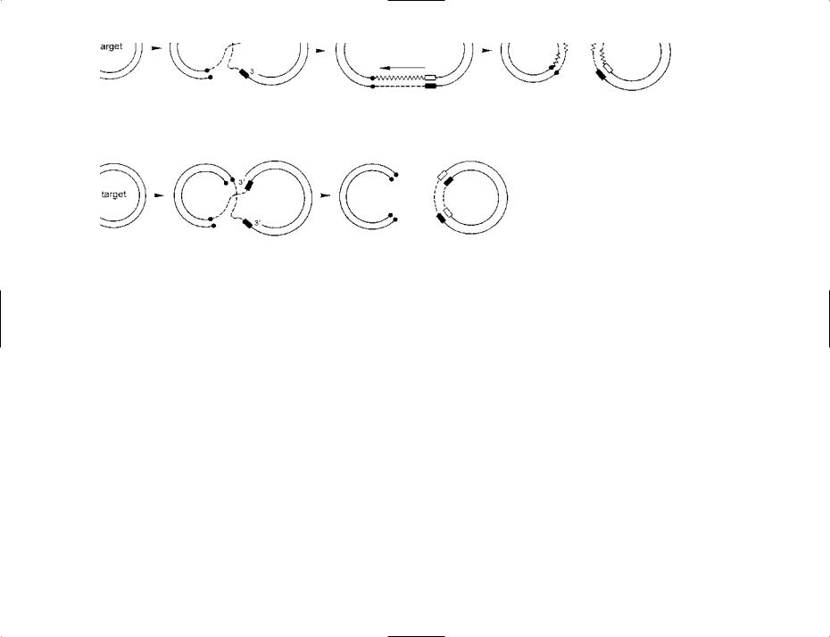
TRANSPOSABLE ELEMENT A simplified, diagrammatic scheme for the replicative transposition (top) and simple (‘cut-and-paste’) transposition (bottom) of a transposable element (TE).
Replicative transposition. Two circular, double-stranded DNA molecules are shown at the left-hand side. The donor molecule includes a TE (dashed lines), either side of which is an old target site (l); this target site was duplicated when the TE was originally inserted into that molecule (see later). The target molecule has a single target site (n) where the TE element will be inserted.
An enzyme (transposase – not shown) mediates at least the initial stages of transposition. In the target molecule a staggered break has been made at the target site, leaving the 3′ ends recessed. In the donor molecule a nick has been made in each strand of the TE, at opposite ends, and the free ends have been ligated (joined) to the 5′ ends of the target molecule, as shown.
In the next stage, DNA synthesis (zigzag line) has occurred from each 3′ end in the target molecule (arrows). Such synthesis first copies the target site ( ) and then continues beyond the target site – using each strand of the TE as template; that is, the TE has been replicated. The end of each newly synthesized strand has been ligated to a free strandend in the donor molecule. The resulting structure is called a cointegrate.
The final stage of replicative transposition involves a resolvase – an enzyme, encoded by the TE, which resolves the cointegrate by mediating site-specific recombination at a site in each TE – forming the two molecules as shown.
The donor and target molecules now both contain the sequence of the TE. Note that each of these two sequences contains parts of the original TE (dashed lines) as well as newly synthesized DNA (zigzag lines). Note also that the target site in the target molecule has been duplicated – each of these target sites consisting of one newly synthesized strand ( ) and one original strand (n).
Simple (‘cut-and-paste’) transposition. The initial stages are similar to those shown for replicative transposition. However, in this case, DNA synthesis (from the 3′ ends of the target molecule) is required simply to duplicate the target site. The remaining strand-ends of the TE have been cut and ligated to the freshly duplicated target sites, as shown. The phrase donor suicide is used if the remainder of the donor molecule is non-viable.
Figure reproduced from Bacteria in Biology, Biotechnology and Medicine, 6th edition, Figure 8.4, pages 202–203, Paul Singleton (2004) John Wiley & Sons Ltd, UK [ISBN 0-470- 09027-8] with permission from the publisher.