

Dictionary of DNA and Genome Technology
.pdfPart II
SDA (strand displacement amplification) – part II: the amplification phase (diagrammatic).
(a)A double-stranded product derived from the target-generation phase. In this product the antisense (3′-to-5′) strand contains a nick-
able HincII restriction site (indicated by the arrowhead); this nickable site was derived from the tag in primer S2 (see part I (g)). When this restriction site is nicked, DNA synthesis proceeds from the 3′ end at the nick site; during synthesis, the polymerase dis-
places the downstream section of the strand, producing an antisense copy of the target sequence which is flanked, on each side, by half a restriction site. Moreover, DNA synthesis from the 3′ end of the nick site regenerates the original duplex. Both of these
238

Seamless® cloning kit
For the determination of molecular weights, proteins are denatured with SODIUM DODECYL SULFATE: an anionic detergent that binds cooperatively to proteins, forming rod-like SDS–polypeptide complexes in which surface charges on the protein are masked by SDS. Most proteins bind SDS in proportion to their molecular weight, so that the electrophoretic mobility of a given protein will be related to its molecular weight. Electrophoresis is carried out with the proteins under study together with one or more proteins of known molecular weight which act as standards for comparison.
For separating proteins in a mixture, maximum resolution can be achieved by using the Laemmli system in which the proteins in a sample are first compressed into a thin band by electrophoresis within a stacking gel before entering the socalled resolving (= running or separating) gel in which they are subjected to the sieving action of a gel structure with a smaller pore size.
Seamless® cloning kit A kit (Stratagene, La Jolla CA) which can be used e.g. for expression cloning, for the preparation of chimeric genes, and for site-specific mutagenesis; it involves the facilitated introduction of a fragment of interest into a circular vector.
Initially, the fragment of interest, and the functional region of the vector, are both amplified (separately) by PCR with the use of specialized primers. Each primer’s 5′ terminal region
contains the recognition sequence of the (type IIS) restriction endonuclease Eam1104I (see table in the entry RESTRICTION ENDONUCLEASE); this enzyme cuts at a fixed distance from its recognition sequence – a feature that allows the fragment to be cloned without introducing additional nucleotides (see later).
The PCR reaction mixture contains a (high-fidelity) DNA polymerase which minimizes the introduction of mutations.
The final five cycles of the PCR reaction are carried out with 5-methyl-dCTP, thereby modifying, and thus protecting from subsequent cleavage, any Eam1104I sites which happen to be within the fragment or vector sequence.
After PCR the (linear) products from both the fragment and vector are mixed and digested with Eam1104I. Eam1104I binds to the termini of each product and cleaves each portion of the product derived from the 5′ flap of a primer – i.e. that part which contains the enzyme’s own recognition and cutting sites; hence, these sequences are not included in the final products.
The Eam1104I-cleaved products are then ligated with T4 DNA ligase. Circularized molecules, containing both vector and fragment components, can be used to transform cells of
Escherichia coli.
Chimeric genes may be constructed by using two different fragments, each fragment contributing to a chimeric product.
SDA (continued )
products – the antisense copy and the regenerated duplex – are shown at (b). Notice that the restriction site in the regenerated duplex is still nickable; this is because the regenerated site contains only one modified nucleotide (in the 5′-GAC-3′ part of site) and this does not affect nickability (see entry).
(b)Products formed from the double-stranded structure at (a) by nicking, strand displacement and regeneration of the duplex. This process is repeated cyclically, with continual regeneration of the original duplex and the production of many copies of the antisense strand.
(c)One of the antisense strands from (b). Each of these strands can bind primer S1. Extension of S1 – and of the (recessed) 3′ end of the antisense strand – produces the product shown at (d). During extension of the (recessed) 3′ end of the antisense strand, a
modified, non-nickable HincII restriction site is formed (nn); the site is non-nickable because the three terminal nucleotides in this strand (3′-CAA-5′) include two modified nucleotides (dATPaS).
(d)This product has a nickable restriction site (arrowhead) derived from primer S1. Like the product at (a), it can undergo a cyclical process of nicking, strand displacement and regeneration of the duplex, displacing many copies of a single-stranded product. Note that this single-stranded product is a sense strand of the target sequence (whereas the strand formed from (a) is an antisense strand). The sense strand and regenerated duplex are shown at (e).
(e)The products from (d). The sense strand will be able to bind primer S2.
(f)The sense strand from (e) has bound primer S2. DNA synthesis (i.e. extension from S2, and also extension from the recessed end of the sense strand) gives rise to the product shown at (g). (DNA synthesis at the 3′ end of the sense strand produces a non-
nickable restriction site for the reason given under (c), above.)
(g)This product is equivalent to the one shown at (a).
Summarizing, double-stranded products from the target-generation phase undergo cyclical nicking, strand displacement and regeneration of the original duplex, forming numerous copies of antisense and sense strands of the target duplex. Antisense strands bind primer S1 and form the (double-stranded) products that yield sense strands; sense strands bind primer S2 and form the (double-stranded) products that yield antisense strands. Nickable HincII restriction sites are provided by the 5′-tags on S1 and S2 primers (which are present in large numbers in the reaction mixture).
Parts I and II are modified from Figures 5.3 and 5.4, pages 134–137, in DNA Methods in Clinical Microbiology [ISBN 07923-6307-8], Paul Singleton (2000), with the kind permission of Springer Science and Business Media.
239

second strand
second strand (of cDNA) See fiRST STRAND.
secondary homothallism (homoheteromixis) (fungal genetics) In some heterothallic fungi: a seemingly homothallic state, involving a self-fertile thallus, which can occur e.g. when a given spore incorporates nuclei of two compatible mating types – leading to a self-fertile thallus. In such cases the compatibility of nuclei may be based on a one-locus two-allele system (the process then being termed homodimixis) or based on a oneor two-locus multi-allele system (homodiaphoromixis).
segmented genome Any viral genome that consists of two or more pieces of nucleic acid. Segmented genomes occur e.g. in the bunyaviruses (negative-sense/ambisense ssRNA); in the influenzaviruses A and B (negative-sense ssRNA); in reoviruses (dsRNA); and in bacteriophage ϕ6 (dsRNA).
segregation (of plasmids) (syn. partition) See PLASMID. segregation lag A PHENOTYPIC LAG due to the time needed for
segregation of a newly acquired allele. For example, a mutant allele in a bacterial cell that contains multiple chromosomes may not be expressed in the presence of the wild-type alleles; an opportunity for expression may occur when the mutant chromosome has been segregated (by cell division) to a cell which contains no wild-type alleles.
selectively amplified microsatellite polymorphic loci (selective amplification of microsatellite polymorphic loci) See
SAMPL.
selector See SELECTOR-BASED MULTIPLEX PCR.
selector-based multiplex PCR A form of PCR in which a range of different target sequences can be amplified in one assay with a single pair of primers (cf. conventional MULTIPLEX
PCR).
For each of the targets to be amplified the reaction mixture contains a target-specific selector. A selector comprises two oligonucleotides: one (the selector probe) of ~70 nt and the other of 34 nt; the shorter oligonucleotide is hybridized to the central region of the selector probe – so that the selector probe contains two terminal (~18 nt) single-stranded targetcomplementary regions. The central, i.e. duplex, region of the selector incorporates a primer-binding motif; this motif is the same in all the selectors (regardless of their target specificity) and is designed for a common (‘universal’) primer pair.
Sample DNA is first digested with appropriate restriction enzymes. Digested DNA is mixed with a pool of selectors (of different target specificities) and denatured. The subsequent circularization of target sequences with selectors is followed by ligation. (Any non-circularized DNA is degraded by exonucleolysis.) The collection of circularized templates is then amplified by PCR using the universal primer pair. [Method: Nucleic Acids Res (2005) 33(8):e71.]
SELEX See APTAMER.
self-activation (syn. autoactivation) (two-hybrid systems) See
e.g. BACTERIOMATCH TWO-HYBRID SYSTEM.
self-reporting primer A 5′-fluorophore-labeled primer hybridized, via a 5′ tag, to a quencher-labeled PNA (Q-PNA) probe; the PNA probe is displaced when double-stranded amplicons
are formed, permitting fluorescence from the primer’s fluorophore [Genome Res (2001) 11(4):609–613].
self-sustained sequence replication (3SR) An early system for the in vitro amplification of nucleic acids; it was based on the retroviral mode of replication [see: Proc Natl Acad Sci USA (1990) 87:1874–1878].
This system is reflected in current methods such as NASBA and TMA.
SENP The designation given to a number of SUMO proteases – SENP1, SENP2, SENP3 etc. – homologous to the Ulp family of proteases: see SUMOYLATION.
sense strand (of DNA) See CODING STRAND.
separate-component-stabilization system (SCS system) See
CCD MECHANISM.
Sequenase® A modified form of the DNA polymerase from bacteriophage T7 which lacks 3′→5′ exonuclease activity but which has strand-displacement activity. The processivity of the enzyme is promoted by complexing it with THIOREDOXIN from Escherichia coli.
sequence capture An approach used e.g. for the isolation of sequence-specific fragments of single-stranded nucleic acid in solution. In this method, a BIOTINylated probe hybridizes to the fragments; the probe is then captured by streptavidincoated DYNABEADS that are held immobilized by a magnetic field while the other, unwanted fragments are removed by washing.
sequence conversion A phrase sometimes used to refer to the treatment of genomic DNA with BISULfiTE (an initial stage in procedures used to detect methylated cytosine residues in the DNA); such treatment is used to convert non-methylated cytosine residues to uracil residues.
sequence-tagged site (STS) A unique, known sequence of nucleotides (several hundred base-pairs) in a given fragment of genomic DNA. STSs can be useful in genomic mapping; thus, for example, they may help in aligning cloned, tagged fragments into an overlapping series – e.g. if a given STS is found in two different fragments these fragments may share an overlapping sequence.
A given STS may be identified by means of an EXPRESSED
SEQUENCE TAG.
sequencing (of DNA) See DNA SEQUENCING.
SEQUEROME See BLAST.
SERE Salmonella enteritidis repetitive element: a repetitive sequence found in the genomic DNA of S. enteritidis; it has been used for typing (see REP-PCR).
serial analysis of gene expression See SAGE.
serine proteinase Any proteinase which has a serine residue at the active site. Examples of serine proteinases: e.g. ENTERO-
KINASE and THROMBIN.
serine recombinase See RECOMBINASE.
SET SOLUBILITY ENHANCEMENT TAG.
sex chromatin Syn. BARR BODY.
sex determination assay See entry DNA SEX DETERMINATION
ASSAY.
sex-linked chromosome Syn. HETEROSOME.
240

sickle-cell anemia
sex-linked disorder Syn. X-LINKED DISORDER. sex pilus Syn. PILUS.
sexual PCR Syn. DNA SHUFflING.
Sf9 cells Ovarian cells from the lepidopteran insect Spodoptera frugiperda which are widely used for baculovirus-mediated overexpression of recombinant proteins. These cells can be grown in monolayers or in suspension, and growth occurs in serum-free media.
(See also MIMIC SF9 INSECT CELLS.) sfiA gene See SOS SYSTEM.
SFM-adapted Refers to any cell line which has been adapted to growth in serum-free media.
Sgal See BLACK–WHITE SCREENING.
Shine–Dalgarno sequence See 5′-UTR.
SHOM Sequencing by hybridization to oligonucleotide microchips: a method used e.g. for sequencing PCR-amplified DNA by hybridization to an array of oligonucleotides immobilized on gel-based microchips.
The method was used for diagnostic investigations, e.g. the detection of mutations in chromosomal DNA of patients with β-thalassemia.
short hairpin RNA (shRNA) A short, engineered molecule of RNA (~60 nucleotides in length) which forms a stem–loop structure and which can be cleaved in vivo (within cells) to an SIRNA able to induce gene silencing (RNA INTERFERENCE).
shRNAs can be synthesized by in vivo transcription from an appropriate DNA target sequence within a plasmid or viral vector; the DNA target sequence may be synthesized chemically as two single-stranded complementary oligonucleotides that are hybridized before ligation into the vector.
In vivo transcription of shRNAs from an appropriate DNA target sequence often employs the promoter of RNA polymerase III. The use of RNA polymerase III for this purpose was reported to be advantageous in that transcription ends predictably at the second thymidine residue in a sequence of more than four thymidine residues in the template – avoiding the addition of a poly(A) tail to the transcript (which would inhibit the gene-silencing activity).
A further advantage of the RNA polymerase III promoter is that it supports transcription in many types of cell; shRNAs can be expressed constitutively in such cells. However, the RNA polymerase II promoter can also be used for expressing shRNAs, and a system based on RNA polymerase I was used for a species-specific expression vector for shRNA [Nucleic Acids Res (2007) 35(2):e10].
Once synthesized in vivo, an shRNA is cleaved and the product (siRNA) enters the RNA interference pathway.
A method for generating shRNA libraries involves random enzymatic digestion of relevant cDNA molecules by DNase I. The resulting fragments of cDNA are ligated to hairpin adaptor molecules (one adaptor at each end) which contain the recognition sequence of RESTRICTION ENDONUCLEASE MmeI. This restriction enzyme cleaves at a site 20 nt in the 3′ direction from the binding sequence (see table in the entry RESTRICTION ENDONUCLEASE), i.e. in each case it cuts the
fragment at a fixed distance from the (closed) end of the hairpin adaptor. The resulting pieces – all of the same length, and consisting of both fragment and adaptor DNA – therefore contain a hairpin adaptor at one end and a 2-nt 3′ overhang at the cut end of the fragment.
Another adaptor is then ligated to the sticky end of each fragment. PCR amplification of the resulting structure yields dsDNA containing the original hairpin sequence and flanking regions of target DNA. Appropriate target DNA is inserted into vectors at a site downstream of an RNA polymerase III promoter.
A ligand-controlled aptamer–shRNA fusion transcript has been used to regulate gene expression in mammalian cells [RNA (2006) 12:710–716].
shRNAs have been used to knock down the expression of several target genes within the same cell in an arrangement using a tetracycline-regulated RNA polymerase II promoter [BioTechniques (2006) 41(1):64–68].
[An shRNA vector for high-efficiency RNAi in embryonic stem cells: BioTechniques (2007) 42(6):738–743.]
short patch repair Syn. UVRABC-MEDIATED REPAIR.
short sequence repeats (SSR) A non-specific term that may apply e.g. to MICROSATELLITE DNA or minisatellite DNA.
(See also SNR.)
short tandem repeat See STR.
shotgun sequencing A method used for sequencing genomes (or large molecules of DNA). A sample of the DNA is cut extensively with a RESTRICTION ENDONUCLEASE and a large number of the fragments are cloned and sequenced. Because of overlap among the fragments, sequence data obtained from individual clones can be amalgamated to reveal long, continuous sequences within the genome. However, usually there are gaps in the genome’s sequence because some parts of the genome may not have been cloned. A missing sequence may be found by using PCR to copy that sequence from genomic DNA – see e.g. PACE.
shRNA SHORT HAIRPIN RNA.
shuffling (of DNA) See DNA SHUFflING.
shufflon In e.g. the IncIα plasmid R64: a set of DNA sequences which can invert, singly or in groups, to form complex rearrangements that may influence the specificity of cell–cell contact in conjugation.
shuttle vector (bifunctional vector) A CLONING VECTOR, or other type of vector, which is capable of replication in more than one type of organism – for example, in (the bacterium)
Escherichia coli and (the yeast) Saccharomyces cerevisiae; the vector must include an appropriate origin of replication for each type of organism.
(See also BACMID.)
sickle-cell anemia An inherited disorder involving the β-chain of hemoglobin; the mis-shaped erythrocytes (red blood cells) are eliminated, leading to (sometimes fatal) anemia.
Sickle-cell trait refers to the generally less serious condition in those heterozygous for the gene; it may be asymptomatic, and it confers a degree of resistance to falciparum malaria.
241

sickle-cell trait
[Correction of the sickle-cell mutation in embryonic stem cells: Proc Natl Acad Sci USA (2006) 103(4):1036–1040.]
(See also THALASSEMIA.)
sickle-cell trait See SICKLE-CELL ANEMIA.
SIDD Stress-induced duplex destabilization: destabilization at a particular region in duplex DNA (a SIDD site), promoting strand separation, in response to a given level of superhelicity. SIDD appears to be frequently involved in the regulation of promoters and origins of replication.
In Escherichia coli, transcription from the ilvPG promoter is apparently preceded by destabilization at an upstream SIDD site; following the binding of integration host factor (a multifunctional E. coli heterodimeric protein) near the SIDD site, superhelical energy may be transmitted to the promoter’s –10 sequence, facilitating the formation of an open complex and the initiation of transcription. In the E. coli genome, SIDD sites appear to be closely associated with promoters [Genome Res (2004) 14(8):1575–1584].
In Saccharomyces cerevisiae the autonomously replicating sequences (ARSs) appear to be much more susceptible than other regions to the influence of superhelically driven duplex destabilization [PLoS Comput Biol (2005) 1(1):e7].
SIGEX Substrate-induced gene expression screening: a method in which induction of gene expression by substrates is used as an approach for the isolation of catabolic genes from metagenome libraries [Nat Biotechnol (2005) 23:88–93].
signal amplification An approach used for detecting and/or quantitating a specific DNA or RNA target sequence within a given sample; essentially, each target sequence in the sample is linked to an enzyme which, with a suitable added substrate, generates an amplified signal whose intensity indicates the number and/or location of target sequences in the sample.
Signal amplification avoids a major problem associated with TARGET AMPLIfiCATION methods (such as PCR), i.e. the contamination of reaction mixtures with AMPLICONS from earlier assays. One disadvantage is that this method provides no copies of the target sequence for analysis.
One example of signal amplification is the BDNA ASSAY;
another is TYRAMIDE SIGNAL AMPLIfiCATION. signal peptide Syn. SIGNAL SEQUENCE.
signal sequence (signal peptide) In certain exported/secreted proteins: a specific N-terminal sequence of amino acid resides that directs the protein into, or through, the cytoplasmic membrane; typically, the signal sequence is cleaved during translocation in the membrane. Signal sequences are found in prokaryotic and eukaryotic cells, although the translocation of proteins occurs by distinct processes in the two types of cell.
In Escherichia coli (and in other Gram-negative bacteria) signal-sequence-dependent translocation is involved e.g. in the type II mode of export/secretion of proteins. In some cases a protein may also include other sequence(s) that help to guide it to the correct destination; for example, a protein targeted to the membrane (as opposed to a secreted protein) may include a membrane anchor sequence which binds the
mature protein on or within the membrane. In the Braun lipoprotein (which links peptidoglycan to the outer membrane in E. coli) the signal sequence is cleaved and replaced by a fatty acid residue; this post-translational modification appears to target the protein to its correct destination.
In the so-called Tat system, which is involved in exporting folded proteins across the cytoplasmic membrane, the signal sequence characteristically includes two consecutive arginine residues (hence Tat: twin arginine translocation).
Some other modes of protein export/secretion in the Gramnegative bacteria are not characterized by an N-terminal signal sequence. For example, secretion via the type I system in E. coli (involving a so-called ABC exporter) is characterized by a C-terminal secretion sequence.
signal transducers and activators of transduction (STATs) In mammalian cells: a family of cytoplasmic proteins which are activated by phosphorylation when certain types of signaling molecule (e.g. interferons, interleukins) bind to cell-surface receptors. The binding of a signaling molecule initially activates a tyrosine kinase of the JAK (Janus kinase) family which then activates a STAT.
There are at least seven different types of STAT. When activated, STATs form homoor heterodimers, according to the type of STAT, which then promote transcription of relevant gene(s).
signal transduction pathway Any pathway which involves the sequential transmission of a signal along a series of two or more components – often involving activation of particular components e.g. by phosphorylation.
For some examples of methods used for investigating signal transduction pathways see e.g. CYTOTRAP TWO-HYBRID
SYSTEM and PATHDETECT SYSTEMS.
(See also TWO-COMPONENT REGULATORY SYSTEM.)
signature-tagged mutagenesis (STM) An approach used e.g. for detecting those genes (in a pathogen) which are essential for the pathogen’s growth in a host organism.
The principle of STM is shown (diagrammatically) in the figure.
The method has been used successfully e.g. for identifying a PATHOGENICITY ISLAND in Salmonella typhimurium. It was also used to identify genes, in Mycobacterium tuberculosis, involved in parasitism of human macrophages [Infect Immun (2007) 75(1):504–507].
The possibility of a false-positive result in STM may arise e.g. if (during the random mutagenesis) a transposon inserts into a non-virulence gene in a cell which (by chance) already contains a mutation in a virulence gene; with this scenario, the given tag would be absent (or low level) in the ‘recovered’ pool, thus giving a false indication for the particular gene inactivated by the transposon.
STM has also been employed to generate attenuated strains of Salmonella choleraesuis as candidates for a live vaccine [Infect Immun (2005) 73:8194–8203].
In a non-medical context, the STM principle has been used to study the survival of certain bacteria (e.g. Desulfovibrio) in
242
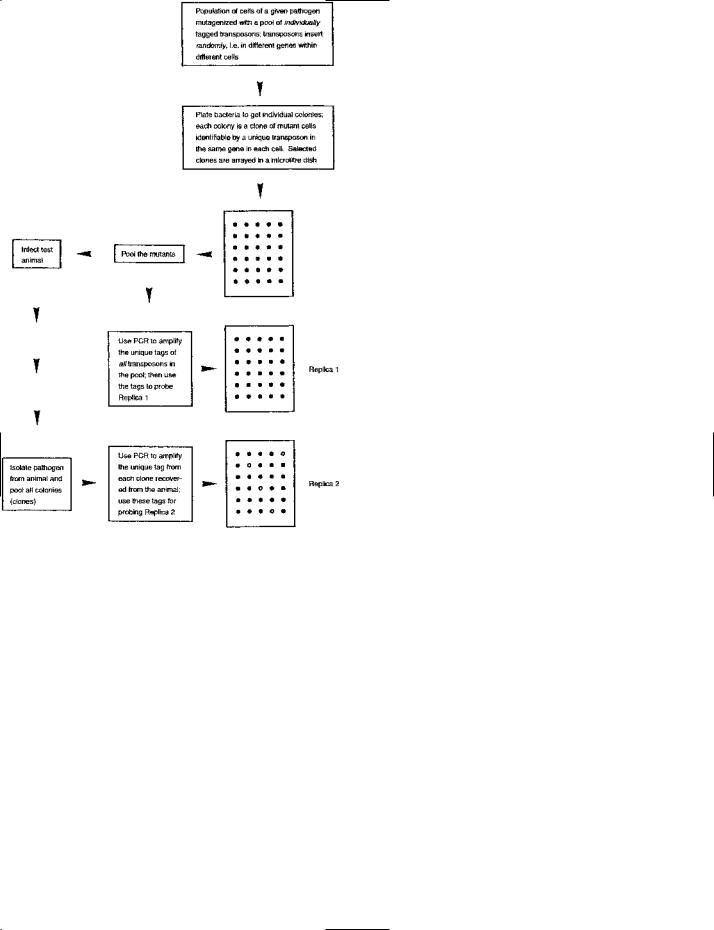
SIGNATURE-TAGGED MUTAGENESIS: a method used e.g. for detecting virulence-associated genes in a bacterial pathogen. The figure shows the general principle diagrammatically.
A unique sequence (‘tag’), about 40 base-pairs in length, is inserted into each of a population of transposons, i.e. the tag in a given transposon is different from that in other transposons. Each tag is flanked, on both sides, by a primer-binding site; these primer-binding sites are the same in all transposons. The transposons are used to mutagenize a population of cells of the pathogen, and they insert – randomly – into different genes in different cells.
The mutagenized cells are plated to form individual colonies. In a given colony of mutant cells, each cell contains the same uniquely tagged transposon in the same gene. A number of colonies are chosen, and an inoculum from each colony is arrayed, separately, in a
243

silencing
anaerobic sediment [Appl Environ Micobiol (2005) 71:7064– 7074].
Another method – IVET – is used e.g. to detect those genes in a pathogen which are induced (i.e. which become active) during infection of the host animal. Like STM, this method is also used in a non-medical context.
silencing (of genes) See GENE SILENCING. silencing (of miRNAs) See ANTAGOMIR.
silent mutation Any mutation lacking a phenotypic effect.
(cf. SAME-SENSE MUTATION.)
Note that these mutations may not be ‘silent’ in DNA-based identification tests (see e.g. SSCP ANALYSIS).
silent origin See e.g. ARS.
simian foamy virus See SPUMAVIRINAE. simian virus 40 See SV40.
simple sequence repeats A non-specific term which may apply
e.g. to MICROSATELLITE DNA.
The term has also been used to include single-nucleotide repeats (SNRs) [J Clin Microbiol (2006) 44(3):777–782].
simple transposon (class II transposon) See TRANSPOSON. SINE Short interspersed element: any one of a number of rep-
etitive elements, each typically <500 nt long, dispersed in the genomes of at least some mammalian species (including the human genome). The SINEs include ALU SEQUENCES.
(cf. LINE.)
single-base extension (SBE) A method used to test for the presence of a given base at a specific location. Essentially, the presence of the base, at a specific location in a strand, is determined by hybridizing a primer (or probe) whose 3′ end can be extended with a nucleotide that base-pairs with the particular base in question. Extension of the primer or probe
with a known nucleotide argues for the presence of the given, complementary nucleotide at the given location. The primer or probe is extended by a single dye-linked chain-terminating dideoxyribonucleoside triphosphate (ddNTP); such extension is detected via the labelled product or, as in QEXT, by a FRET- based reaction.
This procedure can be used e.g. to detect a point mutation at a specific location.
(See also
ION.)
single-base repeat Syn. single-nucleotide repeat: see SNR. single-cell electroporation See ELECTROPORATION. single-cell in vivo fluorescence See DFRS PLASMID. single-chain antibody (intrabody) See GEME THERAPY. single-chain variable fragment (scFV) A type of engineered
antibody which has been used e.g. in IMMUNO-PCR.
[Use of scFV in phage display libraries for studying a tumor antigen: PNAS (2006) 103(4):1041–1046.]
single-nucleotide polymorphism See SNP. single-nucleotide repeat See SNR.
single-strand binding protein (SSB protein; helix destabilizing protein) Any of various proteins that bind specifically and co-operatively to single-stranded DNA in processes such as DNA replication and repair; they may stabilize and protect ssDNA but apparently do not unwind a duplex.
single-strand conformation polymorphism analysis See SSCP
ANALYSIS.
single-strand rescue See HELPER PHAGE. single-strand-specific nuclease Any of a category of nucleases
that degrade single-stranded DNA or single-stranded regions in double-stranded DNA; this kind of enzyme is used e.g. to
SIGNATURE-TAGGED MUTAGENESIS: (continued ) microtiter dish. (The dish shown in the figure has 30 wells, but larger dishes are normally used.) Two replica ‘blots’ of the array are made on membranes (for subsequent DNA hybridization studies); in these blots the cells are lysed and their chromosomal DNA is exposed and fixed to the membrane.
Cells are taken from each of the wells and pooled. The pool is used in two ways. First, it provides an inoculum for infection of the test animal. Second, cells from this (‘input’) pool are lysed, and PCR (polymerase chain reaction) is used – with labeled primers – to amplify the unique tags of all transposons in the pool. The amplified, labeled tags are then used as probes on one of the replica blots (Replica 1); in this blot, each unique tag should hybridize with the DNA from cells containing the corresponding transposon. This ‘pre-screening’ process on Replica 1 checks for efficient amplification of the unique tags.
The pathogen is then recovered from the test animal by plating an appropriate specimen. The resulting colonies are pooled (forming the ‘recovered’ pool), and PCR is used (with labeled primers) to amplify the tag from each clone in this pool. The amplified, labeled tags are then used to probe the second replica blot (Replica 2).
Considering the original, mutagenized cells (in the ‘input’ pool), the cells of interest are those which, through a (transposon-mediated) mutation, are unable to grow within the test animal, i.e. cells whose virulence has been lowered. Such cells will be absent, or few in number, in the ‘recovered’ pool – compared with cells which have grown normally in the test animal. Hence, the signature tags of these ‘virulence-attenuated’ cells will be present in the input pool but absent (or rare) in the recovered pool; consequently, such cells can be identified by hybridization in Replica 1 but an absence of (or weak) hybridization in Replica 2 (see figure).
In a given, ‘virulence-attenuated’ clone, the relevant gene can be identified from the inserted transposon. This gene can be isolated and sequenced for further study.
Figure reproduced from Bacteria in Biology, Biotechnology and Medicine, 6th edition, Figure 8.22, pages 256–257, Paul Singleton (2004) John Wiley & Sons Ltd, UK [ISBN 0-470-09027-8] with permission from the publisher.
244

site-specific recombination
remove terminal overhangs in fragments of DNA which have been cut by certain types of restriction endonuclease (such removal being achieved without significantly affecting the double-stranded regions). (See also NESTED DELETIONS.)
Another useful feature of these enzymes is their ability to nick one strand at the site of a single-base-pair mismatch in (heteroduplex) DNA; this is exploited e.g. in ECOTILLING.
The category of single-strand-specific nucleases includes enzymes that have similar zinc-dependent active-site motifs; they include endonuclease CEL I (from celery), endonuclease P1 (from the fungus Penicillium citrinum), ENDONUCLEASE S1 (obtained originally from the fungus Aspergillus oryzae), and the mung bean nuclease (from Phaseolus aureus).
siRNA Small interfering RNA: an effector molecule involved
in RNA INTERFERENCE (q.v.).
For experimental purposes, siRNAs can be generated in a number of ways, for example:
(a)Chemical synthesis (commercially available) according to specified target sequences.
(b)In vitro transcription of target sequences using e.g. T3 or T7 polymerase: enzymes which can work with simple transcription signals without incorporating (an unwanted) poly(A) tail in transcripts; poly(A) tails can inhibit the silencing effect in RNAi. Relevant cDNAs can be initially amplified by PCR
with primers that incorporate a T3 or a T7 promoter sequence at the 5′ end; the resulting amplicons are then transcribed.
(c)Cleavage of dsRNA with bacterial RNase III.
(d)In vivo transcription with cellular RNA polymerase III. By using a promoter at each end of the target sequence it is possible to obtain sense and antisense transcripts. The use of RNA polymerase III may be facilitated by means of an inducible minimal RNA polymerase III promoter activated by a transactivator in the presence of doxycycline; induction in a dose-dependent manner is achieved by the administration of doxycycline [Nucleic Acids Res (2006) 34(5):e37].
(e)RNAi may be induced in target cells by in vivo expression of short hairpin RNAs (see SHORT HAIRPIN RNA) which are then cleaved, in situ, to siRNAs.
Using siRNAs
siRNAs modified with 2′-deoxy-2′-fluoro-β-D-arabinonucleo- tide units had improved activity and serum stability, suggesting possible use in therapy [Nucleic Acids Res (2006) 34(6): 1669–1675]. [siRNAs for distinguishing between genes that differ by one nucleotide: PLoS Genetics (2006) 2(9):e140; an assessment of siRNA potency by a secreted luciferase assay: BioTechniques (2007) 42(5):599–606.]
In a novel approach to the intracellular delivery of siRNAs, an siRNA was linked, via a streptavidin bridge, to an aptamer that was known to bind to prostate tumor cells; the conjugate (i.e. siRNA plus aptamer) was taken up by cells within 30 minutes following simple addition of conjugate to the cells, and siRNA-mediated inhibition of gene expression was found to be as efficient as that produced by lipid-based internalization reagents [Nucleic Acids Res (2006) 34(10):e73].
Single-stranded siRNA molecules may also induce RNAi
but are generally less effective. However, boranophosphatemodified single-stranded siRNA was reported to have high potency [Nucleic Acids Res (2006) 34(9):2773–2781].
Transcriptional gene silencing (TGS)
siRNAs can also mediate a process known as transcriptional gene silencing (TGS) in mammalian and other cells (e.g. cells of the small plant Arabidopsis and the fission yeast Schizosaccharomyces pombe).
In some cases, TGS involves siRNA-mediated methylation of DNA as the silencing mechanism (so-called RNA-directed DNA methylation, RdDM).
In some cases the mechanism of TGS apparently involves methylation of histones.
sis (SIS) An ONCOGENE identified in simian sarcoma virus. The c-sis product is the B chain of platelet-derived growth factor (PDGF). The viral gene encodes an almost-identical B chain which is the transforming agent in virus-infected cells.
site-directed mutagenesis (or site-specific mutagenesis) (DNA technol.) Any form of engineered MUTAGENESIS involving change(s) in target molecules at pre-determined site(s). Older methods (e.g. D-loop mutagenesis) have been superseded by much simpler ones – see e.g. QUIKCHANGE SITE-DIRECTED
MUTAGENESIS KIT and MEGAPRIMER MUTAGENESIS; see also EXSITE PCR-BASED SITE-DIRECTED MUTAGENESIS KIT and GENETAILOR.
A deletion can be introduced by using INVERSE PCR to copy all except an internal sequence of the target; the amplicons are ligated (making a shortened form of the target sequence), and the product is subjected to second-round amplification. This technique can also be used for making point mutations, frameshift mutations, truncations etc. [BioTechniques (2005) 38:864–868].
In PCR, insertions can be introduced e.g. via 5′ entensions on the primers.
Site-directed insertion in vivo (within cells) may be carried out e.g. by using a targeting vector to prepare an insertion site
and subsequently using RECOMBINASE-MEDIATED CASSETTE
EXCHANGE to exchange the insert for target DNA [see e.g. Figure 1 in Proc Natl Acad Sci USA (2005) 102(18):6413– 6418].
site-specific biotinylation See BIOTIN.
site-specific cleavage (of proteins) Cleavage of proteins at sites between specific amino acid residues. Some of the enzymes that can carry out site-specific cleavage are exploited in DNA technology.
(See e.g. EKMAX, SUMO protease in SUMOYLATION, TEV protease in TEV; see also ENTEROKINASE and THROMBIN; see
also SORTASE.)
site-specific mutagenesis Syn. SITE-DIRECTED MUTAGENESIS. site-specific recombination (SSR) A form of RECOMBINATION between specific dsDNA sequences in the same molecule, or in different molecules, in which there is neither synthesis nor degradation of DNA; this (ATP-independent) process is mediated by a site-specific RECOMBINASE. The two sequences of dsDNA that are recognized by a given recombinase are not
245

six-histidine tag
necessarily identical.
Examples of SSR: (i) integration of the PHAGE LAMBDA genome into the bacterial chromosome, and excision of the phage genome – the phage attachment site, attP, not being identical to the bacterial site, attB; (ii) PHASE VARIATION in the Salmonella filament protein; (iii) the CRE–LOXP SYSTEM; and (iv) insertion of a cassette into an INTEGRON.
six-histidine tag (6xHis tag) A sequence of six consecutive histidine residues, incorporated into some recombinant proteins, which facilitates detection (by anti-His antibody) or purificat-
ion (by a NICKEL-CHARGED AFfiNITY RESIN).
size fractionation (DNA technol.) Any procedure in which a population of DNA molecules/fragments – present in a range of sizes – is treated so as to obtain physical separation of the molecules/fragments according to size.
For example, a population of fragments, in a range of sizes, may be subjected to electrophoresis in an agarose gel; a particular band of fragments, of appropriate size, can be excised, purified (i.e. agar removed), and then used for the required purpose.
Size fractionation may be used e.g. for selecting fragments of appropriate size(s) from a restriction-digested genome in order to use such fragments in cloning vectors which accept inserts only within a certain range of sizes.
Skyline (DNA notation) See DNA SKYLINE.
sliding A movement made by certain DNA-binding proteins: translocation lengthwise along a DNA duplex until the appropriate binding site is reached. For example, sliding of the lac repressor protein along the duplex occurs until it reaches the operator.
slipped-strand mispairing (SSM) Mispairing between strands during DNA replication in susceptible regions of the genome; regions susceptible to SSM are characterized e.g. by multiple tandemly repeated short sequences of nucleotides – such as
MICROSATELLITE DNA.
SSM can apparently result in an increase or a decrease in the (normal) number of repeated subunits in a given sequence of microsatellite DNA. In humans, ‘microsatellite expansion’ (i.e. an increase in the number of subunits at a given locus) can cause certain neurological diseases when associated with particular genes (e.g. fragile X disease, Huntington’s disease).
Among bacteria, changes in such sequences are reported to have various effects. For example, they can promote PHASE VARIATION in the opa genes of Neisseria. In Helicobacter pylori, the mutY sequence (encoding a DNA excision-repair function) has a region of eight consecutive adenine residues which appears to be subject to slipped-strand mispairing – the occurrence of SSM being able to cause frameshift mutations that eliminate gene function; this has been interpreted as a mechanism which contributes to phase-variable base excision repair in H. pylori [J Bacteriol (2006) 188(17):6224–6234]. In Escherichia coli strain O157:H7, slipped-strand mispairing may be responsible for a proportion of the instability in the VNTR (variable number of tandem repeats) loci [J Bacteriol (2006) 188(12):4253–4263].
slippery sequence In an mRNA molecule: a sequence of nucleotides – downstream of the initiation codon – which causes slippage of the ribosome along the mRNA. Slippage, which may occur in an upstream (5′) or a downstream (3′) direction, is an integral part of the mechanism of FRAMESHIFTING.
slot blot See DOT BLOT. |
|
small interfering RNA (siRNA) |
See RNA INTERFERENCE; see |
also SIRNA. |
|
small nuclear ribonucleoprotein particles See SNRNAs. |
|
small nuclear RNAs See SNRNAs. |
|
small ubiquitin-like modifier |
See SUMOYLATION. |
smart probe A (singly-labeled) PROBE based on a principle similar to that of the MOLECULAR BEACON PROBE. In this (stem–loop) probe the 5′ end carries a fluorophore that (in the unbound probe) is quenched by guanosine residues which are present in the (3′) complementary stem region.
[Example of use: Nucleic Acids Res (2006) 34(13):e90.] smear-positive specimen See AMTDT.
Smith–Lemli–Opitz syndrome An autosomal recessive syndrome, involving (e.g.) mental retardation, reported to be due to defective cholesterol metabolism resulting from a mutation in gene DHCR7 (encoding 3β-hydroxysterol-∆7 reductase).
(See also GENETIC DISEASE (table).)
SMT3 The SUMO protein in Saccharomyces cerevisiae: see
SUMOYLATION.
Smt4 (Ulp2) See SUMOYLATION.
snip-SNP Any SNP that affects RESTRICTION ENDONUCLEASE
recognition site(s).
[Method for detecting snip-SNPs: BMC Genomics (2005) 6:118. Program for identifying snip-SNPs within reference sequences: BMC Genetics (2006) 7:27.]
SNP Single-nucleotide polymorphism: in the genome of an individual, a variant nucleotide which differs from that at the corresponding site in most other members of the population – but which occurs in at least 1% of other individuals in the population; for example, if 1% of the population has the sequence ...TTCCAT... but the majority have ...TTTCAT... then the minority group have an SNP at the third nucleotide in this sequence. SNPs are common; in the human genome they are found approximately once every 100–300 bases, making a total of several million SNPs in the whole genome. Most of the recorded SNPs in humans involve a C-to-T change.
(See also SNP GENOTYPING.)
SNPs that occur with frequencies below ~5% have been classified as ‘rare’ while those that occur with frequencies >5% are said to be ‘common’.
SNPs are found in coding and non-coding sequences. Many SNPs have no known effect, but some may influence the response e.g. to infectious agents (bacteria, viruses etc.) and chemicals, including drugs; this characteristic is helping to shape pharmaceutical/biomedical research: the comparison of SNP patterns in groups of drug-resistant and drug-sensitive patients may reveal important links between specific SNPs and the efficacy of a given drug.
SNP patterns may also be helpful in elucidating the poly-
246

SOC medium
genic nature of certain diseases in which a given gene is only partly responsible for the pathology.
[Database of >1 million (human) SNPs: Nature (2005) 437: 1299–1320.]
Because of their potential uses in biomedicine, SNPs are being actively characterized and recorded in public databases by various government and commercial organizations. However, one report suggested that some of the SNPs recorded in public databases may be associated with editing sites rather than genuine SNPs [Nucleic Acids Res (2005) 33(14):4612– 4617].
SNPs are also found in prokaryotic and eukaryotic microorganisms. For example, SNPs in the inlB gene of Listeria monocytogenes were used for classification [Appl Environ Microbiol (2001) 67(11):5339–5342], and SNP-based phylotyping has been carried out in Escherichia coli [Appl Environ Microbiol (2005) 71:4784–4792]. The genome of Candida albicans has been mapped on the basis of SNPs [Eukaryotic Cell (2004) 3(3):705–714]. SNP analysis of Mycobacterium tuberculosis (and other microorganisms) is reported to be facilitated by a modified ARMS assay [J Clin Microbiol (2004) 42:1236–1242]. [Global phylogeny of Mycobacterium tuberculosis based on SNP analysis: J Bacteriol (2006) 188 (2): 759–772.]
SNP genotyping The process of examining samples of genomic DNA with the object of detecting and recording the presence of specific SNPs. (cf. SNP MAPPING.) Large collections of validated SNPs are needed in order to facilitate socalled ‘association studies’ in which particular traits (such as susceptibility to a given disease, or resistance to a drug) may be linked to established patterns of SNPs.
Various methods have been proposed for the simultaneous genome-wide typing of thousands of SNPs. One such method
is MOLECULAR INVERSION PROBE GENOTYPING (q.v.).
For detecting small numbers of SNPs (e.g. in studies on specific candidate genes) use can be made of a modified form of ALLELE-SPECIfiC PCR which uses Tm-shift primers. In this method [BioTechniques (2005) 39(6):885–893], the alleles of a given gene are assayed using two allele-specific forward primers, each primer containing a 3′ terminal base which is complementary to a given SNP in one of two allelic variants of the gene; the same reverse primer is used with both of the forward primers.
Each of these two forward primers (one for each allelic variant) has a 5′ GC-rich tag. However, the two types of primer have tags of different length; hence, the products (i.e. dsDNA amplicons) formed from one primer are characterized by a melting temperature (Tm) which differs from that of the products formed from the other primer. Consequently, the melting curve characteristics of a given product will indicate which of the two (allele-specific) primers gave rise to that product. An earlier version of this method, in which only one primer was tagged, was found to be problematic in that, in some cases, the primers were amplified unevenly.
A novel approach to the detection of SNPs was suggested
by the finding that linear dsDNA fragments and oligonucleotides form highly stable RecA-mediated terminal/subterminal triple-stranded structures specifically when the single strand is complementary to the 5′-phosphate end of a strand in the dsDNA; the stability of the structure is drastically reduced by a single mismatched base, regardless of its location. In this scheme, sample dsDNA fragments can be effectively probed by sequence-specific oligonucleotides [DNA Res (2005) 12 (6):441–449].
A number of rare SNPs have been detected by the method known as Ecotilling [Nucleic Acids Res (2006) 34(13):e99].
Detection and quantitation of SNPs can also be achieved by multiplex QEXT.
The term call has been used to refer to a positive identification of a specific SNP in a given sample of genomic DNA. Call rate was used to refer to the number of calls recorded, for a given SNP, in a range of genomic samples, divided by the total number of genomic samples examined (expressed as a percentage).
SNP mapping A phrase used to refer to a procedure in which a given gene is mapped by using SNPs as genetic markers (cf. SNP GENOTYPING). In so-called interval mapping, the object is to locate the gene of interest in the interval between two consecutive SNPs.
SNR Single-nucleotide repeat: a sequence of nucleotides of one type, present at a given genetic locus, which may vary in length (i.e. in number of constituent nucleotides) in different individuals; variation in length may result e.g. from SLIPPED- STRAND MISPAIRING. Mutation rates at such loci are reported to be high, apparently reflecting the ease with which events such as slipped-strand mispairing may occur.
SNRs may be found at various loci in a given genome. SNRs in the genome of Bacillus anthracis (causal agent of anthrax) have been exploited for the TYPING of this pathogen (an organism for which various other typing procedures have proved to be inadequately discriminatory) [J Clin Microbiol (2006) 44(3):777–782].
SNRs are also called ‘mononucleotide repeats’ and ‘singlebase repeats’.
snRNAs (small nuclear RNAs) RNA molecules, <300 nt, rich in uridylic acid residues and capped (see CAP), present in the eukaryotic nucleus. There are six types, designated U1–U6. snRNAs complex with proteins to form small nuclear ribonucleoprotein particles (snRNPs; snurps) which are involved in the SPLICING of pre-mRNAs; they function e.g. as external guide sequences (EGSs) in the (human) splicing reaction, i.e. helping to select a particular splice site.
snRNPs See SNRNAS. snurps See SNRNAS.
SOB system The equivalent of the SOS SYSTEM in the Grampositive bacterium Bacillus.
SOC medium (S.O.C. medium) A medium used for incubating bacteria followiug transformation. The medium contains (per liter): tryptone 20 g, yeast extract 5 g, glucose 3.6 g, together with sodium chloride (10 mM), potassium chloride (2.5 mM),
247