

Dictionary of DNA and Genome Technology
.pdfpBAD-DEST49 vector
natant is transferred to a fresh tube and ethanol is added. Subsequently the supernatant is added to a PAXgene spin column for abstraction of the RNA. Following several washing steps with buffers the RNA is eluted.
The procedure generally removes most of the DNA. For a more rigorous elimination of DNA it is possible to include DNase treatment in the protocol.
Examples of use of the PAXgene™ system: studies on the time course of proinflammatory/immunomodulatory cytokine mRNAs [Infect Immun (2006) 74(7):4172–4179]; studies on expression of genes encoding e.g. the glucocorticoid receptor in children with asthma [Proc Natl Acad Sci USA (2006) 103(14):5496–5501].
(See also NUCLEIC ACID ISOLATION.)
pBAD-DEST49 vector A destination vector in the GATEWAY
SITE-SPECIfiC RECOMBINATION SYSTEM.
pBLOCK-iT™3-DEST vector See table in the entry GATE-
WAY SITE-SPECIfiC RECOMBINATION SYSTEM.
pBluescript® Any of a set of ~3-kb PHAGEMIDS (marketed by Stratagene, La Jolla CA). Derived from plasmid pUC19, the phagemids contain: (i) the replication origin from plasmid ColE1, (ii) the replication origin from bacteriophage f1, (iii) an ampicillin-resistance gene, (iv) a sequence that encodes α- PEPTIDE (part of the lacZ gene), (v) the lac promoter, and (vi) a POLYLINKER, situated between the lac promoter and the α- peptide sequence. The polylinker is flanked by promoter sites for T3 and T7 RNA polymerases (allowing transcription of an insert in either direction).
The f1 origin can be inserted in either orientation – so that either the sense or antisense strand can be amplified if the host cell contains a helper phage.
These vectors can be replicated in strains of Escherichia coli (e.g. JM105 or JM109) which contain a chromosomal lacZ∆M15 mutation that deletes part of the N-terminal region of the enzyme β-galactosidase. Such bacteria produce inactive β-galactosidase – but if the (phagemid-encoded) α- peptide is synthesized it complements the mutant enzyme, by non-covalent binding, to form an active β-galactosidase; the presence of the functional enzyme can be determined e.g. by growing the bacteria on plates containing X-GAL – bacteria containing a functional enzyme form blue colonies.
If the phagemid contains a gene/fragment (inserted in the polylinker), the α-peptide cannot be formed and the cells will therefore lack a functional β-galactosidase; these cells form colorless (white) colonies on X-gal media. Hence, a colony’s appearance indicates whether or not the (intracellular) phagemids are recombinant, that is, whether or not they contain an insert; this is blue–white screening. (See also BLACK–WHITE
SCREENING.)
PBMCs Peripheral blood mononuclear cells. PBP 2a (PBP 2′) See MECA GENE.
PBPs Penicillin-binding proteins: see β-LACTAM ANTIBIOTICS. pBR322 A recombinant PLASMID (~4360 bp) constructed from several naturally occurring plasmids. Small plasmids like this are useful as VECTORS in genetic engineering; they are taken
up by TRANSFORMATION more efficiently (as compared with larger plasmids) and they are also less susceptible to damage during manipulation.
pCAL vectors A set of vectors used in the AFfiNITY PROTEIN
EXPRESSION AND PURIfiCATION system.
pcDNA™6.2/nLumio™-DEST™ |
A destination vector in the |
GATEWAY SITE-SPECIfiC RECOMBINATION SYSTEM. |
|
pcnB mutant (in Escherichia coli) |
See MULTICOPY PLASMID. |
PCR Polymerase chain reaction: primarily a method for copying (amplifying) specific sequences (<100 to >1000 nucleo-
tides) in DNA (or in RNA: REVERSE TRANSCRIPTASE PCR);
millions of copies are produced within a few hours.
[The development of miniaturized on-chip PCR (review): Nucleic Acids Res (2007) 35(13) 4223–4237.]
Patents on PCR are held by Hoffmann–La Roche. Essentially, PCR depends on the ability of a thermostable
DNA polymerase to extend a PRIMER – bound to its target sequence (the AMPLICON) – and to do this, repeatedly, in a process involving temperature cycling: see the accompanying figure. (The use of a thermostable DNA polymerase avoids the need to add fresh polymerase to the reaction mixture after each high-temperature stage to replace the heat-inactivated enzyme.) See notes on temperature cycling (below) and also
the entry THERMOCYCLER.
Alternative methods for amplifying nucleic acids include the LIGASE CHAIN REACTION (LCR) – which also involves temperature cycling. Other methods, e.g. NASBA and SDA, are conducted isothermally, i.e. they do not involve temperature cycling.
Volume of sample, choice of enzyme
PCR is often carried out in a total volume of approx. 25–50 µL, although smaller volumes have been used.
The choice of enzyme (i.e. DNA polymerase) depends on specific requirements; a wide range of thermostable enzymes is available from commercial sources. For example, some DNA polymerases are designed to amplify ‘difficult’ target sequences (e.g. GC-rich sequences: see e.g. ACCUPRIME GC- RICH DNA POLYMERASE); others are designed specifically to introduce errors into the products (in studies on mutagenesis) (see e.g. GENEMORPH).
The TAQ DNA POLYMERASE is used when the products are
required for TOPOISOMERASE I CLONING.
Temperature cycling
As indicated in the figure (legend), temperature cycling often follows the pattern:
→ 95°C → 45–65°C → 72°C → 95°C →
These temperatures are designed for the cyclical repetition of
(i) strand separation (denaturation of the sample dsDNA), (ii) binding (annealing) of primers, on opposite strands, at sites which bracket the amplicon, and (iii) extension (synthesis of DNA from the 3′ terminus of each primer) to form products – i.e. amplicons. Following the synthesis of amplicons, a rise in temperature separates the amplicons from their templates.
176
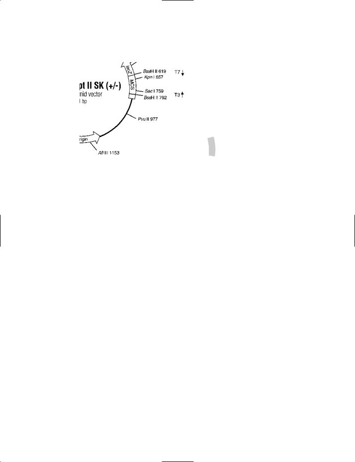
pBluescript® II SK (+ /−) PHAGEMID VECTOR One of a range of phagemid vectors in the pBluescript® series. SK indicates that the orientation of the polylinker is such that transcription of lacZ proceeds in the direction SacI to KpnI.
A vector containing the f1(+) origin (the origin of replication of the filamentous phage f1) permits recovery of the sense strand of the lacZ gene when a strain containing the pBluescript® vector is co-infected with a helper phage. The f1(−) origin permits recovery of the antisense strand of the lacZ gene.
The ColE1 origin of replication can be used in the absence of a helper phage.
The inclusion of a portion of the lacZ gene permits the identification of recombinant phagemids by the blue–white selection procedure (see entry for details).
Courtesy of Stratagene, La Jolla CA, USA.
Although the temperatures for denaturation and extension are usually ~94/95°C and ~72°C, respectively, the annealing temperatures reported in the literature vary significantly. One reason is that GC%-rich primers require higher annealing temperatures in order to avoid ‘mispriming’ – i.e. binding of primers to inappropriate (i.e. non-complementary) sequences in the template strand. (Mispriming leads to the formation of spurious products.) Optimization of the annealing temperature may be achieved e.g. by TOUCHDOWN PCR.
A different approach to avoiding mispriming is HOT-START
PCR.
In some cases, depending e.g. on the primers involved, a two-temperature protocol has been used – e.g. 94°C for the denaturation stage and a combined annealing/extension temp-
erature of 68°C.
Theoretically, temperature cycling could be carried out by manual control, but in routine laboratory conditions it is conducted in an instrument called a THERMOCYCLER (q.v.).
Stringency
The physicochemical conditions (e.g. temperature, electrolyte concentrations) under which the primers hybridize to their respective sites on the template strands affect the accuracy (specificity) of the polymerase chain reaction. For example, if a primer is allowed to hybridize with a template strand at a temperature which is too low, binding may occur aberrantly at a site which is not complementary to the primer’s sequence (and which may be outside the amplicon); thus, extension of the 3′ end of the primer may give rise to a product which is
177
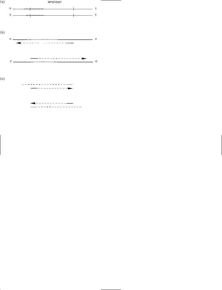
PCR (polymerase chain reaction): the basic method (principle, diagrammatic).
The reaction mixture includes: (i) The sample of (double-stranded) DNA – which may be, for example, genomic DNA or fragments of genomic DNA; (ii) millions of copies of the two types of primer, each primer being about 20–30 nucleotides in length; (iii) a thermostable DNA polymerase (e.g. the Taq DNA polymerase); (iv) deoxyribonuleoside triphosphates of all four types (ATP, CTP, GTP, TTP); and (v) appropriate buffer.
For PCR, the reaction mixture undergoes a cyclical series of changes in temperature, such changes being controlled automatically in an instrument called a thermocycler. The cycle is repeated about 20–40 times, amplification of the target sequence occurring at each cycle.
(a)Part of the sample double-stranded DNA: two antiparallel strands held together by base-pairing. The sequence to be amplified – the amplicon – is shown between the short vertical bars.
(b)Initial heating to about 95°C has separated the two strands of DNA. Subsequently, transient cooling to about 45–65°C has allowed the primers – shown as short, solid lines – to bind (anneal) to their complementary sites on the two template strands. Importantly, notice the positions of the bound primers in relation to the two ends of the amplicon. The temperature is then increased to e.g.
72°C, and DNA synthesis (mediated by the thermostable DNA polymerase, and shown here as a dashed line with an arrowhead) proceeds from the 3′ end of each primer. Each of these newly synthesized strands is longer than the amplicon.
(c)The second round of replication begins with initial heating to ~95°C – separating the new daughter strands (synthesized in the first round of replication) from their templates. Each of the new daughter strands (as well as the two parent strands) can act as a template in the next round of replication. In the diagram, a primer (short, sold line) has bound to each new daughter strand and has been extended (dashed line with arrowhead). Note that the strand synthesized on a new daughter strand is the same length as the amplicon; such strands rapidly become dominant in subsequent rounds of replication.
Reproduced from Figure 4.1 on page 57 of DNA Methods in Clinical Microbiology [ISBN 07923-6307-8], Paul Singleton (2000), with the kind permission of Springer Science and Business Media.
178

PCR (polymerase chain reaction): some of the many variant forms and their uses (see separate entries for each variant form)
Variant form |
Uses (e.g.) |
|
|
Allele-specific PCR |
Amplification of a particular allele of a given gene (in the presence of other alleles of that gene) |
Anchored PCR |
Amplification of an unknown sequence flanking a known sequence |
Asymmetric PCR |
Preparation of probes and the preparation of ssDNA e.g. for sequencing |
Bubble-linker PCR |
See Vectorette PCR (below) |
Competitive RT-PCR |
Estimation of the number of RNA target sequences in a given sample |
Headloop suppression PCR |
Protocol arranged to promote suppression of amplification of sequences related to the given |
|
target sequence (without affecting the amplification of the intended target sequence) |
Hot-start PCR |
Cycling delayed until the temperature of the reaction mixture is sufficient to inhibit non- |
|
specific binding of the primers. The object is to increase the specificity and the sensitivity of |
|
the assay |
IAN-PCR |
Inverse affinity nested PCR: a modification of inverse PCR used for the recovery of flanking |
|
sequences from low-copy-number fragments: see entry METAGENOME |
Immuno-PCR |
Ultrasensitive detection of antigen; the antigen-binding antibody is linked to a DNA fragment |
|
containing a sequence which is amplified by PCR; the amplicons (visualized by gel |
|
electrophoresis) provide an indication of the presence of specific antigen |
Inverse PCR |
Amplification of a sequence that flanks a known sequence |
Island rescue PCR |
Amplification of CpG islands in cloned fragments |
LMPCR |
Amplification of fragments produced in an in vivo footprinting assay (see entry LIGATION- |
|
MEDIATED PCR) |
Methylation-specific PCR |
Monitoring methylation status of CpG islands in genomic DNA |
Quantitative PCR (QPCR) |
Absolute or relative quantitation of DNA or RNA target sequences (see entry QUANTITATIVE PCR) |
Real-time PCR |
Determination of the number of specific target sequences in the reaction mixture prior to cycling |
Relative RT-PCR |
Comparison of the numbers of RNA targets, e.g. transcripts of a given gene, in samples from |
|
different cells/tissues etc. |
Sexual PCR |
See entry DNA SHUFflING. |
Splice overlap extension PCR |
Fusion of two sequences of DNA (present either on the same molecule or on separate |
|
molecules) without the use of restriction enzymes |
Terminal transferase-dependent |
Detection of the presence of any obstruction or barrier to DNA polymerase during replication |
PCR |
|
Touchdown PCR |
A protocol designed to optimize stringency and specificity of the assay by avoiding mispriming |
|
at the annealing stage |
Vectorette (bubble-linker) PCR |
Amplification of an unknown sequence adjacent to a known sequence |
|
|
not the one required. Hence, such low-stringency conditions are not generally used – although they are used e.g. for the initial phase of AP-PCR (q.v.).
Normally, PCR uses high-stringency conditions (which are dictated primarily by temperature). Under such conditions the
binding (annealing) of primers to the target DNA occurs only when they are exactly complementary, or very nearly so, to their appropriate binding sites; thus, the higher the stringency the greater will be the degree of matching required between primer and target sequence for successful binding and, hence,
179

PCR
extension, to occur.
Multiplex PCR
With appropriate experimental arrangements, more than one target sequence can be amplified in a single reaction: see the
entry MULTIPLEX PCR.
Contamination by extraneous DNA
Extraneous DNA that contaminates a reaction mixture may contain (i) sequence(s) to which the primers can bind, and/or (ii) sequence(s) that are able to prime irrelevant sites in the sample DNA. In either event, unwanted sequences may be amplified, resulting in a troublesome mixture of products; in addition, the efficiency of the legitimate amplification may be reduced owing to sequestration of polymerase and nucleotides.
Successful PCR needs rigorous exclusion of contaminating DNA – which may be derived from the environment and/or from the apparatus and reagents.
An important source of contaminating DNA consists of the amplicons from previous reactions in the same laboratory.
There are two basic approaches to this problem: AMPLICON
CONTAINMENT and AMPLICON INACTIVATION (q.v.).
Inhibitors of PCR
In some cases the sample DNA is derived from a source that may include substance(s) inhibitory to PCR; this may give rise to a false-negative result from a sample which, in fact, contains the sequence of interest.
Inhibitors of PCR are found e.g. in various types of clinical specimen – including bone marrow aspirates, CSF (cerebrospinal fluid), feces and urine; specific inhibitory substances include hemoglobin (in red blood cells), lactoferrin (in white blood cells) and various polysaccharides (in feces).
Other inhibitors include AGAR, hemin, and the blood anticoagulants heparin and sodium polyanetholesulfonate (SPS).
Detection of inhibitors can be approached in several ways. For example, if an aliquot of the sample is ‘spiked’ with the target DNA, prior to assay, then failure to amplify the target in this aliquot may suggest the presence of inhibitor(s). This technique, however, increases the risk of contamination.
Alternatively, an internal control can consist of molecules of nucleic acid which (i) include binding sites for the targetspecific primers, but which (ii) can be distinguished from the actual target by a unique PROBE-binding sequence. In the absence of inhibitors the control and target sequences should both be amplified; failure to obtain any amplification could indicate the presence of inhibitor(s), while amplification of the control sequence (only) would suggest the absence of the target sequence in the sample DNA.
The effects of an inhibitor may be reduced by diluting the sample, but this concomitantly reduces the number of copies of the target sequence (if present) and may therefore lower the sensitivity of the assay. An alternative approach uses a magnetic separation technique (see e.g. DYNABEADS): copies of the target sequence are captured, and held, on magnetic beads while any inhibitory substances are removed by washing.
Facilitators of PCR
Certain agents can enhance the efficiency of a PCR assay in the presence of inhibitory substances. These agents include bovine serum albumin (BSA) and betaine; for example, BSA has been reported to improve the performance of PCR in assays on fecal specimens (which characteristically contain a range of inhibitory substances). Some of these facilitators are effective against a variety of inhibitors, while others are more selective in their activity.
It was reported that the accuracy of PCR may be promoted by the presence of the RecA protein.
PCR with damaged/ancient DNA
The amplification of poor-quality DNA, for forensic or other purposes, may be facilitated by the use of DNA polymerases of the so-called Y FAMILY (q.v.). Minute quantities of DNA
may be recovered e.g. by CHARGESWITCH TECHNOLOGY.
Detection of PCR products
Given that the products of PCR (amplicons) are of a precise length, i.e. an exact number of nucleotides delimited by the primer pair, they can be readily detected and identified by gel electrophoresis. Thus, a given product forms a single band at a precise location in the gel; the size of the product forming such a band can be verified by using a so-called DNA ladder (see entry GEL ELECTROPHORESIS). Bands of fragments in a gel may be further examined e.g. by SOUTHERN BLOTTING followed by hybridization of a target-specific, labeled PROBE.
To verify that an amplicon of a specific size and sequence has been amplified, the amplicons can be subjected to a particular RESTRICTION ENDONULEASE directed at a known cutting site in the amplicon; the products of restriction can then be checked by gel electrophoresis to see whether fragments of the expected sizes were produced.
Gel electrophoresis is a slow process. Hence, in some cases use is made of instrumentation that permits monitoring of the amplification (i.e. increase in amplicons) during the reaction: so-called real-time PCR.
Real-time PCR
Real-time detection of PCR products can be achieved with a dye-based approach, a PROBE-based approach, or a combined probe-and-dye-based approach: see the entry REAL-TIME PCR.
Quantitative PCR
PCR can be used to give an estimate of the number of copies of the target sequence which were present in the sample prior to amplification: see QUANTITATIVE PCR.
Some modified protocols
The flexibility of PCR can be illustrated by some examples:
• ALLELE-SPECIfiC PCR: use of an allele-specific primer (and a gene-specific primer) to amplify a particular allele among a mixture of alleles.
• ASYMMETRIC PCR: a procedure in which the concentration of one primer is much lower than that of the other; it is used for obtaining a particular strand of the template dsDNA.
• DNA SHUFflING: a primer-less form of PCR in which fragments of DNA, in the reaction mixture, prime each other; this technique is used to promote in vitro recombination and the
180

PCR-RFLP analysis
development of a product with new properties.
• EXSITE SITE-DIRECTED MUTAGENESIS KIT: a commercial
system in which mutations (including point mutations, additions or deletions) can be introduced into plasmids by using appropriately designed primers in PCR.
• GENEMORPH: a commercial system in which an error-prone DNA polymerase is used for promoting mutagenesis in the amplicons.
• HEADLOOP SUPPRESSION PCR: use of modified primers that selectively suppress amplification of unwanted sequence(s) without affecting amplification of the wanted sequence.
• IMMUNO-PCR: a technique used for ultrasensitive detection of antigens.
• INVERSE PCR: used e.g. for amplifying DNA which flanks a known region, and for the preparation of deletion mutations.
• ISLAND RESCUE PCR: used e.g. for amplifying CpG islands from inserts in yeast artificial chromosomes.
• LIGATION-MEDIATED PCR: a procedure used for analyzing the products obtained from an in vivo footprinting assay.
• MEGAPRIMER MUTAGENESIS: a technique for site-directed mutagenesis.
• MULTIPLEX PCR: amplification (simultaneously, in a single assay) of more than one target sequence.
• PCR CLAMPING: use of a PNA probe to favor amplification of a given sequence by blocking amplification of other sequence(s).
• SELECTOR-BASED MULTIPLEX PCR: a procedure in which a
number of different target sequences can be amplified with a single pair of primers (cf. conventional MULTIPLEX PCR).
• SOLID-PHASE PCR (on-chip PCR): the extension of primers on immobilized target sequences.
• SPLICE OVERLAP EXTENSION PCR: a procedure which can
be used to fuse two sequences of DNA (either on the same molecule or on different molecules) without using restriction endonuleases.
• SUICIDE POLYMERASE ENDONUCLEASE RESTRICTION: a
procedure for enhancing amplification of a low-copy-number template (in the presence of so-called dominant templates) – an alternative to PCR clamping.
• TERMINAL TRANSFERASE-DEPENDENT PCR: a procedure
used for detecting an obstruction or barrier (in a sample of dsDNA) which may inhibit the activity of DNA polymerase during replication.
• VECTORETTE PCR: a modified form of ANCHORED PCR –
involving a dsDNA linker with a central mismatched region – used for copying an unknown sequence adjacent to a known sequence.
PCR-assisted contig extension See PACE.
PCR clamping In a PCR reaction: an arrangement by which the amplification of one or more specific sequences is facilitated by concomitant blocking of amplification of other specific sequence(s). PCR clamping has a range of uses, one of which is the detection of rare (low-copy-number) mutant templates in the presence of a great excess of the corresponding wildtype templates; in such cases, without clamping, a dominant
template may preclude detection, or reduce the possibility of detecting, minority template(s) e.g. by the early depletion of reagents.
There are two main types of PCR clamping (see later), each of which involves the use of an oligomer of peptide nucleic acid (see PNA) as a clamp. The properties of PNA that make it particularly useful for this role are:
•duplexes formed from complementary strands of PNA and DNA have greater thermal stability than the corresponding
DNA:DNA duplexes (the Tm of a PNA:DNA duplex is higher than that of the corresponding DNA:DNA duplex);
•a PNA:DNA duplex is destabilized (Tm significantly lower) by a single mismatch;
•a PNA oligomer, bound to its complementary sequence, is not extendable by any DNA polymerase and therefore cannot act as a primer.
Elongation arrest
This type of PCR clamping uses a PNA oligomer which is complementary to an internal (non-terminal) sequence in the amplicon. If the oligomer has a wild-type sequence it will hybridize strongly to its binding site in wild-type templates; accordingly, the wild-type templates will not be amplified because primer extension is blocked by the oligomer. Under these conditions, a mutant template (in which one or more mutant bases occur in the oligomer’s binding site) will be amplified because the absence of a bound PNA oligomer will
allow primer extension.
Primer exclusion (= competitive clamping)
In this type of PCR clamping one of the primers is targeted to a binding site that includes a particular (required) mutant sequence. If this sequence occurs in a given template then that template will be amplified (through extension of primers). If the PNA oligomer is complementary to the wild-type version of the primer’s binding site it will block amplification of the wild-type templates but will not interfere with amplification of the mutant templates. With this scheme, point mutations at different sites in the amplicon sequence can be investigated by using primers with appropriate complementarity.
In one study, rare K-ras DNA was detected by a clamping procedure in which a fluorophore-labeled PNA oligomer acted as both the PCR clamp and sensor probe, the latter enabling detection of specific mutations by melting curve analyses [Nucleic Acids Res (2006) 34(2):e12].
An alternative approach to handling dominant templates is
SUICIDE POLYMERASE ENDONUCLEASE RESTRICTION.
In an analogous context, dominant RNA templates may be dealt with e.g by the use of COMPETIMERS.
PCR cloning See CLONING.
PCR-RFLP analysis A form of RflP ANALYSIS in which the sample DNA is copied from genomic DNA (or from another source) by PCR. One advantage of this approach is that the amplicons (copies of the target sequence) differ from source DNA in being non-methylated; this absence of methylation permits cutting by those restriction enzymes that are inhibited by the methylation of bases in DNA isolated from cells. The
181

PCR-ribotyping
PCR-RFLP procedure is widely used e.g. for typing bacteria and for detecting mutations/polymorphisms.
PCR-ribotyping An alternative approach to the standard form of RIBOTYPING in which PCR is used to amplify the intergenic spacer region between the 16S rRNA gene and the 23S rRNA gene; one of the PCR primers is complementary to a conserved sequence in the 16S rRNA gene while the other is complementary to a conserved sequence in the gene for 23S rRNA. The amplicons are subjected to gel electrophoresis in order to generate a fingerprint.
PCR-SSCP See SSCP ANALYSIS.
pDONR™ See GATEWAY SITE-SPECIfiC RECOMBINATION SYS- TEM.
PDR See DNA POLYMORPHISM DISCOVERY RESOURCE. PEG See POLYETHYLENE GLYCOL.
PEGfilgrastim See Neulasta® in entry BIOPHARMACEUTICAL (table).
pegylated Refers to any in vitro construct prepared with polyethylene glycol (PEG). A molecule (e.g. an enzyme) may be conjugated to PEG in order e.g. to stabilize it.
PEI Polyethyleneimine. [Use in gene delivery: BioTechniques (2007) 42(3):285–288.] (See also MELITTIN.)
Peltier system See THERMOCYCLER.
penicillin Any of a class of β-LACTAM ANTIBIOTICS characterized by a thiazolidine ring fused to the β-lactam ring. As in all β-lactam antibiotics, the penicillins (which act only on growing cells) kill or inhibit bacteria by interfering with the function of the so-called penicillin-binding proteins (PBPs) – proteins which have enzymic roles in the synthesis of the cell wall polymer peptidoglycan (present in both Gram-positive and Gram-negative species); cells lyse when they are killed by penicillins (cf. STREPTOZOTOCIN).
Some penicillins, e.g. penicillin G (= benzylpenicillin), are active primarily, or solely, against Gram-positive bacteria as they cannot penetrate the outer membrane of Gram-negative species; others (e.g. ampicillin, amoxycillin and carbenicillin) have activity against both Gram-negative and Gram-positive species.
Penicillins (and cephalosporins) are synthesized by fungi; they include semi-synthetic antibiotics such as AMPICILLIN, cloxacillin, flucloxacillin, METHICILLIN and oxacillin.
Some of the other β-lactams (e.g. carbapenems and monobactams) are produced by bacteria.
In some cases, bacteria are resistant to penicillins because they produce enzymes (β-lactamases) capable of cleaving the β-lactam ring. Some bacteria contain the mecA gene which encodes a PBP that is resistant to the action of a number of β-
lactam antibiotics. |
|
penicillin-binding protein 2a |
See MECA GENE. |
penicillin-binding proteins |
See β-LACTAM ANTIBIOTICS. |
PEP Phosphoenolpyruvate. |
|
(See also PTS.) |
|
peplos (virol.) See CAPSID. |
|
α-peptide The N-terminal 146 amino acid residues of β-galac- tosidase (an ezyme encoded by the lacZ gene in Escherichia
coli). This peptide can complement a mutant (inactive) form of the enzyme that lacks part of the N-terminal region; that is, the α-peptide can associate (non-covalently) with the mutant enzyme to form an active enzyme. Such association is called α-complementation and is exploited in a method for selecting recombinant vectors (see e.g. PBLUESCRIPT).
(See also ENZYME FRAGMENT COMPLEMENTATION.)
peptide nucleic acid See PNA.
perfloxacin See QUINOLONE ANTIBIOTICS.
permeaplast A cell-wall-deficient cell obtained by treating a cyanobacterial cell with LYSOZYME and EDTA. Permeaplasts have been used e.g. for TRANSFORMATION in certain species.
permease See PTS.
pertussis toxin An exotoxin, formed by the Gram-negative bacterial pathogen Bordetella pertussis, which is a virulence factor in whooping cough (pertussis); the toxin has an AB5 arrangement of subunits, i.e. a central A subunit surrounded by a pentameric ring of (heterogeneous) B subunits – the A subunit being to one side of the plane of the ring.
The A subunit of this toxin (~26 kDa) has ADP-ribosyl- transferase activity (see ADP-RIBOSYLATION); within target cells it ADP-ribosylates the G1 protein, thereby stimulating ADENYLATE CYCLASE and leading to an increase in the intracellular concentration of cyclic AMP (cAMP).
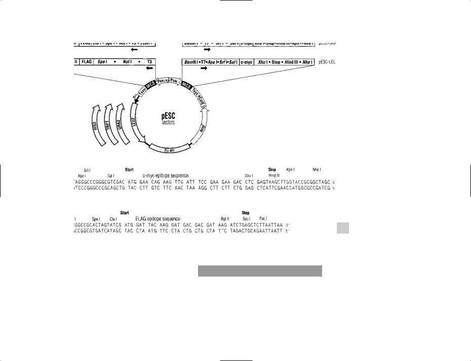
pESC vectors Plasmid vectors used e.g. for expression of eukaryotic genes within yeast cells (Saccharomyces cerevisiae); these vectors include sequences for EPITOPE TAGGING which facilitate detection/isolation of the expressed proteins.
Two different genes can be expressed in each vector (transcription occurring in opposite directions).
Four different yeast selective markers are available.
A series of pESC vectors marketed by Stratagene (La Jolla CA) contain sequences encoding flAG and c-myc epitopes (used for tagging target proteins). These vectors also include GAL promoters and one of several selectable markers (see figure).
pESC vectors were used e.g. for investigations on gene regulation in yeast [Genetics (2006) 173(1):75–85] and on the suppression of viral RNA recombination by cellular exoribonuclease [J Virol (2006) 80(6):2631–2640].
Petri dish A round, shallow, flat-bottomed and vertically sided dish, made of plastic or glass, which has a flat, loosely-fitting lid.
A Petri dish is commonly about 10 cm in diameter. (See also PLATE.)
Peutz–Jeghers syndrome (PJS) A rare autosomal dominant condition characterized by intestinal hamartomatous polyposis and pigmentation in the oro-facial region; complications include intestinal obstruction, and there is an increased risk of gastrointestinal malignancy.
Mutation in a coding region of the serine-threonine kinase gene STK11/LKB1 (chromosomal location 19p13.3) appears to account for most – but not all – cases of PJS. A study of the promoter region of STK11/LKB1 concluded that changes in the sequence in this region are unlikely to contribute to the
182
183
pESC VECTORS A versatile system of vectors for expression of proteins in the yeast Saccharomyces cerevisiae (see entry for details). The polylinker (MCS) sequence downstream of each of the GAL promoters is shown at the bottom of the figure.
Courtesy of Stratagene, La Jolla CA, USA.

pEXP-DEST vectors
PJS syndrome [BMC Genomics (2005) 6:38].
pEXP-DEST vectors See CELL-FREE PROTEIN SYNTHESIS.
pEXP3-DEST vector A destination vector in the GATEWAY
SITE-SPECIfiC RECOMBINATION SYSTEM.
(See also CELL-FREE PROTEIN SYNTHESIS.)
pEXP4-DEST vector A destination vector in the GATEWAY
SITE-SPECIfiC RECOMBINATION SYSTEM.
pFastBac™ dual vector kit A kit (Invitrogen, Carlsbad CA) containing a vector (pFastBac™ Dual, 5.2 kb) for use in the BAC-TO-BAC expression system; the pFastBac vector contains two promoters (polyhedrin and p10), in opposite orientations (and with appropriate polylinkers), allowing the simultaneous expression of two proteins (for details of this vector see the diagram in the entry BAC-TO-BAC).
The pFastBac dual vector kit has been used e.g. in studies on generating murine heavy-chain-only antibodies [Proc Natl Acad Sci USA (2006) 103(41):15130–15135].
PFGE Pulsed-field gel electrophoresis: a specialized form of GEL ELECTROPHORESIS which can be used to separate large pieces of nucleic acid of up to ~10–15 million base pairs. The fragments are exposed, alternately (in pulses), to two separate electric fields that are arranged at different angles to the gel.
The ability to separate fragments of linear DNA depends on several factors that include pulse time, total running time (of the experiment), the applied field strength and the positions of the electrodes. The pulse length affects the size range of the fragments separated: short pulses can be used to separate the relatively smaller fragments, while long pulses are needed to separate the larger fragments.
Disadvantages of PFGE include the time factor (the process may take 2–3 days) and the susceptibility of the isolated genomic DNA to endogenous nucleases.
Variant forms of PFGE differ e.g. in the arrangement of the electrodes; they include:
CHEF (contour-clamped homogeneous electric field electrophoresis). The electrodes are arranged on sides 2, 3, 5 and 6 of a hexagonal chamber.
Double inhomogeneous field electrophoresis. Two, long negative electrodes (cathodes) are on one side of the gel, and two short positive electrodes (anodes) are on the other side.
Field inversion electrophoresis. Two electrodes of equal size are present, one on each side of the gel; one acts as an anode, the other as a cathode – but periodically (depending on the pulse length) the polarity is switched.
PFGE has been widely used e.g. for TYPING bacteria, and is regarded by some as the ‘gold standard’ among molecular typing methods.
[PFGE for typing MRSA (methicillin-resistant Staphylococcus aureus): J Clin Microbiol (2005) 43(10):5069–5073; J Clin Microbiol (2005) 43(11):5642–5647.]
pfu PLAQUE-FORMING UNIT.
PfuTurbo® The trade designation (Stratagene, La Jolla CA) for certain types of DNA polymerase which are formulated for high-fidelity replication in PCR.
(See also ARCHAEMAXX.)
The enzyme PfuTurbo® Cx Hotstart DNA polymerase has been found useful for replicating DNA on uracil-containing templates [Nucleic Acids Res (2006) 34(18):e122].
pGEX-2T plasmid See TEV.
Ph1 The Philadelphia chromosome: see ABL.
phage A common abbreviation for BACTERIOPHAGE (q.v.); see also entries below for details of specific phages.
phage Bxb1 A BACTERIOPHAGE that infects a rapidly growing species of Mycobacterium: M. smegmatis. An integrase from phage Bxb1 (a serine recombinase) was reported to mediate efficient SITE-SPECIfiC RECOMBINATION in mammalian cells [BioTechniques (2006) 40(4):460–464].
phage conversion See LYSOGENY.
phage CTXΦ A filamentous BACTERIOPHAGE whose ssDNA genome encodes e.g. cholera toxin; CTXΦ infects the Gramnegative bacterium Vibrio cholerae. The manner of insertion of (double-stranded) versions of the phage genome into the host cell chromosome was reported to affect the ability of the prophage to give rise to phage virions [J Bacteriol (2000) 182 (24):6992–6998].
(See also (specialized) TRANSDUCTION.)
phage display A method used for selecting a ligand that binds to a given target molecule with high-level affinity/specificity.
Initially, molecules of the given target are immobilized and exposed to a large population of recombinant phages. These phages have been genetically modified, in vitro, so that their coat proteins (i.e. surface proteins) differ from wild-type coat proteins; genetic modification of the phages is carried out on a random basis so that, collectively, the population of phages exhibits an enormous diversity of recombinant coat proteins, each having a potential binding site for a particular type of target molecule. This kind of genetically modified phage population is called a phage library (see later).
After the given target molecules have been exposed to the phage library, any phage particles which do not bind to the target molecules are removed, by washing, and discarded. The phages which do bind are subsequently eluted (removed from the target molecules) by physical means (e.g. adjusting the pH); these phages are used to infect their host bacteria – within which they are replicated to produce vast numbers of progeny phages. These progeny phages are therefore an expanded population of those phages which were selected from the phage library on the basis of their ability to bind to the given target molecule; they usually consist of a number of different strains with various forms of modified coat protein.
Purification of phage particles in the phage display process may be carried out by cesium chloride equilibrium density gradient centrifugation or, more easily, by chromatography with e.g. Sephacryl™ [BioTechniques (2005) 38(2): 194–198] or hydroxyapatite [BioTechniques (2005) 39(6): 879–884].
The target molecules are exposed to the progeny phages under more stringent conditions – i.e. conditions under which only those phages with the highest level of affinity/specificity will bind to target molecules. Unbound phages are removed, by washing.
184

phage M13
The entire selective process is again repeated.
The technique described above for selecting phages from a phage library by exposing the library, and successive batches of progeny phages, to the target ligand is called biopanning (or simply panning). The basic principle involved in this process is similar to that used in the selection of aptamers by SELEX (see APTAMER).
The final batch of phages, characterized by strong, selective binding to the target molecules, are examined to determine the nature of the phage’s binding site and the coding sequence of the binding site in the phage genome.
Phage display technology makes use of filamentous phages (e.g. M13) which are typically very long and thin and which (compared with e.g. phage T4) have a relatively high ratio of surface protein to nucleic acid.
Phage libraries
Phage libraries are prepared e.g. by inserting a gene library (consisting of unrelated genes/fragments) into the DNA of unmodified phages. Many of the genes/fragments will fuse with a phage gene that specifies a coat protein (e.g. protein pIII), and the resulting fusion protein will replace the phage’s normal coat protein. Each hybrid gene will encode a different type of fusion protein – so that the coat proteins of the phage population, as a whole, will display a wide range of potential binding sites.
Another procedure for making a phage library involves in vitro randomization of a given sequence of nucleotides in the gene of a phage coat protein. The result is an immense range of different combinations of nucleotides, and the coat protein gene in a given phage particle will contain only one of these combinations; consequently, the coat proteins of the phage population collectively display an immense range of potential binding sites.
The target ligands for phage display technology include e.g. those involved in gene therapy techniques and various other mammalian cell-surface receptors (such as receptors for cytokines, and ligands relating to receptor-mediated endocytosis). The need to optimize receptor–ligand binding in the context of gene therapy is clearly important as this can help to avoid wasteful delivery of genes to untargeted cells and, therefore, help to reduce the required dosage for the patient.
Examples of use of phage display: studies on induction of an immune response by a peptide mimotope [Infect Immun (2005) 73(1):325–333]; study of in vitro protein evolution to optimize binding of an interleukin 13-neutralizing antibody [PNAS (2006) 103(20):7619–7624]; generating Epstein–Barr virus antigen mimotopes for diagnosis of EBV infection [J Clin Microbiol (2006) 44(3):764–771]; use of scFv antibody phage display libraries in studies on a tumor antigen [PNAS (2006) 103(4):1041–1046].
phage display-mediated immuno-PCR See IMMUNO-PCR. phage f1 A filamentous ssDNA BACTERIOPHAGE that is related
to PHAGE M13.
phage fd A filamentous ssDNA BACTERIOPHAGE that is related
to PHAGE M13.
phage L54a A BACTERIOPHAGE which infects Staphylococcus aureus, causing phage conversion (see LYSOGENY).
phage lambda (λ) A temperate BACTERIOPHAGE of the family Styloviridae; host: Escherichia coli. The virion has an icosahedral head (55 nm diameter) and a non-contractile tail (~150 × 10 nm).
The genome (within the virion) is linear dsDNA (~48.5 kb) with single-stranded 12-base 5′ STICKY ENDS; circularization occurs intracellularly, the sticky ends hybridizing to form the so-called cos site.
During transcription of the phage early genes, the result of infection may be either lysogeny or lysis of the cell. The outcome depends e.g. on the activity of two mutually antagonistic products of the early genes: the cI protein (a repressor of transcription from certain operators) and the Cro protein – which inhibits transcription of the gene encoding cI. Factors that affect the lysogeny/lysis decision also include starvation (favoring lysogeny) and the multiplicity of infection (MOI) – a high MOI (i.e. each bacterium infected with a number of phages) favoring lysogeny.
Lysogeny involves integration of the circularized prophage into the host cell chromosome. Integration occurs by sitespecific recombination between one ‘attachment site’ (which is designated attP) in the prophage and another (attB) in the bacterial genome (located between the gal gene and the bio gene in the chromosome).
The bacterial and phage attachment sites are not identical, although there is a central ‘core’ sequence (O) that is common to both sites. The bacterial (attB) site contains two ‘arms’ (B and B′) either side of the central O sequence, i.e. the structure of attB is BOB′. Analogously, the structure of attP is POP′.
Integration of the prophage into the chromosome involves a phage-encoded integrase (int gene product) and the bacterial
INTEGRATION HOST FACTOR (IHF).
The recombination event involves strand breakage in the O region of each att site, strand exchange, and ligation. Hence, the integrated prophage is flanked by hybrid att regions BOP′ (= attL) and POB′ (= attR).
In the lytic cycle of phage λ, the prophage excises from the bacterial chromosome. This requires the product of the phage xis gene: an excisionase which recognizes the attL and attR sites.
The att sites are exploited in commercial systems: see e.g.
GATEWAY SITE-SPECIfiC RECOMBINATION SYSTEM.
Phage λ also encodes another recombination system which is exploited as the LAMBDA ( λ) RED RECOMBINATION system.
The phage λ cos site is used in COSMIDS and PHASMIDS – while modified forms of phage λ are used as cloning vectors
(see e.g. LAMBDA fiX II VECTOR and ZAP EXPRESS VECTOR).
[The impact of phage lambda (from restriction to recombineering): Biochem Soc Trans (2006) 34(2):203–207.]
phage library See LIBRARY.
phage M13 A filamentous, non-lytic BACTERIOPHAGE able to infect strains of Escherichia coli containing an F PLASMID (or a related plasmid). When infecting cells of E. coli, phage
185