

Dictionary of DNA and Genome Technology
.pdfFRT
occurs as a low-copy-number vector.
FPET Formalin-fixed paraffin-embedded tissue.
Fpg protein Syn. MutM: see 8-OXOG.
fps (FPS) An ONCOGENE in strains of avian sarcoma virus. The v-fps product has TYROSINE KINASE activity.
fragile X disease A hereditary disease characterized by mental retardation; the defect, on the X chromosome, arises through
instability of MICROSATELLITE DNA.
(See also GENETIC DISEASE (table).)
frame-shift mutation (phase-shift mutation) Any mutation in which the addition or deletion of nucleotide(s) results in an out-of-phase message (altered reading frame) in the sequence downstream of the mutation. A frame-shift mutation may be
induced e.g. by INTERCALATING AGENTS. (cf. FRAMESHIFTING.)
frameshifting (ribosomal frameshifting; programed ribosomal frameshifting; translational frameshifting) A mechanism in which the precise product of translation is determined by an induced (programed) movement of the ribosome at a specific site in the mRNA: either a movement of one nucleotide upstream (i.e. in the 5′ direction) or one nucleotide downstream (in the 3′ direction) – translation continuing in the new −1 or +1 reading frame, respectively.
The ribosome’s movement is induced by signals which are incorporated within the nucleotide sequence of the mRNA. In general, two kinds of signal have been reported. First, a socalled slippery sequence, downstream of the initiator codon, which contains groups of repeated nucleotides. Second, a socalled stimulator sequence – a short distance downstream of the slippery sequence – which appears to be often in the form of an RNA pseudoknot. It appears that, in at least some cases, frameshifting can occur through the influence of the slippery sequence alone, i.e. without assistance from a stimulator.
Frameshifting has been reported in relation to genes from viruses, bacteria, insects and animals. One of the functions of frameshifting is apparently to confer on a given coding sequence the ability to yield more than one type of product.
Studies on the translation of the 3a protein from the SARS (severe acute respiratory syndrome) virus have indicated that heptauridine and octouridine stretches in the mRNA are the sole signals needed for frameshifting in this instance [Nucleic Acids Res (2006) 34(4):1250–1260].
framycetin See AMINOGLYCOSIDE ANTIBIOTICS.
FRET (fluorescence resonance energy transfer) A phenomenon in which energy (that would otherwise be dissipated as fluorescence) is transferred from one molecule or moiety (donor) to another, closely adjacent molecule or moiety (acceptor) by a mechanism that does not involve emission of a photon; the energy received by the acceptor molecule or moiety is typically converted to fluorescence, but in some cases is absorbed (quenched) without the production of fluorescence.
(Compare BRET; see also IFRET.)
FRET can be exhibited between two regions on the same molecule, one region acting as the donor and the other as the acceptor. One example is the CCF2 substrate (which includes
both coumarin and fluorescein moieties): see pcDNA™6.2/ GeneBLAzer™-DEST vector in the table in GATEWAY SITE-
SPECIfiC RECOMBINATION SYSTEM.
In general, FRET requires that donor and acceptor molecules be separated by no more than ~100 Å; the separation in experimental protocols is often e.g. 30–70 Å. The donor and acceptor dipoles must also be orientated correctly for effective transmission of energy between the molecules.
In a TAQMAN PROBE (q.v.), energy received by the donor is quenched by the acceptor molecule that is bound adjacently on the oligonucleotide. When the probe is degraded (by the exonuclease activity of the DNA polymerase), the donor and acceptor molecules are separated; the donor molecule (i.e. reporter dye) can therefore exhibit fluorescence because its energy is no longer quenched.
The probes used in the LightCycler™ instrument involve a different arrangement (see REAL-TIME PCR). Fluorescence from the reporter dye (at a wavelength of 640 nm) is detected only if both halves of the probe bind correctly to the target sequence. When this happens, FRET-based excitation of the reporter dye, LightCycler™ Red 640 (present on one half of the probe) occurs by transfer of energy from the fluorescein molecule (on the other half of the probe) because, under these conditions, the donor and acceptor molecules are within the correct distance of one another for effective energy transfer.
In some donor–acceptor pairs the acceptor molecule is a non-fluorescent quencher such as DABCYL or one of the QSY dyes. The QSY dyes are derivatives of diarylrhodamine; their absorption spectra are largely insensitive to a wide range of pH values. The absorption maxima of QSY dyes include e.g. ~660 nm (QSY 21) and ~472 nm (QSY 35). Non-fluorescent acceptors are particularly useful in that they avoid the background fluorescence produced by non-specific excitation of fluorescent acceptor molecules.
The FRET principle has been used in a wide variety of studies involving e.g. detection of hybridization of nucleic acid molecules, the intracellular localization and distribution of specific molecules, the conformation of proteins, enzyme– substrate interaction etc. Note that, in all of these studies, the object is to determine whether or not the donor and acceptor molecules are located sufficiently close to one another in the experimental material to permit FRET-based interaction.
Examples of FRET-based studies include: determination of cell-cycle-dependent localization of proteins in the bacterium Bacillus subtilis [EMBO J (2002) 21:3108–3118]; studies on interactions between enzyme and substrate [Proc Natl Acad Sci USA (2002) 99:6603–6606]; studies on quadruplex and Watson–Crick DNA equilibrium [Nucleic Acids Res (2005) 33(21):6723–6732]; the detection of SNPs [BioTechniques (2006) 40(3):323–329]; studies on DNA bending [Proc Natl Acad Sci USA (2006) 103(49):18515–18520]; and studies on homodimer formation of the enzyme signal peptide peptidase [Mol Neurodegener (2006) 1:16].
(See also DARK QUENCHING.)
FRT The recognition site of the flP recombinase (q.v.).
89

ftsZ gene
ftsZ gene In Escherichia coli: a gene encoding the FtsZ protein, molecules of which form the Z RING.
fucosidosis An autosomal recessive disease involving a defective lysosomal enzyme, α-fucosidase, and the accumulation of glycoproteins.
(See also GENETIC DISEASE (table).) furocoumarins See PSORALENS.
fusidic acid A steroidal antibiotic, synthesized by various fungi (e.g. some species of Acremonium and Cylindrocarpon), that is active against a range of Gram-positive bacteria, including staphylococci; it inhibits protein synthesis by preventing the dissociation of the elongation factor EF-G from the ribosome following translocation.
Resistance to fusidic acid (in e.g. Staphylococcus aureus) can arise through mutations that affect EF-G. Such mutations in S. aureus are associated with a marked decrease in the biologic fitness of the bacterial cells – e.g. reduced growth in comparison to wild-type strains; this effect is reported to be
compensated for by another mutation (designated S416F) in the gene for EF-G [Antimicrob Agents Chemother (2005) 49: 1426–1431].
fusion protein (1) In some viruses (e.g. paramyxoviruses such as the Sendai virus): a protein that promotes fusion between the virus envelope and the membrane of target cells; these proteins can also promote fusion between host cells. (Another example is the GP64 protein of baculoviruses: see BACULO-
VIRIDAE.)
(See also VP22.)
(2) In recombinant DNA technology/genetic engineering: a protein consisting of amino acid residues specified by coding sequences from two different proteins – i.e. a hybrid protein. Fusion proteins can be prepared by the techniques of GENE
FUSION.
(See also ENZYME FRAGMENT COMPLEMENTATION.) fusogen See CELL FUSION.
90

G
G Glycine (alternative to Gly).
G-less cassette Essentially, a DNA template which, on transscription, yields RNA that contains no guanosine residues. G- less cassettes are used in in vitro transcription assays e.g. for studying transcriptional initiation; in such assays, unwanted transcripts (which contain guanosine residues) are eliminated by treatment of the reaction mixture with RNASE T1.
G loop A loop of unpaired ssDNA in the G region of phage Mu observed when DNA from an induced lysogenic population of the phage is first denatured and then re-annealed; a G loop reflects a local region of non-homology caused by inversion of the G region in the lysogenic population.
G-quadruplex See QUADRUPLEX DNA.
G-quartet A square, planar group of four guanine molecules held together by hydrogen bonds. A stack of G-quartets, with their planes parallel, forms the core of QUADRUPLEX DNA – in which each of four strands of DNA is associated with the N9 position of the aligned guanine molecules that form one corner of the stack of quartets.
Note that some authors [Nucleic Acids Res (2006) 34(Database issue):D119–D124] have used G-quartet as synonymous with G-quadruplex (quadruplex DNA); these authors refer to a G-quartet as a G-tetrad.
G-tetrad See G-QUARTET.
G418 sulfate An antibiotic, related to gentamicin (see AMINO- GLYCOSIDE ANTIBIOTIC), active against prokaryotic, yeast, plant and mammalian cells; cell expressing the gene for an aminoglycoside phosphotransferase – such as a neomycinresistance gene – exhibit resistance to G418 sulfate.
[Uses (examples): PLoS ONE (2007) 2(3):e269; PLoS Biol (2007) 5(3):e64.]
(See also HYGROMYCIN B.) gag See RETROVIRUSES.
GAL4 A eukaryotic transcription activator. In yeast (Saccharomyces cerevisiae), GAL4 is involved in the utilization of galactose; in this role it binds to the corresponding upstream activator sequence (UAS).
(See also PATHDETECT SYSTEMS.) Galacton™ See CHEMILUMINESCENCE.
β-galactosidase An enzyme (EC 3.2.1.23), encoded by the lacZ gene in Escherichia coli, which cleaves lactose to glucose and galactose.
(See also LAC OPERON and ONPG.)
The gene for β-galactosidase is widely used as a REPORTER GENE; it is used in various indicator systems such as BLACK–
WHITE SCREENING, blue–white screening (see PBLUESCRIPT) and ENZYME FRAGMENT COMPLEMENTATION.
(See also CHEMILUMINESCENCE, α-PEPTIDE, Q TAG and X- GAL.)
gamete A type of cell involved in sexual reproduction – e.g. an egg or a sperm.
γ δ (gamma delta) Syn. IS1000.
ganciclovir A nucleoside analog which inhibits DNA synthesis
after it has been phosphorylated intracellularly.
gap (1) (in dsDNA) A single-stranded region produced by loss of one or more nucleotides. (cf. NICK; see also AP SITE.)
(2) Following SHOTGUN SEQUENCING of a genome (or other large molecule of DNA): a region for which no cloned fragments are available.
gapped vector A linear VECTOR molecule prepared e.g. by introducing a double-strand break in a ccc DNA vector [see e.g. Proc Natl Acad Sci USA (2005) 102(18):6413–6418].
Gateway® site-specific recombination system Various types of product (Invitrogen, Carlsbad CA) which can be used e.g. to move a given sequence into, or between, vector molecules for expression or directional cloning, and which are based on a site-specific recombination system of PHAGE LAMBDA that involves the ATT SITES.
Thus, e.g. a segment flanked by attP sites can be replaced by a segment flanked by attB sites. During this process, the inserted sequence becomes flanked by hybrid (attB–attP) sites referred to as attL sites; the displaced segment is also flanked by hybrid sites (referred to as attR sites). This procedure is mediated by BP CLONASE.
Similarly, a segment flanked by attR sites can be replaced with one flanked by attL sites; this results in reconstitution of the attB and attP sites (attB sites then flanking one of the segments, and attP sites flanking the other). This procedure is mediated by LR CLONASE.
Entry clones and destination vectors
Entry into the Gateway® system (for cloning, expression) can be achieved e.g. via PCR. Thus, the sequence of interest can be amplified with primers whose 5′ ends incorporate an attB sequence. The amplicons can be inserted (in vitro) e.g. into a pDONR™ vector to form a so-called entry clone; by inserting into a pDONR™ vector, an amplicon displaces the ccdB gene (see CCD MECHANISM) that is flanked by attP sites (thus permitting subsequent recovery of recombinants). The vectors are inserted (e.g. by transformation) into Escherichia coli; following growth, cells containing recombinant vectors are selected on a medium containing kanamycin or Zeocin™ (pDONR™ vectors contain either a kanamycin-resistance or a ZEOCIN-resistance gene).
The sequence of interest, within the entry clone, can then be transferred to any of a range of destination vectors (see accompanying table) in order to carry out a particular task.
Also available are so-called entry vectors (containing att sites) which include a polylinker for the insertion of a DNA sequence with restriction enzymes and ligase; the resulting entry clone can then be used to create an expression clone by transferring the sequence to a given destination vector.
(See also MULTISITE GATEWAY TECHNOLOGY and TOPO- ISOMERASE I CLONING.)
gatifloxacin See QUINOLONE ANTIBIOTICS.
GC-profile See ISOCHORE.
GC type See BASE RATIO.
91

Gateway® DESTINATION VECTORS: examples of some of the wide range of vectors to which a gene or fragment of interest can be transferred from a Gateway® entry clone. Every destination vector contains attR sites which can be used for recombination with any sequence that is flanked by attL sitesa
Vector |
Size |
Properties/notes |
|
|
|
Cell-free protein expression systems |
|
|
pEXP3-DEST |
4.6 kb |
T7 promoter and terminator for high-level expression. N-terminal SIX-HISTIDINE |
|
|
TAG and LUMIO tag to assist purification and detection; cleavage site for efficient |
|
|
removal of tags. (See also entry CELL-FREE PROTEIN SYNTHESIS.) |
pEXP4-DEST |
4.4 kb |
T7 promoter and terminator for high-level expression. C-terminal SIX-HISTIDINE |
|
|
TAG and LUMIO tag for purification and detection |
Expression of proteins in Escherichia coli
Champion™ pET104-DEST 7.6 kb T7 promoter and terminator for strong expression of recombinant proteins. Transcription regulation via a LacO operator. N-terminal BIOEASE tag permitting
IN VIVO BIOTINYLATION of fusion proteins
pBAD-DEST49 |
6.2 kb araBAD promoter providing tight regulation of expression of toxic proteins. |
|
Improved solubility of protein product provided by N-terminal thioredoxin fusion |
|
partner. SIX-HISTIDINE TAG to facilitate detection/purification of product |
Expression of proteins in Saccharomyces cerevisiae
pYES-DEST52 7.8 kb High-level expression of protein product provided by the GAL1 promoter. Detection and purification of the product facilitated by a C-terminal
SIX-HISTIDINE TAG
Expression of proteins in insect cells
BaculoDirect™ Insertion of gene/fragment of interest generates a recombinant baculovirus genome. Any non-recombinant vectors are negatively selected by expression of the thymidine kinase gene in the presence of ganciclovir. The lacZ gene (excised during insertion of the gene/fragment) permits checking of viral stock purity. C-terminal or N-terminal SIX-HISTIDINE TAG for purification. One version of the vector includes a MELITTIN secretion signal to promote secretion of the expressed protein
pMT-DEST48 |
5.3 kb Designed for expression in Drosophila S2 cells. Transcription from a |
|
metallothionein promoter can be induced e.g. with copper sulfate or with |
|
cadmium chloride. SIX-HISTIDINE TAG for purification |
Expression of proteins (and shRNAs) in mammalian cells
pAd/CMV/V5-DEST™ 36.7 kb The plasmid includes a replication-deficient part of the adenovirus genome (lacking sequences in the E1 and E3 regions). A population of the recombinant molecules, consisting of the gene of interest integrated in the plasmid, is linearized (by restriction enzyme PacI), leaving the ITRs (inverted terminal repeats) of the viral genome as the flanking sequences. This construct is used to transfect specialized cells (cell line 293A) within which replication-incompetent (but infectious) virus particles are produced; these particles can be used to infect target cells and, hence, to deliver the gene of interest.
The CMV promoter provides a high level of expression
pBLOCK-iT™3-DEST |
5.9 kb |
A vector designed for long-term expression of shRNAs (see entry SHORT |
|
|
HAIRPIN RNA). Expression may be made either constitutive or inducible, |
|
|
depending on the type of entry vector used. Geneticin®-selectable (neomycin- |
|
|
resistance gene) |
pBLOCK-iT™6-DEST |
5.5 kb |
Similar to pBLOCK-iT™3-DEST but smaller, and blasticidin-selectable |
pcDNA™6/BioEase™-DEST |
7.0 kb |
High-level expression from the CMV promoter. Blasticidin-selectable. The |
|
|
BIOEASE tag facilitates purification of in vivo biotinylated recombinant proteins; |
|
|
the fusion tag can be excised via an adjacent enterokinase cleavage site |
(continued)
92

Gateway® DESTINATION VECTORS: (continued)
Vector |
Size |
Properties/notes |
|
|
|
pcDNA™6.2/GeneBLAzer™-DEST |
|
A fusion vector in which the gene of interest is fused (in N- or C-terminal |
|
|
orientation) to a mammalian-optimized β-lactamase gene (bla) which acts as a |
|
|
reporter gene. Reporter activity depends on the FRET (q.v.) principle. Cells are |
|
|
permeated with a FRET substrate (CCF2) – the molecule of which incorporates |
|
|
two fluorophores: coumarin and fluorescein. Cells in which bla is not expressed |
|
|
emit green light (520 nm) on excitation (409 nm) due to FRET-based interaction |
|
|
between coumarin and fluorescein. If bla is expressed, CCF2 is cleaved; |
|
|
excitation (at 409 nm) then causes simple (non-FRET-based) fluorescence from |
|
|
the coumarin moiety: blue light at 447 nm. |
|
|
The CMV promoter provides high-level expression of the fusion protein. |
|
|
Blasticidin-selectable. |
|
|
A related vector includes the option of topo cloning (see TOPOISOMERASE I |
|
|
CLONING) as well as the facility of a Gateway® destination vector; it also |
|
|
contains a CMV promoter and is blasticidin-selectable |
pcDNA™6.2/nLumio™-DEST™ |
6.8 kb |
High-level constitutive expression provided by the CMV promoter. The LUMIO |
|
|
tag facilitates detection and/or localization of the protein of interest either in vivo |
|
|
(in living cells) or in vitro |
pEF5/FRT/V5-DEST™ |
7.5 kb |
For inserting a gene of interest into cells of an flP-IN CELL LINE. EF-1α |
|
|
promoter for use in those parental cells (e.g. BHK) in which the CMV promoter |
|
|
is not optimal. Bovine growth hormone (BGH) polyadenylation signal and |
|
|
transcription termination to promote stability of transcript. Hygromycin- |
|
|
resistance gene for selection of recombinant vectors |
pLenti6/V5-DEST™ |
8.7 kb |
A lentiviral expression vector incorporating the 5′-LTR and 3′-LTR sequences |
|
|
for insertion into the target cell’s genome. The vector, carrying the gene of |
|
|
interest, is co-transfected into a producer cell line (293FT) together with |
|
|
plasmids encoding (i) the lentiviral packaging proteins, and (ii) the VSV-G |
|
|
envelope protein (the latter protein confers on a viral particle the ability to infect |
|
|
a broad range of types of mammalian cell). The recombinant (replication- |
|
|
incompetent) lentiviral particles, produced in the 293FT cells, can be used to |
|
|
infect target cells, within which the gene of interest is expressed. The CMV |
|
|
promoter normally provides high-level expression. The pLenti6/UbC/V5-DEST™ |
|
|
vector contains the (human) ubiquitin C (UbC) promoter (instead of CMV) for |
|
|
efficient expression when CMV is inappropriate. A blasticidin-resistance gene is |
|
|
incorporated for selection of stable cell lines |
pT-REx™-DEST31 |
7.5 kb |
Strong, tetracycline-regulated CMV promoter, two copies of the TetO2 operator |
|
|
being downstream of the promoter. Geneticin®-selectable. SIX-HISTIDINE TAG to |
|
|
facilitate purification |
pVAX™200-DEST |
|
A small vector designed specifically for use in the development of DNA |
|
|
VACCINES. In order to minimize the possibility of integration into the |
|
|
chromosome, the plasmid includes only the minimal amount of eukaryotic DNA |
|
|
necessary for expression. The polylinker permits insertion of DNA from PCR- |
|
|
generated fragments etc. as well as from Gateway® entry clones |
|
|
|
aDescriptions of products are derived from data provided by courtesy of Invitrogen Corporation, Carlsbad CA, USA.
GC% (GC value; %GC; mol% G + C etc.) Of bacterial DNA: the ratio (G + C)/(A + T + G + C) in which A, T, G and C are the relative molar amounts of adenine, thymine, guanine and cytosine residues, respectively, in the DNA of a given organism; the ratio is expressed as a percentage. (cf. BASE RATIO.)
The value of GC% depends on genus and species. It is one of the criteria used in taxonomy, although similarity in GC%, in itself, is not an indicator of taxonomic affinity. In bacteria,
GC% values range from ~25 to ~75; for example, the GC% of Staphylococcus aureus is ~33, and that of Mycobacterium tuberculosis is ~66.
GC% is useful as a descriptor e.g. for chromosomal DNA, for specific sequences of nucleotides in chromosomal DNA, and for plasmid DNA etc. In some pathogenic bacteria the genome includes certain sequences of nucleotides (so-called pathogenicity islands) in which the GC% differs from that of
93
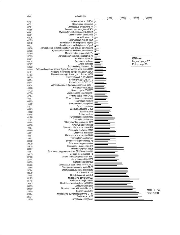
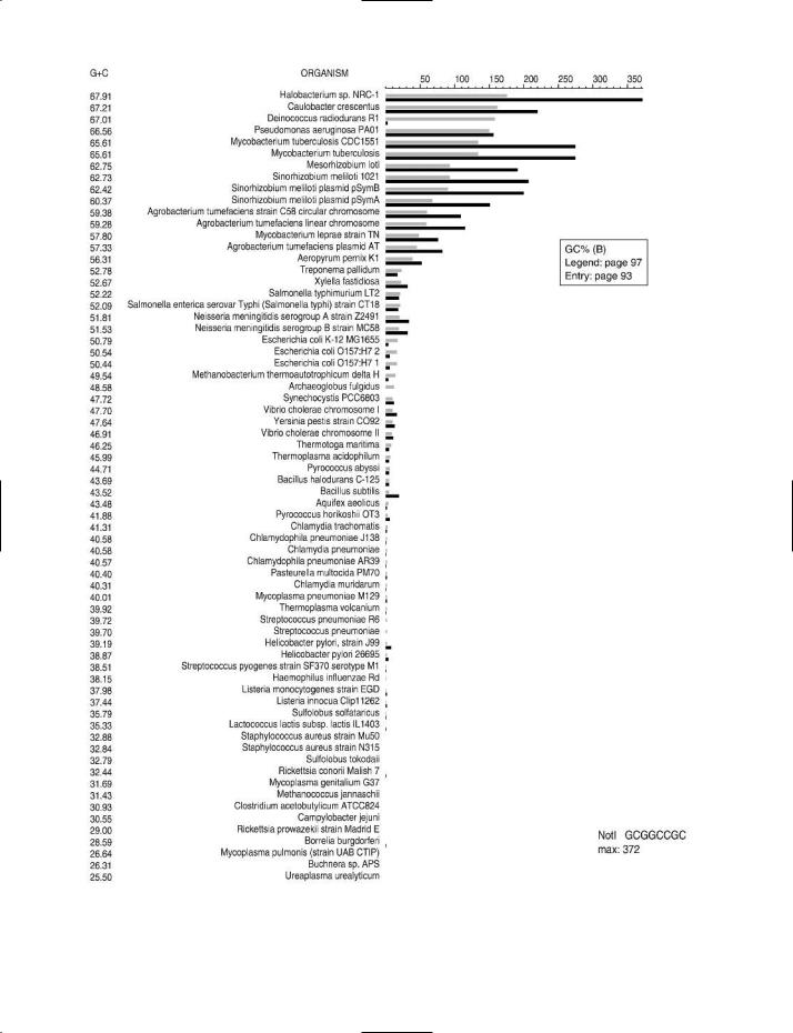


gel electrophoresis
the remainder of the chromosomal DNA; this has been taken to indicate that such sequences of nucleotides were imported into the genome from an extracellular source.
A knowledge of the GC% of an organism’s DNA may be useful for assessing the susceptibility of the DNA to a given RESTRICTION ENDONUCLEASE. For example, the restriction enzyme NotI (recognition site GC↓GGCCGC) lacks cutting sites in the genomic DNA of Staphylococcus aureus but has more than 250 cutting sites in Mycobacterium tuberculosis. Charts which show the cutting frequency of various types of restriction endonuclease in the genomic DNA of a range of bacteria and archaeans can be found on the Internet (see entry REBASE for details of access); some examples of these charts are shown in the figure. The choice of restriction enzyme is important e.g. in RflP ANALYSIS.
An indirect indication of the GC% of a given sample of DNA may be obtained from its ‘melting point’ (see THERMAL MELTING PROfiLE) owing to the linear relationship between Tm and GC% (a high Tm correlating with a high GC%). It is also possible to assess GC% by ultracentrifugation in a CsCl density gradient.
In any determination of genomic GC%, the presence of a plasmid (especially a high-copy-number plasmid) may affect the observed value because the GC% of the plasmid may not be similar to that of the chromosomal DNA. When isolating chromosomal DNA it is therefore necessary to use a method that discriminates against extrachromosomal DNA.
GC% in eukaryotic/mammalian DNA
G + C content is also used to describe regions in eukaryotic/ mammalian DNA. For example, human genomic DNA can be divided into >3000 regions called isochores, each isochore characterized by a uniform or near-uniform G + C content; the isochores in human genomic DNA fall into five groups on the basis of their G + C values. [Isochore mapping of the human genome: Genome Res (2006) 16(4):536–541.]
The DNA of CPG ISLANDS is typically characterized by a value higher than ~60%. This high value of G + C content is associated with susceptibility to certain types of ‘rare-cutting’
RESTRICTION ENDONUCLEASE; for example, more than 85%
of the recognition sites of the restriction endonuclease NotI in the human genome are found in CpG islands.
G-CSF Granulocyte colony-stimulating factor – see e.g. entry
GEL ELECTROPHORESIS: examples of concentrations of agarose and acrylamide in gels used for separating nucleic acid fragments of different sizes (from various sources)
Nucleic acid fragment |
Agarose gel |
Polyacrylamide gel |
(linear dsDNA, in bp) |
(% agarose) |
(% acrylamide) |
|
|
|
20000–100000 |
0.3 |
|
1000–30000 |
0.5 |
|
800–12000 |
0.7 |
|
500–10000 |
1.0 |
|
400–7000 |
1.2 |
|
200–3000 |
1.5 |
|
100–1000 |
|
3.5 |
50–2000 |
2.0 |
|
60–400 |
|
8.0 |
50–200 |
|
12.0 |
10–500 |
4.0 |
|
5–100 |
|
20.0 |
|
|
|
BIOPHARMACEUTICAL (table).
gefitinib An anticancer agent which inhibits oncogenic (EGF receptor) TYROSINE KINASE activity.
(cf. IMATINIB.)
gel electrophoresis A form of ELECTROPHORESIS used e.g. for separating fragments of nucleic acid, of different sizes and/or conformations, in a gel medium. (cf. ISOTACHOPHORESIS.)
Following electrophoresis, the separated fragments may be stained in situ (i.e. in the gel), extracted from the gel (for ongoing study), or transferred to another medium (see e.g. SOUTHERN BLOTTING). In some cases, fragments are labeled (e.g. with a fluorophore) before electrophoresis; in such cases the label is read from the bands of fragments that develop in the gel during electrophoresis.
In methods such as NASBA and PCR, gel electrophoresis is
GC% (opposite) The effect of GC% on the cutting frequencies of three restriction endonucleases in the genomic DNA of various bacteria and archaeans.
The organisms are arranged in order of the GC% of their genomic DNA – the highest value (67.91) at the top, the lowest (25.5) at the bottom.
For a given organism, the solid black bar represents the observed number of recognition sites (see scale at top of chart) in the genomic DNA of that organism. The adjacent gray bar represents the expected number of cutting sites.
(A) Restriction endonuclease MseI. |
Cutting site: T↓TAA |
(B) Restriction endonuclease NotI. |
Cutting site: GC↓GGCCGC |
(C) Restriction endonuclease XmnI. |
Cutting site: GAANN↓NNTTC |
Charts supplied by, and reproduced by courtesy of, Dr Janos Posfai and Tamas Vincze, New England Biolabs, Ipswich MA, USA.
97

gel shift assay
used e.g. to check that amplicons of the expected length are produced in the reaction. To facilitate measurement of length, use can be made of a ‘DNA ladder’; this consists of a set of DNA fragments, in a range of known sizes, which undergo electrophoresis in a lane parallel to the lane(s) in which test sample(s) are being examined. Following electrophoresis, the size of the fragment in a given ‘experimental’ band is assessed by comparison with those bands in the DNA ladder that have migrated at a similar rate.
For nucleic acids, the matrix used in gel electrophoresis is commonly AGAROSE or POLYACRYLAMIDE; agarose is used for separating large fragments, while polyacrylamide is used for the smaller fragments (see table).
(See also NOVEX GELS.) gel shift assay Syn. EMSA.
geldanamycin A benzoquinoid compound, produced by strains of Streptomyces, that inhibits heat-shock protein 90 (Hsp90) and which was reported to inhibit the expression of c-myc in murine lymphoblastoma cells.
gellan gum A bacterial product (a linear polymer containing residues of glucose, rhamnose and glucuronic acid) that has been recommended as a gelling agent for bacterial growth media used in the context of PCR (agar inhibits PCR) [J Med Microbiol (2001) 50:108–109].
Purified gellan gum is available commercially (Gelrite®; Kelco, San Diego CA).
Gelrite® See GELLAN GUM.
gemifloxacin See QUINOLONE ANTIBIOTICS.
GenBank A major DATABASE forming part of the INTERNAT-
IONAL NUCLEOTIDE SEQUENCE DATABASE COLLABORAT- ION.
(See also gene ACCESSION NUMBER.)
gender determination assay See
ASSAY.
gene A specific sequence of nucleotides in DNA (in cells and certain viruses), or in RNA (in other viruses), that encodes a given polypeptide or an RNA molecule with a biosynthetic or control function.
(See also ALLELE.)
gene cassette See CASSETTE. gene cloning See CLONING.
gene conversion Conversion of one allele into another. Gene conversion can occur e.g. following hybridization between one strand from each of a pair of non-identical alleles; during the repair of this heteroduplex (in which one of the strands is used as template) the sequence of one strand is converted to a sequence which is complementary to that of the other.
(See also MATING TYPE.)
gene-delivery system Any system used for inserting gene(s) (or other fragments of DNA) into target cell(s) – often with the object of expression within those cells.
There are various types of gene-delivery vehicle – such as e.g. plasmids, cosmids and viruses (including animal viruses, phages, phagemid particles); these generally involve circular molecules of nucleic acid into which target DNA is inserted
prior to TRANSFECTION (sense 1). (Compare LAMBDA ( λ) RED
RECOMBINATION and BIGEASY LINEAR CLONING SYSTEM.) (See also SPINOCULATION.)
Viral vectors (in which the target DNA is incorported in a recombinant or modified viral genome) internalize this DNA when they infect cells. Commercial gene-delivery systems
include e.g. the AAV HELPER-FREE SYSTEM, the ADEASY XL ADENOVIRAL VECTOR SYSTEM and the VIRAPORT RETRO- VIRAL GENE EXPRESSION SYSTEM.
Plasmids, and other molecules of naked nucleic acid which contain target DNA, are often inserted into cells by trans-
formation or by ELECTROPORATION.
Other modes of gene delivery include LIPOFECTION, MAG-
NETOFECTION and MICROINJECTION. (See also PEI.)
(See also GENE GUN.)
gene expression profile The pattern of expression (i.e. active, non-active, level of activity) of the genes in a given tissue, cell or organism at a particular time under given conditions. gene-for-gene concept ( plant pathol.) An early concept – relating to interactions between plants and plant pathogens – in which e.g. a specific avirulence gene in a given pathogen interacts with the corresponding resistance gene in one or more strains of the host species. Such interaction enables the host strain(s) to give a defensive response (hypersensitivity reaction) and hence exhibit resistance to the given pathogen. Interaction between the products of avirulence and resistance genes was reported e.g. in rice plants challenged with the pathogen which causes rice blast disease, Magnaporthe grisea (Pyricularia oryzae) [EMBO J (2000) 19:4004–4014]. gene fusion In recombinant DNA technology/genetic engineering: any process in which sequences of nucleotides from two distinct genes are joined together, with their reading frames in phase, so that transcription from a single promoter region produces an mRNA transcript that encodes a hybrid fusion protein; the fusion protein may or may not have biological activity – or functionality may be limited to particular part(s)
of the protein.
Gene fusion may be carried out in various ways – e.g. by deleting a sequence of nucleotides between two contiguous genes so as to provide a single, continuous coding sequence controlled via the first (upstream) gene. More generally, it can be achieved e.g. by using a plasmid vector to insert a given gene, in frame, with a partner gene; such a procedure may be carried out in vitro or in vivo.
Fusions of the type described above are sometimes referred to as translational fusions because they give rise to a fusion protein whose synthesis requires both transcription and translation signals from the upstream fusion partner. By contrast, a transcriptional fusion is one in which, through engineering, the genes of interest are subjected to a common mode of regulation; in this case the genes themselves are not fused, and no fusion protein is formed.
Uses of gene fusion
• To facilitate detection/isolation of a specific gene product. If a gene product is difficult to detect (or to assay), the gene
98