

Лекция Неотл ЭХОКГ / Emergency Echocardiography
.pdfFIG. 1.19. End-diastolic frame in apical 5-chamber view. This view is obtained from the apical 4-chamber view position by tilting the transducer anteriorly. The outflow tract area is thus opened, allowing cardiac output measurements by combined two-dimensional and pulse wave Doppler imaging (see Appendix A). LVOT left ventricular outflow tract.
FIG. 1.20. End-diastolic frame in apical 2-chamber view. The LV walls are identified with the corresponding coronary supply. The descending aorta is occasionally visualized in long-axis in this view. LVAW left ventricular anterior wall, LVIW left ventricular inferior wall.

1.10 STANDARD VIEWS IN TRANSTHORACIC ECG 25
FIG. 1.21. End-diastolic frame in apical 3-chamber (long-axis) view. AO ascending aorta.
FIG. 1.22. Mid-diastolic frame in subcostal view. Chambers size and contractility and any significant pericardial effusion can all be quickly assessed in this view.
26 GETTING READY FOR THE STUDY
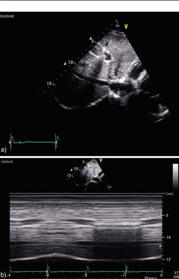
FIG. 1.23. IVC visualized by mild counterclockwise rotation and rightwards tilting of the transducer from the subcostal position. The diameter and the respiratory variations are assessed at about 1 cm from the junction with the right atrium. a two-dimensional image. b M-mode imaging demonstrating complete IVC collapse during inspiration, characteristic for low right atrial pressures. IVC inferior vena cava.

1.10 STANDARD VIEWS IN TRANSTHORACIC ECG 27
FIG. 1.24. Suprasternal view. The arch, left-side neck vessels, and the proximal descending aorta are clearly demonstrated. This view is not a routine one in emergency echocardiography unless aortic dissection is suspected. LCA left carotid artery, LSA left subclavian artery.
visualization of the inferior vena cava and, thus, assessment of right heart pressures (Fig. 1.23).
1.10.4 Suprasternal View (Fig. 1.24)
This is not a mandatory routine view but should be attempted whenever aortic enlargement or dissection is suspected. The patient should lie on his back with some neck hyperextension to allow proper transducer positioning.
References
1. de Jong N. Mechanical index. Eur. J. Echocardiogr. 2002;3(1):73–74.
2. Lang, RM, Mor-Avi V, Zoghbi WA, Senior R, Klein AL, Pearlman AS. The role of contrast enhancement in echocardiographic assessment of left ventricular function. Am. J. Cardiol. 2002;90(10A):28J–34J.
3. Stewart MJ.Contrast echocardiography. Heart 2003;89(3):342–348.

Chapter 2
Emergency Echocardiography
This chapter provides an overall view of the increasing contribution of echocardiography in the management of acutely ill patients and of its impact on outcome. Emphasis is put on the role of echocardiography in typical critical care situations and a diagnostic algorithm is suggested for immediate echocardiographic triage and monitoring of unstable patients. Techniques such as transesophageal and hand-held echocardiography are discussed in detail and a step-by-step practical guide of transesophageal imaging is provided, supported by relevant figures from real-life studies.
2.1 GENERAL OUTLOOK
2.1.1 Modalities
Echocardiographic studies in critically ill patients are generally performed bedside or in the acute assessment bay either as:
•Transthoracic study using:
–Mainframe machine
–Portable unit
–Hand-held unit
•Transesophageal study using:
–Mainframe machine
–Portable unit
The information to be derived and the “logic of the study” are similar irrespective of the technique used. Specific indications, limitations and technical issues for transesophageal and handheld echocardiography are detailed below.
29
A. Chenzbraun, Emergency Echocardiography, DOI: 10.1007/978-1-84882-336-5_2, © Springer-Verlag London Limited 2009

30 EMERGENCY ECHOCARDIOGRAPHY
2.1.2 Clinical Scenarios and Indications for Use
of Echocardio-graphy in Critically Ill Patients
An urgent echocardiographic study in an unstable patient may be required in the following clinical scenarios:
1. Hemodynamic instability and/or hypoxemia of unclear aetiology.
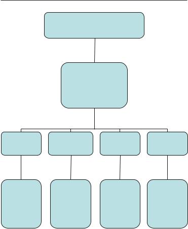
(a)As a general rule, a hemodynamically unstable patient should be promptly assessed with echocardiography since this may provide an immediate understanding of the case. Mainly, a targeted echo assessment will allow the assignment of the patient to a broad pathophysiological category, which will guide subsequent management. This information is frequently more useful and comprehensive than the data obtained with invasive measurements, which often offer little on the mechanism of patient’s deterioration. A general workflow approach for Immediate Echocardiographic Triage (IMET) is outlined in Fig. 2.1
(b)Within this category there are two clinical situations which are recognized as indication for “emergency echocardiography.”1
(i)Suspected tamponade
(ii)Pulseless electrical activity (PEA) can be the presenting symptom for:
•Tamponade
•Massive pulmonary embolism
•Acute massive internal hemorrhage (e.g., ruptured aortic aneurysm)
•Tension pneumothorax
2. Critically ill patients due to non-cardiac illness where, however, cardiac function and left ventricular (LV) filling status assessment may be crucial to guide fluid resuscitation, for example:
(a)septic shock and
(b)diabetic ketoacidosis.
3. Relatively stable patients with known or suspected acute cardiac pathology where echocardiography is necessary for further management and risk stratification.
The degree of “urgency” for an echocardiographic assessment in these cases may vary from emergency to scheduled pre-discharge evaluation. Typical examples in this category include:
1. Stable patients after successful resuscitation
2. Selected cases of acute coronary and chest pain syndromes 3. Aortic dissection
2.1 GENERAL OUTLOOK 31
Unstable patient with unexplained:
•Shock
•Pulmonary edema
Quick, targeted ECHO
•Hand-held device: acceptable
•Minimum set of views:
o Subcostal
o Apical
moderate PE |
Severely enlarged, |
Severely hypokinetic |
Small hyperdynamic |
|
Compressed RV/RA |
heart |
|||
akinetic RV |
LV |
|||
Dilated IVC |
Collapsed IVC |
|||
|
|
Assume: |
Assume: |
|
Assume: |
Assume: |
|
ACUTE PUMP |
|||||
PULMONARY |
SEVERE |
||||
TAMPONADE |
|||||
|
FAILURE |
||||
EMBOLISM |
|
HYPOVOLEMIA |
|||
|
|
||||
|
|
|
|||
|
|
• |
MI? |
• Major |
|
|
|
• |
Myocarditis? |
internal |
|
|
|
• |
Toxic? |
bleeding |
|
|
|
|
|||
SEPSIS
OTHER?
FIG. 2.1. Immediate Echocardiographic Triage (IMET) algorithm. Diagnostic criteria for specific scenarios including use of echocardiography during resuscitation are discussed further in the text.
4. Massive pulmonary embolism
5. Infective endocarditis
6. Acute native valves pathology
7. Prosthetic valves malfunction
8. Acute embolic events
A review of various studies on the use of echocardiography in critically ill patients2–5 shows a quite consistent pattern, with the main conditions to benefit from an echo-guided approach listed below:

32 EMERGENCY ECHOCARDIOGRAPHY
•Hemodynamic instability
•Sepsis
•Source of emboli
•Suspected aortic dissection
•Various
There is a wide consensus on the impact of the echo studies on the further management of these patients with most authors reporting an extremely positive input which translates in a change in therapy in 30–60% of cases even in patients who had invasive hemodynamic monitoring.
Although the clinical picture can be complex and different pathologies can coexist in a given patient, from a practical standpoint, rather standard scenarios can be expected (Table 2.1). Close review of the listed situations reveals that most of them have specific and occasionally divergent therapeutic approaches which would be very difficult to decide upon without echocardiographic information.
“Pre-echo” clinical knowledge of the case is essential in order to know what to look for and what to do with the echocardiographic results. The importance of familiarizing oneself with the case and understanding it before placing the transducer on the patient’s chest cannot be overemphasized. The final diagnosis has to integrate all data using sound clinical judgment. Finally, a decision based on an echocardiographic evaluation should be taken only if the study quality is deemed satisfactory for diagnostic purposes and the person performing it is trained and competent enough in using echocardiography.1 Flawed information may be more damaging than lack of information altogether.
While an emergency bedside study should aim to be as complete as possible, it is expected to be less detailed, of lesser quality and to include fewer measurements than a routine elective one due to a variety of factors:
•Limited echocardiographic windows
•Inability to position the patient for optimal imaging
•Bright room lighting
•Need to perform a short, “targeted” study due to patient instability or discomfort
•Use of a portable echocardiographic machine of less quality than a big unit
•Limited echocardiographic experience of the operator
However, the information listed below is essential and should be actively sought by operator.

TABLE 2.1. Typical scenarios requiring urgent echocardiographic assessment in critically ill patients.
Clinical scenario |
Diagnostic question |
Management question |
Hypotension/shock |
Hypovolemia? |
Fluid challenge? |
|
Pump failure? |
Vasoconstrictors/inotropes? |
|
Inappropriate vasodilatation? |
CVP/Swan–Ganz monitoring? |
Severe dyspnea |
Cardiac versus noncardiac etiology |
Cardiac or noncardiac case? |
|
• LV function |
|
|
– Systolic and diastolic |
|
|
• Valvular heart disease |
|
|
• Pericardial disease |
|
Severe cardiomegaly |
Cardiac cavities enlargement? |
|
|
Pericardial fluid? |
|
Systemic embolism |
Intracardiac source of emboli |
Anticoagulation? |
Suspected pulmonary embolism |
RV enlargement/failure |
Thrombolysis |
|
Thrombus visualization (by TEE) |
Embolectomy |
Deterioration in a patient with a |
Stuck valve |
|
prosthetic valve |
Endocarditis |
|
|
Regurgitation |
|
Suspected tamponade |
Hemodynamic impact of pericardial fluid |
Need for pericardiocenthesis |
|
|
|
|
|
(continued) |
33 OUTLOOK GENERAL 1.2

TABLE 2.1. (continued)
Clinical scenario |
Diagnostic question |
Management question |
Suspected aortic dissection |
Confirm Dg |
Conservative versus emergent |
|
|
surgical approach |
|
Assess: |
|
|
• involvement of ascending aorta |
Need for AVR |
|
• pericardial/mediastinal fluid |
|
|
• aortic regurgitation |
|
Hemodynamic deterioration |
LV/RV function |
Diuretics/inotropes/vasodilators |
in AMI |
|
|
|
Mechanical AMI complications |
|
|
• MR |
IABP |
|
• AVSR |
Emergency surgery |
|
• Free wall rupture |
|
|
• Dynamic LVOT obstruction |
|
Resuscitated sudden cardiac |
LV function |
|
death |
|
|
|
Pericardial fluid |
|
|
Pulmonary embolism |
|
|
Aortic dissection |
|
AMI acute myocardial infarction, AVR aortic valve replacement, AVSR acute ventricular septal rupture, CVP central venous pressure, MR mitral regurgitation, LVOT left ventricular outflow tract, IABP intraortic balloom pump, RV right ventricular, LV left ventricular, TEE transesophageal echocardiography
ECHOCARDIOGRAPHY EMERGENCY 34