

Лекция Неотл ЭХОКГ / Emergency Echocardiography
.pdf12.1 ECG IN ACUTE NONISCHEMIC LV DYSFUNCTION 127
FIG. 12.2. Sequential echocardiograms in a patient admitted with cardiogenic shock and an ECG picture of extensive anterior myocardial infarction. Emergency angiogram with a view to primary percutaneous coronary intervention (PCI) showed normal coronary arteries and apical ballooning. Upper panel: echocardiogram on admission showing a large apical aneurysm (short arrows), severe, eccentric, posteriorly oriented mitral regurgitation (MR) and severe systolic anterior motion of the mitral valve (SAM) with left ventricular outflow tract (LVOT) obstruction (long arrow). Lower panel: repeat echocardiogram after 4 days shows near normal left ventricular (LV) function, no MR and resolution of the LVOT obstruction. a Transthoracic two-dimensional systolic frame in apical fourchamber view. b Transthoracic color-Doppler apical four-chamber view. c Transesophageal long-axis view of the mitral valve (MV) and LVOT.
12.1.2.3 LV Dysfunction in Acute Noncardiac Medical Conditions
Transient myocardial injury manifested as ECG changes, troponin release, and evidence of LV systolic dysfunction, not related to an acute coronary syndrome, have been described in patients with severe, acute medical conditions, mainly sepsis and acute pulmonary

128 ECHOCARDIOGRAPHY IN SPECIAL SITUATIONS
disease6 and, generally, correlates with a worse prognosis. Evidence of non-acute coronary syndrome (ACS)-related myocardial damage was reported in one-third of the patients admitted in a medical intensive care unit (ICU).8 Echocardiography may demonstrate global LV dysfunction6 but some studies suggest a preponderance of anterior and apical segments involvement, with a typical Takotsubo like appearance.9 Like for other conditions in this section, the LV function tends to normalize in survivors, after the acute event.
12.2 EMERGENCY CARDIOVERSION OF ATRIAL FIBRILLATION
Left atrial thrombi are described in 8–13% of patients with atrial fibrillation (AF) and are more likely to be present in patients with organic heart disease.10,11 Present guidelines12 mandate that elective chemical or electrical cardioversion of either AF or flutter of more than 48-h duration should be preceded by at least 3 weeks of anticoagulation in order to reduce the risk of postcardioversion thromboembolism to ≈ 1% as opposed to up to 7% in the preanticoagulation era.13 In selected instances, patients with critical on-going ischemia or cardiac failure, thought to be secondary to uncontrollable AF, may be cardioverted as an emergency procedure even without prior anticoagulation12 since the immediate benefits obviously outweigh the risks. A less straightforward scenario exists when AF is difficult to control, yet the situation is not considered critical enough to cardiovert immediately taking the risks of a stroke if the patient had not been properly anticoagulated. Cardioversion in a previously not anticoagulated patient after a TEE ruled out atrial and appendage thrombi, has been proved as safe as cardioversion after a 3 weeks anticoagulation period14 in terms of thromboembolic risk. In a critical care setting, this approach, generally referred to as “TEE-guided cardioversion,” can solve the dilemma of an early cardioversion in a patient without previous anticoagulation. However, the documented lack of thrombi by TEE is a “snapshot” image valid at the time of the procedure only, which allows “skipping” the 3 weeks period of anticoagulation but does not in any way invalidate the need for anticoagulation. The patient should be properly anticoagulated at least from the time of the study, at the time of the cardioversion and following it as well, according to accepted guidelines. A simple algorithm to manage this frequent scenario is outlined in Fig. 12.3.
12.2.1 Left Atrial and Left Atrial Appendage Thrombi: TEE Diagnosis
Left atrial thrombi are occasionally encountered, but the typical thrombus location is in the left atrial appendage due to the stagnant flow in this cul de sac cavity. TEE is credited in most

12.2 EMERGENCY CARDIOVERSION OF ATRIAL FIBRILLATION 129
Patient with uncontrollable AF and without proper anticoagulation
START ANTICOAGULATION
Clinical condition: critical |
Clinical condition: not critical |
||
Early restoration of sinus rhythm: highly desirable |
|||
|
|||
Cardiovert |
TEE |
|
|
Continue chronic anticoagulation |
|
||
|
|
||
|
No thrombus |
Thrombus present |
|
|
Cardiovert |
Reconsider |
|
|
Continue chronic anticoagulation |
Avoid early cardioversion |
|
FIG. 12.3. Algorithm for transesophageal echocardiography (TEE)-guided cardioversion in critically ill patients.
studies with an excellent sensitivity, reaching 100%, for LAA thrombus detection.13 This high diagnostic power requires a systematic approach. LAA is a tridimensional structure, usually multilobulated, which has to be carefully interrogated in different plans using transducer angulations of 0°, 40°−60°, and 90°−120° in midand high-esophageal transducer positions (see Sect. 2.2). A thrombus should be identified as a protruding or mural mass with an echogenic texture different from that of the adjacent atrial myocardium (Fig. 12.4). Differential diagnosis from an atypical LAA wall appearance or a pectineate muscle may be occasionally difficult. In borderline cases, the following findings support the diagnosis of thrombus:
•presence of the mass in more than one view
•low fibrillatory waves velocities (<20 cm/s)
•intense spontaneous contrast
In selected instances, a repeat study after anticoagulation may be necessary. As mentioned above, thrombi may form in the left atrium (LA) cavity as well, so careful scanning of the LA, using standard views and a left-to-right scanning at 0° in mid-esophageal position should be a part of the study. Also, though most reports deal with presence of thrombi in the LA or atrial appendage, the right atrium should also be scanned for thrombi as well.

130 ECHOCARDIOGRAPHY IN SPECIAL SITUATIONS
FIG. 12.4. Left atrial appendage (LAA) thrombus in a patient with chronic atrial fibrillation (AF). The thrombus is visualized as a protruding, slightly rounded mass with irregular surface (arrows) and a small mobile component at its lower pole. Moderate spontaneous echocontrast was also apparent in the LAA cavity.
12.3 ECHOCARDIOGRAPHY IN SUSPECTED STUCK
PROSTHETIC VALVE (SEE ALSO SECT. 5.5)
Either acute hemodynamic deterioration or progressive development of otherwise unexplained cardiac failure in a patient with a mechanical valve should raise the suspicion of an obstructed or stuck valve. An alternative presentation, of stroke or peripheral embolism, but without hemodynamic compromise, should also be investigated for nonobstructive valve thrombosis. Florid endocarditis with large vegetations is another possible etiology (Fig. 12.5).
12.3.1Questions to be Answered in an Echo-Based Approach to the Patient with Clinical Suspicion of Stuck Valve (Fig. 12.6)
•Is there hemodynamic evidence of obstruction?
°This is, generally, a transthoracic echo assessment, so it should not be postponed until TEE is performed.

12.3 ECG IN SUSPECTED STUCK PROSTHETIC VALVE 131
FIG. 12.5. Transesophageal echocardiography (TEE) findings in a patient with fungal endocarditis on a bioprosthetic aortic valve. a On admission: two large vegetations are present on the aortic valve leaflets, which are severely restricted in their opening motion. b After antifungal treatment: disappearance of the vegetations and free opening of the aortic leaflets.
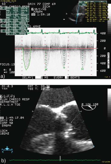
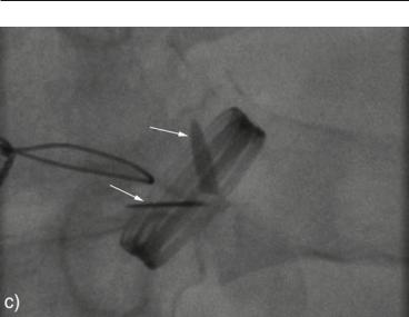
FIG. 12.6. Echocardiographic and fluoroscopic findings in a patient with obstructed mechanical aortic valve. The diagnostic sequence in this case was: TTE→fluoroscopy→TEE. Transthoracic echocardiography (TTE) continuous Doppler interrogation showing high peak and mean aortic transvalvular gradient systolic fluoroscopy frame. Note that the two leaflets (arrows), which should have been parallel to each other, are stuck in partially opened position. Transesophageal echocardiography (TEE) imaging of the aortic valve in long-axis view. The prosthetic valve and its motion were suboptimally visualized but a clear soft echogenic mass (arrows) consistent with thrombus was demonstrated attached to the valve.
12.3 ECG IN SUSPECTED STUCK PROSTHETIC VALVE 133
FIG. 12.6. (continued)
°A Doppler peak and mean gradient above the accepted range for the particular valve type and size support the diagnosis of obstruction. The gradient measurement should be completed by a pressure half-time measurement in mitral position and, ideally, a calculated effective orifice area or a dimensionless index in aortic position.
•Caveats:
°A significant raise in gradients as compared with baseline values (if known) may have more diagnostic information value than absolute numbers.
°Hyperdynamic states, such as sepsis or anemia, can raise the transvalvular gradients in the absence of obstruction.
°Even with obstructed valves, gradients can be spuriously low in patients presenting with shock.
•Is there imaging evidence of reduced valve motion?
°This is usually a TEE assessment.
°Mitral prostheses motion can be generally well evaluated; however, even TEE can be unsatisfactory for aortic prostheses.
134 ECHOCARDIOGRAPHY IN SPECIAL SITUATIONS
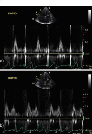
FIG. 12.7. Effect of increasing the atrio-ventricular delay (AVD) in a patient with biventricular pacemaker. a AVD = 110 ms: totally truncated A wave, short diastolic filling time. b AVD = 200 ms: full deployment of A wave with significant increase in the diastolic filling time
°Practical tip:
□If restricted motion is not convincingly demonstrated by echocardiography, fluoroscopy is an excellent imaging aid to assess the occluder opening and closure excursion.

12.4 PACEMAKER OPTIMIZATION IN THE CRITICALLY ILL 135
•Is the obstruction due to thrombus, pannus, or vegetation?
°Thrombus formation is a much more frequent (90%) etiology of valve obstruction than isolated pannus build up (10%).15 With the advent of fibrinolysis as an alternative therapeutic approach for obstructed prosthetic valves, differentiating a thrombus from pannus has clear practical implications. The following clinical and echocardiographic findings support the diagnosis of thrombus over pannus16:
□Shorter period from symptoms onset
□Evidence of inadequate anticoagulation
□Larger and softer masses by echocardiography
□None of the above is an absolute criterion, however, for practical purposes, an obstructing mass on a mechanical valve, in the setup of poor anticoagulation status and without clear evidence of endocarditis should be considered to be a thrombus.
□A large, mobile thrombus is considered a contraindication to thrombolysis due to the risk of embolic complications.
12.3.2 Management of Stuck Valve17,18
Beyond immediate restoration of adequate anticoagulation with heparin, valve replacement is the approach of choice in unstable patients without prohibitive comorbidities. Surgery is also preferred in nonobstructive thrombosed valves with large (>10 mm) thrombi or which do not respond to anticoagulation as demonstrated by serial echocardiographic evaluation. Thrombolysis should be considered as an accepted alternative to surgery in:
•Unstable patients with poor general operative risk
•Lack of appropriate surgical facilities
•Prosthetic tricuspid or pulmonary valves thrombosis.
12.4 PACEMAKER OPTIMIZATION IN THE CRITICALLY ILL
Patients with clinical heart failure, left ventricular ejection fraction (LVEF) <35% and QRS width >120 ms are candidates for cardiac resynchronization therapy with biventricular pacemakers.19 As the use of these devices increases, so does the likelihood of being faced with patients with severe decompensated heart failure requiring inotropic support and intensive care or high-dependency unit setup, who have a biventricular pacemaker. Generally, biventricular pacemakers are programmed at default atrio-ventricular (100–120 ms) and LV–RV delays and optimization of these parameters is not routinely performed. There are no data on the impact of optimizing biventricular pacemakers in patients with cardiac failure decompensation. However, the acute changes in loading

136 ECHOCARDIOGRAPHY IN SPECIAL SITUATIONS
conditions and heart rate may warrant an echocardiographic interrogation to check whether the default pacemaker settings still provide the optimal hemodynamic support in the new conditions. There are two components to an echocardiographic optimization of a biventricular pacemaker:
•Pulsed-wave Doppler interrogation at mitral valve level is used to establish the mitral filling pattern and to find the optimal atrio-ventricular delay (AVD).
•Continuous wave Doppler at aortic level is used to obtain a velocity–time integral (VTI) of the aortic flow as a surrogate for cardiac output to choose the best interventricular delay.
12.4.1 AV Delay Optimization
An optimal AVD allows good diastolic filling with completion of the A wave before the beginning of the ventricular systole. A too short AVD will result in a truncated or even absent A wave, while a too long AVD will result in merging of the E and A waves and, possibly, diastolic MR. Algorithms exist to determine the optimal atrio-ventricular delay,20 but a simpler approach is an iterative one where 10–20 ms steps are used to gradually increase the AVD if the A wave is truncated or to decrease it if there is E and A waves fusion, until an optimal pattern is obtained (Fig. 12.5). Recent data and recommendations suggest that only patients with fused E and A waves or truncated A wave need echoguided optimization of their atrio-ventricular delays.21,22
12.4.2 Interventricular Optimization
There are little data on the benefits of optimization of the LV–RV delay. The reported techniques use the VTI of the aortic flow, obtained with CW Doppler in the apical five-chamber view, as a surrogate for cardiac output. The measurement is obtained at nominal values and then at different LV-RV delays. The LV is generally brought forward using steps of 10–20 ms until the largest VTI is achieved.
12.5 ECHOCARDIOGRAPHY IN THE MANAGEMENT OF PATIENTS WITH MECHANICAL CARDIAC SUPPORT
12.5.1 Intraaortic Balloon Pump
The abrupt deflation of the intraaortic balloon pump (IABP) in systole and its inflation in diastole have the beneficial effects to lower the systolic aortic impedance and thus to augment the antegrade stroke volume and to increase the diastolic aortic pressure