

Лекция Неотл ЭХОКГ / Emergency Echocardiography
.pdf96 ECHOCARDIOGRAPHY IN ACUTE CHEST PAIN
FIG. 6.5. Systolic frame in PSLA view in a patient with a recent large anterior MI and severely decreased LV function. a 2D image shows the two mitral leaflets tethered apically and coapting at a distance (short arrow) from the annular plane (long arrow). b Color Doppler demonstrates severe MR with a central jet.

6.4 MECHANICAL COMPLICATIONS OF AMI 97
FIG. 6.6. Large basal inferior septum ventricular septal rupture in a patient with an inferior MI. a 2D imaging in apical 4-chamber view reveals the lack of tissue continuity (small crosses) at base of the septum. Note the dilated RV, secondary to acute right ventricle overload. b Color Doppler reveals the wide turbulent jet across the defect, consistent with a large left-to-right shunt.

98 ECHOCARDIOGRAPHY IN ACUTE CHEST PAIN
6.4.3 Acute and Subacute Free Wall Rupture
Acute free rupture is a catastrophic and usually fatal event with the patient rapidly developing cardiogenic shock and pulseless electrical activity unresponsive to resuscitation. If the rupture is small, or with a serpiginous path, the slower accumulation of blood in the pericardial cavity may allow time for diagnosis and emergency intervention (subacute rupture). A sudden reoccurrence of pain, possibly with ST re-elevation and rapid hemodynamic deterioration with tamponade features should raise the suspicion of free wall rupture. Emergency echocardiography is mandatory with this clinical scenario. The presence of pericardial fluid >5 mm in this context supports the diagnosis, especially if a dense intrapericardial mass consistent with clot is visualized10,11 (Fig. 6.7).
FIG. 6.7. 2D apical 4-chamber view in a patient with acute MI complicated by subacute free wall rupture. A small pericardial effusion is noted around the apex (arrows) and a thick, dense echogenic mass overlies the RV free wall. (Reproduced from Raposo et al.,11 open publication policy).

6.4 MECHANICAL COMPLICATIONS OF AMI 99
Of note, the finding of pericardial fluid as such, without the typical clinical picture is not diagnostic of cardiac rupture since small amounts are not infrequent with AMI.
6.4.4 Acute Dynamic MR and LVOT Obstruction
Patients with large apical infarctions and hyperdynamic base of the heart can develop in the hyperacute phase systolic anterior motion of the mitral valve (SAM) with resulting severe MR and left ventricular outflow tract (LVOT) obstruction. The clinical suspicion should be raised by the development of severe MR in the first hours of a large anterior MI, as opposed to the typical acute MR which has a later onset and is more common with inferior MI. SAM and LVOT obstruction should be actively sought in these cases since the management is quite specific including beta blockers and avoidance of vasodilators. This complication is reported in the literature,12,13 though it is still not included in the formal list of mechanical complications of acute MI. These patients tend to have single LAD disease and some degree of basal septal hypertrophy or have the transient apical ballooning described as Takotsubo cardiomyopathy in patients with normal coronary arteries who present with a clinical and ECG picture mimicking a large anterior MI (see Sect. 12.1.2).
References
1. Autore C, Agati L, Piccininno M, Lino S, Musaro S. Role of echocardiography in acute chest pain syndrome. Am J Cardiol. 2000;86(4A):41G– 42G.
2. Bholasingh R, Cornel JH, Kamp O, et al. Prognostic value of predischarge dobutamine stress echocardiography in chest pain patients with a negative cardiac troponin T. J Am Coll Cardiol. 2003;41(4):596–602.
3. Buchsbaum M, Marshall E, Levine B, et al. Emergency department evaluation of chest pain using exercise stress echocardiography. Acad Emerg Med. 2001;8(2):196–199.
4. Mohler ER 3rd, Ryan T, Segar SD, et al. Clinical utility of troponin T levels and echocardiography in the emergency department. Am Heart J. 1998;135(2 Pt 1):253–260.
5. Weston P, Alexander JH, Patel MR, Maynard C, Crawford L, Wagner GS. Hand-held echocardiographic examination of patients with symptoms of acute coronary syndromes in the emergency department: the 30-day outcome associated with normal left ventricular wall motion. Am Heart J. 2004;148(6):1096–1101.
6. Birnbaum Y, Chamoun AJ, Conti VR, et al. Mitral regurgitation following acute myocardial infarction. Coron Artery Dis. 2002;13(6):337–344.

100 ECHOCARDIOGRAPHY IN ACUTE CHEST PAIN
7. Ho SY. Anatomy of the mitral valve. Heart. 2002;88(Suppl 4):iv5–iv10. 8. Levine RA, Schwammenthal E. Ischemic mitral regurgitation on the threshold of a solution: from paradoxes to unifying concepts.
Circulation. 2005;112(5):745–758.
9. Birnbaum Y, Fishbein MC, Blanche C, Siegel RJ. Ventricular septal rupture after acute myocardial infarction. N Engl J Med. 2002;347(18):1426–1432.
10.Lopez-Sendon J, Gonzales A, Lopez S, et al. Diagnosis of subacute ventricular wall rupture after acute myocardial infarction: sensitivity and specificity of clinical, hemodynamic and echocardiographic criteria. J Am Coll Cardiol. 1992;19(6):1145–1153.
11.Raposo L, Andrade MJ, Ferreira J, et al. Subacute left ventricle free wall rupture after acute myocardial infarction: awareness of the clinical signs and early use of echocardiography may be life-saving.
Cardiovasc Ultrasound. 2006;4:46.
12.Chockalingam A, Tejwani L, Aggarwal K, et al. Dynamic left ventricular outflow tract obstruction in acute myocardial infarction with shock: cause, effect, and coincidence. Circulation. 2007;116(5):e110–e113.
13.Hrovatin E, Piazza R, Pavan D, et al. Dynamic left ventricular outflow tract obstruction in the setting of acute anterior myocardial infarction: a serious and potentially fatal complication? Echocardiography. 2002;19(6):449–455.

Chapter 7
Echocardiography in Acute
Aortic Syndromes
This chapter covers the excellent diagnostic capabilities and the few limitations of transesophageal echocardiography in the diagnosis of the three presentations of acute aortic syndromes: aortic dissection (AD), intramural hematoma (IMH), and penetrating aortic ulcer (PAU). An algorithm for deciding on the appropriate imaging strategy in patients with suspected acute aortic syndromes is included, as well as a listing of echocardiographic information to be actively sought when assessing a patient with acute aortic dissection.
7.1 INTRODUCTION
Aortic syndromes are a group of conditions characterized by symptoms suggestive of acute aortic pathology and subsequent diagnosis of AD, IMH, or PAU.
7.2 AORTIC DISSECTION
AD is a major emergency with a 1–2% hourly mortality in the first 48 h, without appropriate treatment. The clinical presentation, and the diagnostic and therapeutic approaches, are well described1–3; in intensive care units (ICU) patients, where symptoms are less reliable, a combination of newly enlarged mediastinum on chest X-rays, hemodynamic instability, stroke, peripheral, or visceral ischemia syndrome should raise the suspicion of AD. For practical purposes, a convenient classification is the Stanford classification, whereby all dissections involving the ascending aorta are type A and all dissections where the
101
A. Chenzbraun, Emergency Echocardiography, DOI: 10.1007/978-1-84882-336-5_7, © Springer-Verlag London Limited 2009

102 ECHOCARDIOGRAPHY IN ACUTE AORTIC SYNDROMES
FIG. 7.1. The two widely used classification systems of thoracic aortic dissection. The Stanford one relates only to the involvement or sparing of the ascending aorta, while the De Bakey system also distinguishes between dissections confined to the aortic root or extending more distally. Both type I and II in De Bakey classification would be assigned to type A by Stanford classification (yellow line: intimal flap).
ascending aorta is spared are type B,4 (Fig. 7.1). Noninvasive techniques such as echocardiography, computed tomography (CT), and magnetic resonance imaging (MRI) have replaced aortography as diagnostic method of choice. Transesophageal echocardiography has excellent sensitivity and specificity for both type A (96% and 86%, respectively) and type B aortic dissection (100% and 96%, respectively), thus having a diagnostic accuracy comparable with that of CT and MRI and even transthoracic echocardiography (TTE) fares very well for type A aortic dissection.5 Each medical institution should have an imaging protocol in place for the diagnosis of suspected AD. Which imaging modality is decided upon for a first line approach will necessarily reflect local resources and expertise. While MRI may offer the best sensitivity/specificity balance for all kinds of dissections, it is

7.2 AORTIC DISSECTION 103
neither always available nor is suitable for acutely ill, unstable patients or for patients with mechanical implants. Some of these limitations hold true for CT as well, which in any case has to be completed by at least a TTE scan to assess ventricular function and severity and mechanism of aortic regurgitation, if present. TEE provides a rapid, bedside available, comprehensive diagnosis and is the method of choice when the need for diagnosis is urgent or if the patient is unstable. The safety of TEE in acute AD is excellent. Care should be taken that the patient is appropriately sedated and beta blocked for the procedure to avoid discomfort and possible raise in blood pressure. In stable patients, a combination of two imaging modalities, for example TTE and spiral CT, may be used. By definition, the echocardiographic diagnosis of dissection requires the visualization of the true and false lumen separated by the intimal flap. In some instances, the intimal flap has a typical presentation of a linear echo across the aortic lumen (Fig. 7.2) but, occasionally, its appearance may be puzzling to the
FIG. 7.2. Transesophageal scan in a patient with type B aortic dissection. Descending aorta is visualized at 0° with the scope rotated posteriorly in mid-esophageal position. A thick linear echo (short arrows) demonstrates the intimal flap separating the true lumen with evidence of flow by color Doppler and the false lumen, which has no flow. A small turbulent flow at 3 o’clock (long arrow) reveals the site of an intimal tear. The black, triangular space posterior to the aorta represents left pleural effusion. FL false lumen, TL true lumen.

104 ECHOCARDIOGRAPHY IN ACUTE AORTIC SYNDROMES
FIG. 7.3. Type B aortic dissection with a less common appearance of the flap (short arrows) encircling a compressed true lumen (long arrow). Increased echogenicity in the false lumen reflects blood stagnation.
inexperienced eye (Fig. 7.3). Also, linear artifacts may mimic a dissection flap, especially in the ascending aorta. The main limitation of TEE in this context is the presence of a “blind spot,” due to tracheal and bronchial interposition, which interferes with visualization of the distal ascending aorta and proximal arch. The echocardiographic assessment of AD patients is an elaborate one and is not limited to recognizing or ruling out the intimal flap. As such, TEE assessment of AD should be the provision of an experienced echocardiographer. Essential information to be actively sought and reported includes
•Proximal and distal extent of dissection
•Number and location of intimal tears and amount of communicating flow between the two lumina
•Degree of spontaneous contrast in the false lumen (generally, the false lumen is larger and has lower velocity or no flow as compared with the true lumen which has a brisker flow and is more pulsatile)
•Involvement of neck vessels and coronary arteries
•Severity and mechanism of aortic regurgitation
7.3 INTRAMURAL HEMATOMA 105
•Ventricular function
•Presence of pericardial, pleural, and mediastinal fluid*
*A pleural effusion can develop as part of a local inflammatory reaction; however, the presence of fluid can signal bleeding and imminence of rupture and as such is an ominous finding prompting emergency intervention.
7.3 INTRAMURAL HEMATOMA
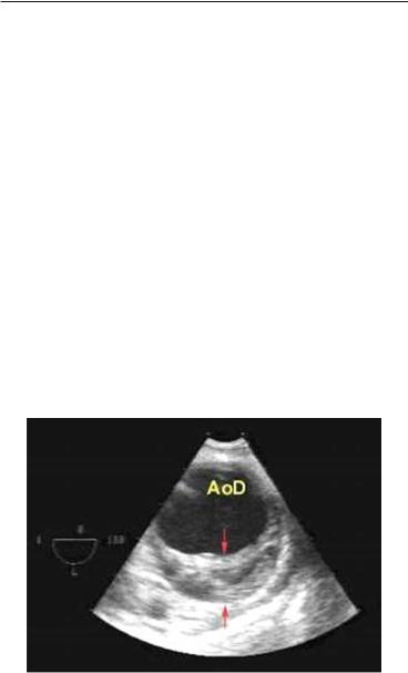
Intramural hematoma is considered to represent a variant of classical AD, possibly a stage in the development of full-fledged dissection. It has been described in 10–30% of patients with suspected AD.2 Pathologically, it is an intramedial bleeding, which did not progress to intimal flap formation and delineation of a true and false lumen. In terms of symptoms, the clinical presentation is similar to that of a typical dissection though evidence of visceral or peripheral ischemia may be less frequent. The prognosis and management are similar to those of completed dissection to which it can evolve in about one-third of cases.2 The echocardiographic diagnosis requires demonstration of a crescentic or circular grossly thickened (>7 mm) aortic wall, frequently with an
FIG. 7.4. Aortic intramural hematoma in the descending aorta. Note the semilunar massive thickening (arrows) of the posterior aortic wall and the echolucent zone between 5 and 6 o’clock. Reproduced with permission from Evangelista et al.6