

Review Test
1.Soon after birth, a term newborn infant presents with increased oral secretions and mild respiratory distress. Which of the following is the most likely diagnosis?
A.Persistent pulmonary hypertension of the newborn
B.Pneumonia
C.Esophageal atresia
D.Respiratory distress syndrome (surfactant deficiency syndrome)
E.Diaphragmatic hernia
2.An abdominal mass is detected on examination of a 2-day-old infant in the newborn nursery. Which of the following is the most likely cause of this abdominal mass?
A.Ovarian cyst
B.Hydronephrosis
C.Wilms tumor
D.Multicystic kidney
E.Hydrometrocolpos
3.The parents of a 5-day-old term infant notice that he is jaundiced. Your physical examination is remarkable only for scleral icterus and jaundice. The infant’s total bilirubin level is
15 mg/dL, with a direct component of 0.4 mg/dL. Which of the following is the most likely diagnosis?
A.Breastfeeding jaundice
B.Choledochal cyst
C.Biliary atresia
D.Neonatal hepatitis
E.Breast milk jaundice
4.You are called to the delivery room to evaluate a newborn infant born at 37 weeks’ gestation who has an abdominal wall defect noted on delivery. Based on your initial physical examination, you diagnose an omphalocele. Which of the following statements is consistent with this clinical diagnosis?
A.To rule out gastroschisis definitively, an abdominal computed tomographic scan is necessary.
B.Compared with gastroschisis, omphalocele is more frequently associated with other congenital malformations.
C.This abdominal wall defect is just lateral to the umbilicus.
D.The incidence of bowel obstruction is higher in this infant than in one with gastroschisis.
E.Omphaloceles may be associated with trisomy 21.
5.You are evaluating a 3-day-old infant with significant respiratory distress. He was delivered by emergency cesarean section at 42 weeks’ gestation because of fetal distress. You note that he has an oxygen saturation of 76% in room air that increases to 95% with administration of 100% oxygen. Which of the following statements most accurately supports your suspected diagnosis of persistent pulmonary hypertension of the newborn (PPHN)?
A.This patient is likely to have an associated cyanotic congenital cardiac defect.
B.PPHN occurs most frequently in premature infants but may occur in postterm infants.
C.PPHN usually resolves spontaneously.
D.This infant is likely to have significant left-to-right shunting.
E.Adequate oxygenation is the best preventive measurement and treatment.
6.A male infant was born at 32 weeks’ gestation via cesarean section because of bleeding from placenta previa. Soon after birth, he developed respiratory distress requiring supplemental oxygen and mechanical ventilation. Chest radiograph shows decreased lung volumes and a
159
diffuse ground glass pattern with air bronchograms. Which of the following is the most likely cause of this condition?
A.Persistent pulmonary hypertension of the newborn (PPHN)
B.Deficient surfactant
C.Fluid retention in the lungs
D.Bronchopulmonary dysplasia
E.Congenital heart disease
7.The parents of a term infant diagnosed with physiologic jaundice are very concerned that their child is at risk for brain damage. Which of the following statements regarding the infant’s hyperbilirubinemia is most accurate?
A.Breastfeeding, compared with formula feeding, is associated with higher peak serum bilirubin levels.
B.Serum conjugated bilirubin concentration is the best predictor of bilirubin encephalopathy.
C.Bilirubin encephalopathy does not occur in healthy term infants.
D.Increased conjugated (direct) bilirubin levels cause neuronal damage, including choreoathetoid cerebral palsy, hearing loss, and opisthotonus.
E.This infant’s jaundice is expected to peak at 10–14 days of life.
8.A 2-day-old term male infant is being evaluated before discharge from the nursery. The parents are concerned about a skin rash on his face. As you perform the physical examination, you contemplate skin disorders that are benign compared with those that may indicate underlying pathology. Which of the following skin findings is most likely to be associated with underlying pathology?
A.Pustular melanosis
B.Nevus simplex
C.Milia
D.Nevus flammeus
E.Erythema toxicum neonatorum (ETN)
9.At a routine health maintenance visit, a 2-week-old infant appears jaundiced. Laboratory evaluation reveals a total bilirubin level of 12.6 mg/dL with a direct bilirubin level of
6.9mg/dL. Which of the following is the most likely diagnosis?
A.Breastfeeding jaundice
B.Breast milk jaundice
C.Crigler–Najjar syndrome
D.ABO incompatibility
E.Choledochal cyst
10.A female infant born at 30 weeks’ gestation develops abdominal distension, abdominal tenderness, and bloody stools on the third day of life. Which of the following statements regarding the most likely diagnosis is correct?
A.The diagnosis is supported by a double-bubble sign on abdominal radiographs.
B.The diagnosis is supported by pneumatosis intestinalis on abdominal radiographs.
C.The diagnosis is supported by a soap-bubble appearance on abdominal radiographs.
D.The infant will ultimately require pancreatic enzyme supplementation.
E.The diagnosis has an increased association with Down syndrome.
Questions 11 and 12: The response options for statements 11 and 12 are the same. You will be required to select one answer for each statement in the set.
A.2
B.3
C.4
160
D.5
E.6
F.7
G.8
In each case, select the infant’s 1-minute Apgar score.
1.At 1 minute of life, a newborn’s respiratory rate is slow and irregular with a heart rate of 120 beats/minute. There is some flexion of her upper and lower extremities; she grimaces
when a catheter is placed into her nose; and she appears to be pink and well perfused, except for some cyanosis of the distal extremities.
2.At 1 minute of life, a newborn’s respiratory rate is slow and irregular with a heart rate of
80 beats/minute. There is some flexion of his upper and lower extremities; he does not respond when a catheter is placed into his nose; and he is blue and pale.
You’re called to the Nursery to evaluate a cyanotic infant with significant respiratory distress. After 100% oxygen is administered to the infant, there is almost no improvement in the PaO2 (only 10 mm Hg). Which of the following is the patient’s most likely diagnosis?
A.Tetralogy of Fallot
B.Pneumonia
C.Respiratory Distress Syndrome (RDS)
D.Meconium aspiration syndrome
E.Transient tachypnea of the newborn
161
Answers and Explanations
1.The answer is C [XII.A.3]. Esophageal atresia in a newborn is characterized by increased oral secretions as a result of the accumulation of saliva in the proximal esophageal pouch. Respiratory distress may occur if the infant aspirates this saliva. The presence of a distal tracheoesophageal fistula may also result in the passage of gastric contents to the trachea and lung, exacerbating the respiratory problem. Half of children with esophageal atresia have other congenital malformations, such as congenital heart disease. Both pneumonia and persistent pulmonary hypertension of the newborn also present with respiratory distress, but without increased oral secretions. Respiratory distress syndrome occurs less commonly in term infants, and increased oral secretions are not expected. Congenital diaphragmatic hernia usually presents with acute respiratory distress soon after birth in a newborn with a scaphoid abdomen. Bowel sounds can be heard on auscultation of the chest.
2.The answer is B [I.G.6 and I.H.1.b]. The most likely cause of an abdominal mass detected during the newborn period is of renal origin, with hydronephrosis being the most common cause. In female infants, an ovarian cyst, which is usually a benign tumor, is not as common as hydronephrosis. Wilms tumor and multicystic kidneys may present as abdominal masses but are also less common causes. Hydrometrocolpos, a retention of vaginal secretions, most commonly presents just after birth as a small cyst located between the labia, although during childhood, it may present as a lower midline abdominal mass.
3.The answer is A [X.C.1]. Breastfeeding jaundice is typically associated with indirect, or unconjugated, hyperbilirubinemia and is caused by suboptimal milk intake during the first week of life, which causes weight loss, poor hydration, and decreased stool output. The treatment of breastfeeding jaundice is hydration, which typically includes increasing the frequency of breastfeeding, along with observation and serial bilirubin assessments. Breast milk jaundice, which occurs later, after the first week of life, is thought to be associated with high levels of lipase and β-glucuronidase within breast milk. Choledochal cysts, biliary atresia, and neonatal hepatitis are more typical causes of direct, or conjugated, hyperbilirubinemia.
4.The answer is B [XII.C]. Omphalocele is more frequently associated with congenital malformations, such as congenital heart defects, and with genetic conditions, such as trisomy 13, and less commonly with trisomy 18, but not with trisomy 21. Omphalocele and gastroschisis are easily distinguished and diagnosed by inspection. An omphalocele occurs centrally through the umbilical ring, whereas gastroschisis is a lateral abdominal wall defect in which the abdominal contents herniate into the amniotic cavity and are directly exposed to amniotic fluid. Because of this difference in clinical presentation, both omphalocele and gastroschisis are diagnosed clinically without the need for radiographic confirmation. In gastroschisis, exposure to the amniotic fluid may cause inflammation of the bowel with subsequent bowel damage and risk of bowel obstruction.
5.The answer is E [VIII.A, VIII.C, and VIII.E]. One of the most common causes of persistent pulmonary hypertension of the newborn (PPHN) is perinatal asphyxia, resulting in increased pulmonary vascular resistance and significant right-to-left shunting through the foramen ovale or the ductus arteriosus. Oxygen is the most potent vasodilator of pulmonary vessels, and in most cases, increases of both alveolar and arterial partial pressures of O2 produce a decrease in pulmonary vascular resistance and reversal of low blood flow to the lungs. By definition, PPHN excludes the presence of congenital heart disease. If left untreated, the hypoxemia caused by PPHN worsens the increased pulmonary vascular resistance, resulting in many cases in irreversible disease and death. In addition, PPHN occurs most commonly in near-term and full-term infants, as well as in postterm infants.
6.The answer is B [VI.A–H]. Respiratory distress syndrome (RDS), which is most common in
162
premature male infants, is caused by a lack or deficiency of surfactant, with alveolar atelectasis and hypoventilation. Chest radiographic findings usually include a diffuse ground glass pattern with air bronchograms. Pneumonia and sepsis should always be included in the differential diagnosis of RDS because their clinical presentations may be quite similar. Persistent pulmonary hypertension of the newborn (PPHN) is more common in term infants than in premature infants and results most frequently from perinatal asphyxia and meconium aspiration syndrome (MAS). Fluid retention in the lungs may cause respiratory distress, but it is usually mild. The chest radiograph usually shows normal or increased lung volume with increased vascular markings. Bronchopulmonary dysplasia (BPD) is a chronic complication of RDS. Some causes of cyanotic congenital heart disease may cause hypoxemia and respiratory distress after birth; however, the chest radiograph does not show a ground glass appearance, nor air bronchograms.
7.The answer is A [X.A–G]. Newborn infants who breastfeed have higher peak serum bilirubin values. However, hyperbilirubinemia alone is not a reason to discontinue breastfeeding. Bilirubin encephalopathy is caused only by unconjugated (indirect) bilirubin because of the ability of unconjugated bilirubin to cross the blood–brain barrier. Encephalopathy caused by indirect hyperbilirubinemia does occur in healthy term newborns, and for this reason, high bilirubin levels in this group of infants should not be ignored. Benign physiologic indirect hyperbilirubinemia is expected to peak in term infants at 3–4 days of life and in preterm infants at 5–7 days of life.
8.The answer is D [I.K.11]. Nevus flammeus or “port wine stain” located over the V-1 branch of the trigeminal nerve may herald Sturge–Weber syndrome, with its associated, and potentially very significant, underlying intracranial vascular malformations and calcifications. Pustular melanosis is a benign rash, characterized by small, dry vesicles over a dark macular base, more frequently seen in African American infants. Nevus simplex is the most common vascular lesion of infancy and is also completely benign and often transient, appearing as a “salmon patch” or “stork bite” on the nape of the neck. Milia are benign very small cysts formed around the pilosebaceous follicles that appear as tiny whitish papules over the nose, cheeks, forehead, and chin. Erythema toxicum neonatorum (ETN) is a benign rash usually present in the first 72 hours of life and seen in approximately 50% of all infants. ETN is characterized by erythematous macules, papules, or pustules on the trunk and extremities.
9.The answer is E [Figures 4-3 and 4-4]. This infant’s presentation with hyperbilirubinemia and a markedly elevated direct bilirubin level is consistent with a choledochal cyst, a disorder that causes obstruction of the biliary tree. Both breastfeeding and breast milk jaundice are characterized by indirect, not direct, hyperbilirubinemia. Crigler–Najjar syndrome, or hereditary deficiency of glucuronyl transferase, would also be expected to result in an indirect,
or unconjugated, hyperbilirubinemia. ABO incompatibility would lead to hemolysis, leading to an elevation of indirect bilirubin.
10.The answer is B [XII.E]. Necrotizing enterocolitis (NEC) is one of the most common surgical conditions in neonates, occurring most commonly in premature infants. Clinical features include abdominal distension, abdominal tenderness, residual gastric contents, bilious vomiting or bilious nasogastric aspirate, bloody stools, and, at times, abdominal wall erythema. Classic radiographic findings include abdominal distension, air–fluid levels, thickened bowel walls, and pneumatosis intestinalis (air within the bowel wall). In contrast, the double-bubble sign on abdominal radiographs is pathognomonic of duodenal atresia, which classically presents with nonbilious emesis and abdominal distension, but not with bloody stools. The presence of a soap-bubble appearance on abdominal radiographs is characteristic of meconium ileus, a presentation of cystic fibrosis during the neonatal period. Infants with meconium ileus would not be expected to pass bloody stools on the third day of life, but may require pancreatic enzyme supplementation if they are ultimately diagnosed
163
with fat malabsorption and cystic fibrosis. There is no known association between Down syndrome and NEC, although there is an association between Down syndrome and duodenal atresia.
11.The answers are E and B, respectively [Table 4.1]. The Apgar scoring system provides a
simple, systematic, and objective assessment of intrapartum stress and neurologic depression. The female newborn earns 2 points for a heart rate >100 beats/minute, 1 point for slow and irregular respirations, 1 point for having some flexion of the extremities, 1 point for reflex
irritability or grimace when a catheter is placed in her nose, and 1 point for her peripheral cyanosis or acrocyanosis, for a total Apgar score of 6 points. The male newborn earns 1 point for a heart rate <100 beats/minute, 1 point for slow and irregular respirations, 1 point for having some flexion of the extremities, 0 points for the absence of reflex irritability when a catheter is placed into his nose, and 0 points for his cyanosis, for a total Apgar score of 3 points.
12.The answer is A [IV.D.2 and V.A] The 100% oxygen test helps distinguish whether cyanosis is caused by cardiac or respiratory disease. When administered 100% oxygen, infants with primary lung pathology, such as neonatal pneumonia, meconium aspiration syndrome, transient tachypnea of the newborn or respiratory distress syndrome, have a very significant increase in PaO2 levels. In contrast, patients with cyanotic congenital heart disease would not be expected to have such a significant rise in their PaO2 level. For example, patients with Tetralogy of Fallot, a cyanotic congenital heart disease associated with reduced pulmonary blood flow, would not be expected to respond with any increase of significance in the PaO2 level when given 100% oxygen. Note that other cyanotic congenital heart diseases with normal or increased pulmonary blood flow, like truncus arteriosus, may have some increase in the PaO2 level, but nowhere near as much of an increase as that seen in infants with primary pulmonary disease.
164

C H A P T E R 5
165

Genetic Disorders and Inborn Errors of
Metabolism
Jessica Tenney, Derek Wong
166
I.Inheritance Patterns
A.Mendelian inheritance (Figure 5-1). The classic patterns of inheritance seen in single-gene disorders
1.Dominant inheritance
a.Autosomal dominant mode of inheritance is observed when an abnormal copy of a gene is located on one of the autosomes (chromosomes 1–22). Males and females have the same chance of being affected. If one parent is affected, the risk of having an affected child is 50% in each pregnancy.
b.X-linked dominant mode of inheritance is observed when an abnormal gene is located on one X chromosome. An affected father will transmit disease to 100% of his female offspring and none of his male offspring, whereas an affected mother has a 50% chance of transmitting disease to any of her offspring.
2.Recessive inheritance
a.Autosomal recessive mode of inheritance is observed when there are no normal copies of a gene. If both parents are carriers, offspring will have a 25% chance of being affected. It is often seen in only one generation in a pedigree. Parental consanguinity increases the risk for autosomal recessive conditions.
b.X-linked–recessive mode of inheritance is observed when there are no normal copies of the gene. Disease occurs when a son inherits the abnormal copy from his mother. There is no male-to-male transmission. Daughters who inherit one abnormal copy of the gene are usually asymptomatic, but some may be affected if there is skewed X-inactivation. For example, because one random X chromosome is inactivated in all cells of females, if the X chromosomes with the normal gene are disproportionately inactivated relative to the X chromosomes with the abnormal
gene, the female may be affected. A female who inherits two abnormal copies will always be affected. A carrier mother has a 50% chance of having an affected son.
B.Mitochondrial inheritance is observed when a mutation occurs in one of the genes in the mitochondrial genome. The mitochondrial genome is housed separately from the nuclear genome and is only inherited from the mother. In a pedigree, offspring of an affected mother can show signs of disease, but an affected father will never have affected offspring.
C.Multifactorial inheritance occurs when a combination of genetic and environmental factors determines whether or not a disorder will manifest. The disorder will be seen in a particular family with increased frequency compared with that of the general population. Examples of multifactorial inheritance include cleft lip and palate, neural tube defects, developmental dysplasia of the hip, and pyloric stenosis.
167
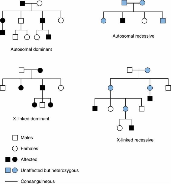
FIGURE 5.1 Pedigrees of different Mendelian inheritance patterns.
Adapted with permission from Sakala EP. BRS Obstetrics and Gynecology. 2nd Ed. Philadelphia: Lippincott Williams & Wilkins, 2000:52.
168
II.Types of Genetic Tests
A.Karyotype analysis is used to determine the number and structure of chromosomes. This may be used to diagnose a chromosomal trisomy, sex chromosome disorders, translocations, and larger deletions or duplications.
B.Microarray is currently the preferred first line test used to diagnose microdeletion/microduplication syndromes, such as 22q11.2 deletion syndrome [see section IV.D.1].
C.Fluorescence in situ hybridization (FISH) is a targeted test that may be also used to look for the presence of a specific sequence of DNA on a chromosome.
D.Methylation studies are performed to help diagnose specific imprinting disorders (e.g., Prader–Willi syndrome). When methyl groups are added to DNA, the activities of the DNA segment can change. This physiologic process plays a role in X chromosome inactivation and imprinting [see section IV.E.1.a], but also plays a role in pathologic processes such as aging, carcinogenesis, and the development of imprinting disorders such as Prader–Willi.
E.Gene sequencing is performed to look for mutations in a specific gene (e.g., fibrillin 1 sequencing to diagnose Marfan syndrome).
F.Exome sequencing looks for potential diseases causing mutations in the protein coding regions (exons) of nearly the entire genome. It is useful when there is a broad differential diagnosis of genetic conditions.
169
III.Fetal Evaluation and Prenatal Diagnosis
A.Maternal serum markers
1.α-Fetoprotein (AFP) is elevated with fetal neural tube defects, multiple gestation pregnancies, underestimated gestation age, ventral abdominal wall defects, fetal demise, or fetal edema or skin defects. Low AFP levels are associated with overestimated gestation age, trisomies 18 and 21, and intrauterine growth retardation.
2.Triple/quadruple markers are used in conjunction with ultrasound findings as a noninvasive method to assess the fetus for the possibility of trisomy syndromes. The three classic second-trimester screening markers are AFP, unconjugated estriol (uE3), and the β-subunit of human chorionic gonadotropin (β-HCG). More recently, the use of inhibin-A has been introduced to increase the detection rate of Down syndrome.
a.Trisomy 21 is suggested by findings of low AFP and uE3 and high β-HCG and inhibin-A.
b.Trisomy 18 is suggested by low levels of AFP, uE3, and β-HCG
B.Ultrasound is used to assess gestation age and fetal growth and to evaluate for major fetal anomalies. The finding of increased nuchal translucency may indicate the presence of trisomies 13, 18, and 21 and Turner syndrome, as well as structural anomalies such as congenital heart disease.
C.Genetic evaluation of the fetus
1.Chorionic villus sampling (CVS) is the use of villus tissue from the chorion of the trophoblast that is collected at 10–13 weeks’ gestation. Karyotype, gene sequencing, and enzyme analyses from CVS can be used to assess for genetic and metabolic disorders.
2.Amniocentesis is the use of amniotic fluid containing sloughed fetal cells collected at 16– 18 weeks’ gestation. This technique can be used to assess for the same genetic diseases as CVS.
3.Percutaneous umbilical blood sampling involves obtaining a sample of fetal blood to assess for hematologic abnormalities, genetic disorders, infections, and fetal acidosis. It can also be used to administer medications or blood transfusions to the fetus.
4.Noninvasive prenatal testing (NIPT) involves the isolation of fetal cells extracted from a cell-free DNA sample of maternal blood. It is used for assessment of fetal trisomies 13, 18, and 21, and has the potential to replace more invasive forms of sampling for all genetic tests in the future.
170
IV. Common Genetic Disorders
A.Terminology
1.Malformations are defects that result from the intrinsically abnormal development of a structure or set of structures (e.g., anencephaly, congenital heart disease).
2.Deformations occur when extrinsic (mechanical) forces impinge on the development of otherwise normal tissue (e.g., clubfoot).
3.Disruptions occur when normal tissue is irreparably destroyed, altering the subsequent formation of the structure (e.g., amniotic bands).
4.Syndromes are recognizable patterns of symptoms or abnormalities that suggest a specific underlying disorder.
B.Trisomy syndromes
1.Trisomy 21 (Down syndrome) is the most common chromosomal disorder. The incidence is about 1:700 live births. The risk increases in advanced maternal age, with a sharper rise in prevalence rates seen every year after the age of 35 years.
a.Clinical features. See Table 5-1 and Figure 5-2.
b.Diagnosis is on the basis of clinical features and genetic testing that demonstrates three copies of chromosome 21. Note that about 3–4% of individuals have Down syndrome due to a translocation. A karyotype is needed to distinguish full trisomy 21 from a translocation. If a parent carries a translocation, the recurrence risk for subsequent children with Down syndrome is higher.
2.Trisomy 18 (Edwards syndrome)
a.Clinical features include severe intellectual disability, prominent occiput, low-set ears, micrognathia, congenital heart disease, rocker-bottom feet, and clenched hands with overlapping digits.
b.Prognosis is poor, as 90% of children die by 1 year of age.
3.Trisomy 13 (Patau syndrome)
a.Clinical features include severe intellectual disability, cutis aplasia, microphthalmia, coloboma, congenital heart disease, polydactyly, and midline defects such as agenesis of the corpus callosum and cleft lip and palate.
b.Prognosis is poor, as 50% die by 1 month of age and 90% die by 1 year of age.
C.Sex chromosome syndromes
1.Turner syndrome (XO) occurs in females with complete or partial absence of a second X chromosome.
a.Epidemiology
1.Monosomy X is found in 45% of affected individuals, whereas the remainder of cases are due to either a structural abnormality of the second X chromosome or mosaicism.
2.The incidence is 1:2500–3000 female live births.
b.Clinical features
1.Short stature is present in 95% of individuals with Turner syndrome. Growth hormone can be used to increase final height.
2.Webbed neck with low posterior hairline
3.Broad chest with widely spaced nipples (shield chest)
4.Congenital lymphedema. May have swelling of the hands and/or feet at birth
5.Cardiac defects: coarctation of the aorta, bicuspid aortic valve, hypoplastic left heart
6.Ovarian dysgenesis leads to primary amenorrhea and lack of secondary sex characteristics in most patients. Estrogen therapy can be used to promote
171
secondary sex characteristics.
7.Most individuals have normal intelligence.
8.Renal malformations, hypothyroidism, diabetes, and hearing loss are also associated with Turner syndrome.
2.Klinefelter syndrome (XXY) is the most common genetic cause of male infertility.
a.Epidemiology
1.The incidence is 1:500–1000 male live births.
2.Risk increases with advancing maternal age.
b.Clinical features
1.Tall stature, thin, with relatively long legs, and gynecomastia
2.Hypogonadism results in small testicles, underdeveloped secondary sex characteristics, oligoor azoospermia, and decreased bone density.
3.Learning disabilities are common, especially in the areas of verbal comprehension and reading. There is an increased risk of psychosocial and behavioral problems.
4.There is also an increased risk for developing mediastinal germ cell tumors (starting in adolescence) and breast cancer.
3.XYY males may be taller than average but have normal sexual development. Intelligence is usually normal, but there is increased risk for learning disabilities and behavioral problems.
D.Chromosomal (micro) deletion syndromes
1.22q11.2 deletion syndrome (DiGeorge, velocardiofacial syndrome) occurs when a portion of the q arm of chromosome 22 is missing. The deletion can be de novo or inherited. The mnemonic CATCH-22 can be used to remember the findings of this disorder (Cardiac defects, Abnormal facies, Thymic hypoplasia, Cleft palate, Hypocalcemia, and deletion on chromosome 22)
a.Epidemiology.
1.The incidence of 22q11.2 deletion syndrome is about 1 in 4000 live births.
b.Clinical features
1.Cardiac: congenital heart disease, especially conotruncal malformations (tetralogy of Fallot, ventricular septal defect)
2.Palatal abnormalities: velopharyngeal incompetence, cleft palate, submucous cleft, and bifid uvula
3.Abnormal facies: hooded eyelids, hypertelorism, overfolded or squared off helices, prominent nasal root, bulbous nasal tip, and micrognathia
4.Thymic hypoplasia can result in immunodeficiency. Parathyroid hypoplasia can result in severe hypocalcemia, and therefore, calcium should be monitored in any newborn with suspected 22q11.2 deletion.
5.Intellectual and learning disability
6.Other associations include renal anomalies, hearing loss, and gastrointestinal anomalies.
2.Williams syndrome results from a deletion on chromosome 7 (7q11.23) that includes the elastin gene.
a.Epidemiology.
1.The incidence of Williams syndrome is about 1 in 20,000 births.
b.Clinical features
1.Distinctive facial features (“Elfin facies”) include prominent forehead, widely spaced eyes, upturned and full nasal tip, long philtrum, distinctive wide mouth, and stellate/lacy iris pattern.
2.Cardiovascular disease (elastin arteriopathy): Supravalvar aortic stenosis is
172
the most common location.
3.Abnormalities of connective tissue may result in a hoarse voice or hernias.
4.Intellectual disability, with a very friendly personality
5.Endocrine problems include hypocalcemia, hypercalcuria, and hypothyroidism.
3.Cri du chat syndrome results from a deletion of the short arm of chromosome 5 (5p). It is characterized by catlike cry in infancy, microcephaly, downslanting palpebral fissures, developmental delay, and intellectual disability.
E. Imprinting disorders
1.Key concepts
a.Genomic imprinting results in differences in gene expression depending on whether the gene is inherited from the mother or father. Disease occurs when the copy from the appropriate parent cannot be expressed. An example occurs in an imprinted region of chromosome 11q. Angelman syndrome occurs when there is a deletion or other mechanism that causes a missing maternal copy of the region, and Prader–Willi syndrome occurs if there is a missing paternal copy of the region. The mnemonic “P is for Prader–Willi and paternal deletion” may be used to remember the molecular basis of these conditions.
b.Uniparental disomy (UPD) occurs when both copies of one chromosome come from the same parent.
2.Prader–Willi syndrome
a.Clinical features
1.In infancy, patients demonstrate hypotonia and feeding difficulties, usually resulting in failure to thrive. In childhood, patients develop hyperphagia leading to obesity.
2.Almond-shaped eyes, strabismus, down-turned mouth, hypopigmentation, small hands and feet, short stature, and hypogonadism
3.Behavioral problems, intellectual disability, and learning disabilities
b.The most common cause is a deletion in a region within the paternally inherited chromosome 15. The second most common cause is maternal UPD of chromosome 15.
c.Diagnosis is by methylation analysis.
3.Angelman syndrome
a.Clinical features
1.Severe developmental delay, speech impairment, and happy demeanor with inappropriate laughter and smiling.
2.Jerky movements and ataxic gait is sometimes described as “puppetlike.”
3.Microcephaly, seizures, large mouth, widely spaced teeth, and prognathia (prominent mandible)
b.The most common cause is a deletion in a region within the maternally inherited chromosome 15.
4.Diagnosis is by methylation analysis.
5.Beckwith–Wiedemann syndrome
a.Clinical features
1.Overgrowth disorder characterized by macrosomia, macroglossia, and visceromegaly
2.Can have hemihyperplasia, ear creases/pits, and omphalocele
3.Increased risk for embryonal tumors (Wilms tumor, neuroblastoma, etc.)
b.This syndrome has several different etiologies, all of which affect imprinting on chromosome 11p15.5.
173
c.Diagnosis can be established by clinical criteria, methylation studies, microarray, or specific gene sequencing.
6.Russell–Silver syndrome
a.Clinical features
1.Intrauterine growth retardation/small for gestational age (SGA), short stature, normal head circumference, and asymmetry (limb, body, or face)
2.Triangular facies, frontal bossing, or prominent forehead. The patient can also have café-au-lait macules.
b.It has been associated with hypomethylation of a region that regulates expression of insulin-like growth factor 2.
c.Diagnosis is largely based on clinical characteristics.
F.Triplet repeat expansion disorders. Certain genes are sensitive to increasing (expanding) the number of nucleotide repeats in a specific gene segment. The number of repeats can increase with each generation, but the disorder only occurs once the number of nucleotide repeats in a gene expands beyond a specific threshold. Once the threshold is reached, this expansion can become even larger, causing earlier onset and more severe symptoms, which is a phenomenon known as anticipation.
1.Fragile X syndrome
a.This is caused by expansion of the number of CGG repeats in the FMR1 gene on the X chromosome. It has an X-linked recessive mode of inheritance. Full
mutation = >200 repeats
b.Clinical features
1.Developmental delay and mild to severe intellectual disability. Behavioral abnormalities including autism and attention deficit/hyperactivity disorder
2.Macrocephaly, long face, prominent jaw, protruding ears. Macroorchidism develops in adolescence.
3.Females with full mutation may have behavioral problems and developmental delays, but most have normal intelligence quotient (IQ).
2.Myotonic dystrophy
a.Genetic mechanism
1.The severe form is caused by expansion of the number of CTG repeats in the DMPK gene.
2.Expansion of the repeats occurs much more frequently in mothers.
b.Clinical features
1.Progressive muscular weakness starting at any time during childhood
2.Other features include cataracts and cardiac conduction abnormalities.
G.Connective tissue disorders
1.Marfan syndrome
a.An autosomal dominant disorder caused by mutations in the gene that codes for fibrillin. Mutations can either be inherited or sporadic.
b.Clinical features. Characteristic findings involve the ocular, skeletal, and cardiovascular systems.
1.Myopia, lens dislocation, and retinal detachment
2.Tall stature with long extremities, long fingers (arachnodactyly), pectus deformity, scoliosis, pes planus, decreased upper-to-lower segment ratio, increased arm span-to-height ratio
3.Aortic root dilatation, mitral valve prolapse, and valvular regurgitation. Note:
Patients with aortic root dilatation are at increased risk for aortic dissection.
β-blockers +/− angiotensin receptor blockers are prescribed to help prevent or slow progressive aortic enlargement.
174
4.Patients are at risk for spontaneous pneumothorax.
2.Ehlers–Danlos syndrome, classic type
a.An autosomal dominant disorders caused by mutations in genes that code for type V collagen. Mutations can be inherited or sporadic.
b.Clinical features. Characterized by skin hyperextensibility, abnormal wound healing, and joint hypermobility. Skin may be described as soft and/or velvety in texture, with bruising caused by fragile blood vessels. Tissue fragility and poor wound healing results in widened atrophic scars.
H.Skeletal disorders
1.Achondroplasia
a.An autosomal dominant disorder caused by mutations in FGFR3. The majority of cases result from a sporadic mutation. Advanced paternal age (>45 years old) increases the risk of having an affected child.
b.Achondroplasia is the most common cause of disproportionate short stature.
c.Clinical features
1.Craniofacial findings include macrocephaly, prominent forehead, low nasal bridge, and midface hypoplasia.
2.Characteristic skeletal findings:
a.Rhizomelic (proximal limb) shortening of the limbs, bowed legs, and trident appearance of the hands.
b.Lumbar gibbus deformity (structural, sharp-angled kyphosis resulting in a prominent “hump” on the back) in infancy that resolves once the patient starts walking. Lumbar lordosis then develops.
3.Normal intelligence with delayed acquisition of motor milestones
4.Recurrent middle ear dysfunction and obstructive sleep apnea
5.Increased risk for foramen magnum stenosis and compression of the craniocervical junction. Patients should therefore be monitored for signs of hydrocephalus and spinal cord compression.
2.Osteogenesis imperfecta (OI)
a.Most cases are due to autosomal dominant mutations in genes that encode for chains of type I collagen. Type I is the most common and the mildest form of OI. Patients with Type II OI are the most severely affected, and rarely survive beyond the first weeks of life. Type III is the most severe type among those who survive the neonatal period.
b.Clinical features of Type I OI
1.Characterized by blue sclera and frequent fractures after minimal or no trauma. Easy bruising. Fractures usually heal without resulting deformity.
2.Normal stature or slightly shorter than the rest of the family
3.May have yellow or grayish brittle teeth (dentinogenesis imperfecta) which are at increased risk for breakage.
4.Progressive hearing loss in adulthood
I.Additional disorders
1.Noonan syndrome
a.Autosomal dominant disorder caused by mutations in certain genes involved in the RAS/MAPK pathway.
b.Clinical features
1.Characterized by short stature, congenital heart defect, pectus deformity, and characteristic facies
2.Craniofacial findings are most prominent in infancy and include low-set, posteriorly rotated ears, wide-spaced eyes (hypertelorism), eyelid ptosis, and
175
downslanting palpebral fissures.
3.Pulmonary valve stenosis is the most common cardiac defect. All patients are at risk for developing hypertrophic cardiomyopathy during their lifetime.
4.Patients can have short, webbed neck with low posterior hairline, and broad chest with widely spaced nipples. Affected females are sometimes initially misdiagnosed with Turner syndrome (which is associated with left-sided heart defects vs. Noonan syndrome with right-sided heart defects).
5.Patients may have developmental delay and/or intellectual disability.
2.VACTERL (VATER) association
a.A group of malformations generally observed as a sporadic occurrence in an otherwise healthy family. The etiology is unknown. Clinical findings overlap with those seen in Fanconi anemia, which should be ruled out in anyone presenting with VACTERL.
b.Clinical features
1.V—vertebral defects
2.A—anal atresia
3.C—cardiac defects
4.TE—tracheoesophageal (TE) fistula
5.R—renal dysplasia
6.L—limb/radial defects
3.CHARGE Syndrome
a.An autosomal dominant disorder most often caused by a spontaneous mutation
b.Clinical features
1.C—colobomas of the iris or retina. Usually results in impaired vision
2.H—heart defects such as conotruncal defects and aortic arch abnormalities
3.A—choanal atresia or stenosis
4.R—retarded growth and development
5.G—genital abnormalities, including genital hypoplasia
6.E—ear anomalies, including abnormal outer ear shape, hearing loss, and ossicular and temporal bone abnormalities
7.Cranial nerve dysfunction is another important feature.
4.Cornelia de Lange syndrome
a.Generally observed as a sporadic occurrence in an otherwise healthy family. Autosomal dominant and X-linked mutations have been found.
b.Clinical features
1.Characteristic facies include synophrys (single eyebrow resulting from both eyebrows growing into one another), long eyelashes, small upturned nose, and microcephaly.
2.SGA, failure to thrive, and small stature
3.Hirsutism
4.Upper limb malformations
5.Developmental delay, intellectual disability, and behavioral problems
5.Neurofibromatosis type 1 (see also Chapter 19, Table 19-3)
a.Autosomal dominant disorder caused by mutations in NF1. Half of the cases are inherited, and the remainder are spontaneous mutations.
b.Clinical features
1.Characteristic findings include multiple café-au-lait macules, axillary and inguinal freckling, cutaneous and/or plexiform neurofibromas, iris hamartomas (Lisch nodules), and osseous dysplasia.
2.Increased risk for malignant peripheral nerve sheath tumors (PNETs), optic
176

nerve gliomas, and central nervous system (CNS) gliomas
3.Learning disabilities are seen in half of patients.
6.Potter syndrome
a.A deformation sequence that results from compressive forces exerted on a developing fetus subjected to prolonged oligohydramnios. The oligohydramnios may have resulted from bilateral renal agenesis, other urinary tract defects, or a chronic leak of amniotic fluid.
b.Clinical features include lung hypoplasia, abnormal limb positioning, and characteristic “Potter facies” (compressed facial appearance, with large ears flattened against the head).
7.Pierre Robin sequence
a.A sequence that can be seen as an isolated finding or as part of a specific multiple malformation syndrome. Clinical findings result from mandibular hypoplasia, which leads to a cascade of other features.
b.Clinical features include micrognathia, glossoptosis (posterior displacement of the tongue), and cleft palate (often a U-shaped cleft).
8.Amniotic band syndrome occurs when rupture of the amniotic sac during pregnancy results in small strands of amnion wrapping around areas of the fetus’s body, causing deformation and/or amputation.
J. Syndromes caused by teratogens(Table 5-2)
1.Diabetic embryopathy
a.Increased risk for congenital malformations occurs in infants born to mothers with type 1 or type 2 diabetes, especially when the Hgb A1C level is >12 just before conception and/or in the early first trimester.
b.The infant may have isolated or multiple malformations, including caudal regression syndrome (sacral agenesis), neural tube defects, renal malformations, cardiac defects, or craniofacial abnormalities.
2.Fetal alcohol syndrome (FAS)
a.FAS is caused by maternal alcohol consumption during pregnancy. Prenatal exposure to alcohol can cause a wide range of findings, with FAS being the most severe. No amount of alcohol is considered safe, but the risk for FAS is higher with binge drinking or chronic alcohol consumption during pregnancy.
b.Clinical features
1.Microcephaly, short palpebral fissures, smooth philtrum, and thin upper lip
2.Developmental delay, intellectual disability, and attention deficit hyperactivity disorder
3.Infants may be SGA.
4.Cardiac defects may occur (ventricular septal defects are most common).
3.Fetal hydantoin syndrome
a.Infants born to mothers taking phenytoin during pregnancy are at increased risk for specific malformations.
b.Clinical features
1.Microcephaly, wide anterior fontanelle, hypertelorism, short nose, wide mouth
2.Low hairline, short neck, hypoplastic nails
3.Developmental delays
Table 5-1
Clinical Features and Complications Associated with Down Syndrome, with Recommendations for Screening
Clinical Features |
Complications |
|
|
177

Craniofacial features |
Atlantoaxial instability (1–2%) |
|
Brachycephaly |
Anemia |
|
|
||
|
|
|
Epicanthal folds |
Check hemoglobin annually |
|
|
|
|
Upslanting palpebral fissures |
Leukemia (1% of patients but more common than the general |
|
population) |
||
|
||
|
|
|
Brushfield spots (speckled irides) |
|
|
|
|
|
Protruding tongue |
Celiac disease (5%) |
|
|
||
|
|
|
Hypotonia |
Early onset Alzheimer disease |
|
Intellectual disability |
Obstructive sleep apnea (50–75%) |
|
|
Sleep study by 4 years of age |
|
|
|
|
Musculoskeletal features |
Hearing problems (75%) |
|
Clinodactyly |
Hearing screens needed every 1–2 years |
|
|
|
|
Single palmar creases |
Thyroid disease |
|
|
||
|
|
|
Wide space between first and second toes (sandal gap) |
Annual TSH screening |
|
|
|
|
Gastrointestinal features |
Cataracts, glaucoma, and refractive errors |
|
Duodenal atresia |
Annual ophthalmologic examinations |
|
|
|
|
Hirschsprung disease and omphalocele |
|
|
|
|
|
Pyloric stenosis |
|
|
|
|
|
Cardiac defects (40%) |
|
|
Endocardial cushion defects (most common)— |
|
|
echocardiogram at birth |
|
|
|
|
TSH = thyroid-stimulating hormone.
178
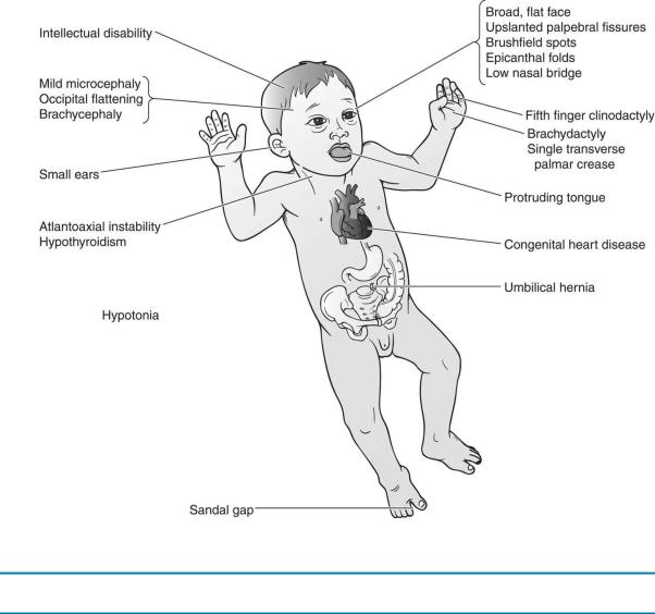
FIGURE 5.2 Clinical features of Down syndrome.
Table 5-2
Teratogens and Associated Anomalies
Drugs |
Associated Anomalies |
Alcohol |
Microcephaly; short palpebral fissures; long, smooth philtrum; variable intellectual disability; and |
|
congenital heart disease |
Cigarette |
Small for gestational age |
smoking |
|
Cocaine |
Premature birth |
Diethylstilbestrol |
Increased risk of vaginal adenocarcinoma and genitourinary anomalies |
(DES) |
|
Isotretinoin |
Small low-set ears, micrognathia, depressed nasal bridge, cardiac defects, thymic hypoplasia, and central |
|
nervous system malformations |
Lithium |
Ebstein anomaly |
Phenytoin |
Wide anterior fontanelle, short nose, wide mouth, microcephaly, low hairline, hypoplastic nails, and |
|
developmental delay |
Thalidomide |
Phocomelia (malformed extremities resulting in flipperlike appendages) |
Valproic acid |
Neural tube defects |
Warfarin |
Hypoplastic nose with depressed nasal bridge, stippled epiphyses, and hypoplastic nails |
179
V.General Concepts of Inborn Errors of Metabolism (IEM)
A.IEM are a heterogeneous group of diseases that can present in a variety of ways. Individually, each disease is rare but causes significant morbidity and mortality.
B.Disorders occur when a specific step in a metabolic pathway is disrupted in some way (defective enzyme, receptor problem, etc.), resulting in the accumulation of toxic metabolites.
C.Each individual disorder may have several subtypes that can vary in level of severity. Unless otherwise stated, the severe/classic form of each disorder is discussed below.
D.An overview of different categories of IEM, including examples of conditions within each category, is summarized in Figure 5-3.
E.Clinical features (Table 5-3)
1.IEM should be considered in patients with the following clinical presentations.
a.An otherwise healthy newborn who develops an acute severe illness within the first few hours to weeks of life
b.Acute and recurrent episodes of altered mental status, vomiting, acidosis, organ failure, or ataxia
c.Chronic and progressive symptoms such as developmental delay or regression of developmental milestones, intellectual disability, or seizures
2.Age of onset. Age of onset is variable depending on the type of IEM. Some disorders that present in infancy have less severe forms that can present later in infancy, childhood, adolescence, or even adulthood.
3.Family history
a.Most IEM are inherited in an autosomal recessive fashion, but several are X-linked recessive. A history of parental consanguinity may be seen in patients with autosomal recessive disorders.
b.There might be a family history of neonatal death.
c.For recessive conditions, there may be an affected sibling, and for X-linked disorders, there may be an affected brother or maternal uncle.
4.Key point: Sepsis is much more common than metabolic disease. However, it is important to keep in mind that the presenting symptoms of IEM may be similar to those of sepsis, and patients with IEM are vulnerable to sepsis.
F.Laboratory evaluation and management(Table 5-4) depends on presenting features and clinical status of patient (acute episode or stable between episodes). Newborn screening programs can diagnose several IEM before symptoms develop; however, the specific disorders that are screened differ from state to state.
180
FIGURE 5.3 Summary chart of inborn errors of metabolism. MELAS = mitochondrial encephalopathy,
lactic acidosis, and strokelike episodes; MERRF = myoclonic epilepsy and ragged-red fibers.
Table 5-3
Typical Clinical Features of Inborn Errors of Metabolism (IEM)
General symptoms:
Lethargy or coma
Poor feeding
Intractable hiccups
Unusual odor (especially when acutely ill):
Odor: |
IEM: |
Mousy/musty |
Phenylketonuria |
Sweet maple syrup |
Maple syrup urine disease |
Sweaty feet |
Isovaleric or glutaric acidemia |
Boiled cabbage |
Tyrosinemia type I |
Neurologic: |
|
181

Hypotonia
Unexplained developmental delay
Unexplained and difficult to control seizures
Ophthalmologic:
Cherry-red macula, cataracts, or corneal clouding
Gastrointestinal:
Unexplained vomiting
Hepatomegaly
Splenomegaly
Metabolic:
Hypoglycemia may be associated with fatty acid oxidation disorders or carbohydrate disease
Elevated NH3 without acidosis is suggestive of urea cycle defects
Elevated NH3, metabolic acidosis, and elevated anion gap are suggestive of organic acidemias
Urinary ketones in a newborn are unusual and may indicate an IEM such as maple syrup urine disease
NH3 = ammonia.
Table 5-4
Initial Evaluation for Inborn Errors of Metabolism
Test |
Reason for Test |
Initial studies |
|
Serum glucose |
Rule out hypoglycemia |
|
|
CBC with differential |
Some organic acidemias may cause pancytopenia |
|
|
Urinalysis |
Assess for ketones: |
|
Presence of ketones is especially suspicious in newborns, because they do not |
|
normally produce ketones well |
|
|
Arterial blood gas and serum |
In older children, absence of ketones with hypoglycemia is suspicious for fatty |
electrolytes |
acid oxidation defect |
|
Assess for urine-reducing substances: |
|
Positive reducing substances with a negative dipstick for glucose is suggestive of |
|
galactosemia |
|
Assess for anion gap metabolic acidosis |
|
|
Plasma NH3 |
Rule out urea cycle defects or organic acidemias |
|
Mild elevations can be nonspecific |
|
|
If metabolic acidosis is present: |
|
Serum lactate and pyruvate |
Rule out lactic acidemias or organic acidemias |
|
|
Urine organic acids |
Rule out organic acidemias |
|
|
|
|
182

If increased ammonia is present:
Plasma amino acids |
If elevated, then suspect aminoacidopathies |
|
|
Urine organic acids |
If elevated, suspect organic acidemias. If elevated orotic acid, suspect ornithine |
|
transcarbamylase deficiency |
|
|
CBC = complete blood count; NH3 = ammonia.
183
VI. Defects in Amino Acid Metabolism
The characteristics of phenylketonuria (PKU), tyrosinemia type 1, and maple syrup urine disease are summarized in Table 5-5. Other examples of defects of amino acid metabolism are described in this section.
A.Homocystinuria. An autosomal recessive disorder caused by cystathionine β-synthase deficiency, resulting in increased levels of homocysteine and methionine
1.Clinical features
a.Marfanoid body habitus (tall, slender), scoliosis, and pes planus
b.Myopia. High risk for lens dislocation. Note that in Marfan syndrome, the lens more often dislocates superiorly, whereas in homocystinuria, the lens more often dislocates inferiorly.
c.Developmental delay, with variable intellectual disability
d.Thromboembolism can occur in any vessel, increasing the risk of stroke and systemic thrombosis, as well as developmental delay.
e.Key point: If a patient is developmentally delayed and has a marfanoid habitus, perform screening tests for homocystinuria. Homocystinuria screening should also be performed in patients who test negative for Marfan syndrome.
2.Diagnosis
a.Plasma amino acids
1.Increased homocysteine in plasma
2.Increased methionine in plasma
b.Genetic testing confirming homozygous mutations
3.Management
a.Special diet (methionine restricted, cystine enhanced)
b.Betaine therapy (lowers homocysteine levels and reduces risk for thromboembolism)
c.Some patients are responsive to treatment with vitamin B6.
B.Urea cycle disorders (UCDs). They result from deficiencies in one of the enzymes involved in the urea cycle. The urea cycle is responsible for the metabolism of excess nitrogen into urea. Defects in the urea cycle result in accumulation of ammonia, which is toxic, especially to the nervous system. The age of onset and severity of the different UCDs varies. All of the UCDs are autosomal recessive, except ornithine transcarbamylase (OTC) deficiency, which has X- linked recessive inheritance.
1.OTC deficiency
a.Epidemiology: the most common of the UCDs
b.Clinical features: Male infants become symptomatic in first 48 hours of life with poor feeding, hypotonia, and hyperventilation, which can rapidly progress to lethargy, coma, and seizures.
c.Diagnosis of OTC deficiency
1.Initial labs usually include a blood gas showing respiratory alkalosis due to hyperventilation.
2.Plasma ammonia concentration >200 µmol/L
3.Low or absent plasma citrulline and high urine ototic acid level
d.Management of OTC deficiency includes a low-protein diet and modalities to decrease ammonia levels. Medications can “scavenge” ammonia (e.g., sodium benzoate binds with ammonia and provides an alternative pathway to excrete nitrogen). For severe episodes of hyperammonemia, hemodialysis and/or liver transplant may be indicated.
184

e.Prognosis of OTC deficiency is variable, depending on the severity of hyperammonemic episodes. In most cases, at least some degree of developmental delay and intellectual disability will be present.
2.Transient hyperammonemia of the newborn is a self-limited disease that may present in premature infants within the initial 24–48 hours of life. Symptoms are nonspecific and can be similar to those of a UCD. Aggressive treatment of hyperammonemia is required to prevent neurologic sequelae.
C.Organic acidemias. The most common organic acidemias are caused by abnormal amino acid catabolism of branched-chain amino acids, and are characterized by urinary excretion of nonamino organic acids.
1.Propionic aciduria (PA) and methylmalonic aciduria (MMA) typically present after the first few days of life with vomiting, poor feeding, hypotonia, and other neurologic symptoms, which will progress if left untreated. Laboratory studies show metabolic acidosis, hyperammonemia, and ketotic hypoglycemia. Hyperammonemia results from inhibition of one of the urea cycle enzymes, and measurement of urine organic acids will diagnose the disorders.
2.Isovaleric acidemia. Presentation is similar to PA and MMA. Patient may also have an odor of sweaty feet.
3.Glutaric acidemia type I. This diagnosis should be considered in any infant with macrocephaly, basal ganglia changes on magnetic resonance imaging (MRI), and a movement disorder exacerbated by intercurrent illness.
Table 5-5
Characteristics of Selected Defects in Amino Acid Metabolism
|
Phenylketonuria (PKU) |
Maple Syrup Urine Disease |
Tyrosinemia Type I |
|
Inheritance |
Autosomal recessive |
Autosomal recessive |
Autosomal recessive |
|
|
||||
|
|
|
|
|
Clinical |
Developmental delay |
Progressive vomiting and |
Episodes of |
|
features |
||||
Infantile hypotonia |
poor feeding |
peripheral |
||
|
||||
|
Mousy or musty odor |
Lethargy, hypotonia, and |
neuropathy |
|
|
Progressive mental retardation |
coma |
Chronic liver disease |
|
|
Eczema |
Developmental delay |
Odor of rotten fish or |
|
|
Decreased pigment (light eyes and |
Maple syrup odor in urine |
cabbage |
|
|
hair) |
Hypoglycemia and severe |
Renal tubular |
|
|
Mild PKU may present in early |
acidosis during episodes |
dysfunction |
|
|
childhood with developmental |
|
|
|
|
delay, hyperactivity |
|
|
|
|
|
|
|
|
Diagnosis |
↑ Phenylalanine: Tyrosine ratio in |
↑ Serum and urine branched- |
Succinylacetone in |
|
|
||||
|
serum |
chain amino acids |
urine |
|
|
|
|
|
|
Management |
Phenylalanine-restricted diet |
Dietary protein restriction |
Dietary restriction of |
|
|
||||
|
|
|
phenylalanine, |
|
|
|
|
tyrosine, NTBC |
|
|
|
|
Liver transplant |
|
|
|
|
|
|
Prognosis |
Near-normal intelligence if diet |
Protein restriction within |
Death by 1 year of age |
|
|
||||
|
restriction begun < 1 month of age |
2 weeks of life may avert |
if disease begins in |
|
|
|
neurologic damage |
infancy |
|
|
|
|
Increased risk of |
|
|
|
|
hepatocellular |
|
|
|
|
carcinoma and |
|
|
|
|
cirrhosis |
|
|
|
|
|
185
NTBC = 2-2 nitro-4-trifluoromethylbenzoyl 1,3-cyclohexanedione.
186
VII. Defects of Carbohydrate Metabolism
A.Galactosemia is an autosomal recessive disorder caused by galactose-1-phosphate uridyltransferase (GALT) deficiency.
1.Clinical features. Patients are unable to metabolize galactose and are therefore unable to tolerate lactose. Symptoms start in newborns after ingesting lactose in breast milk or milkbased formulas.
a.Feeding problems, failure to thrive, vomiting, and diarrhea
b.Jaundice, hypoglycemia, hepatomegaly, liver failure, renal failure, and bleeding
c.Patients are at increased risk of developing Escherichia coli sepsis.
d.Developmental delay
e.Cataracts
f.Even treated patients may have long-term effects:
1.Intellectual disability and neurologic defects, including ataxia
2.Ovarian failure in females
2.Diagnosis
a.Increased red blood cell (RBC) galactose-1-phosphate in blood
b.Absent or markedly decreased GALT activity in RBCs
c.Urinary reducing substances (a nonspecific finding)
3.Management includes restricting galactose intake and a lactose-free diet.
4.Prognosis varies depending on the severity of initial symptoms before diagnosis and compliance with treatment. Even with treatment, patients may demonstrate speech defects and learning disabilities.
B.Glycogen storage diseases (GSDs). A group of autosomal recessive disorders of glycogen metabolism that result in glycogen accumulation in various organs.
1.GSD type I (von Gierke disease) is most commonly caused by glucose-6-phosphatase deficiency.
a.Clinical features
1.Often presents by 3–4 months of age with hypoglycemia, lactic acidosis, hepatomegaly, hyperuricemia, hyperlipidemia, short stature, and cherubic facies.
2.Patients may develop hepatic adenomas that are at increased risk for malignant transformation to hepatocellular carcinoma.
b.Management includes frequent feedings with complex carbohydrates and cornstarch to avoid hypoglycemia.
2.GSD type II (Pompe disease) is caused by a deficiency of the lysosomal enzyme α- glucosidase.
a.Clinical features
1.Often presents in the first two months of life.
2.Should be considered in the differential diagnosis of a profoundly hypotonic infant with failure to thrive, cardiomegaly, and macroglossia (large tongue).
3.Additional findings include hepatomegaly, wide QRS complex, and elevated blood creatine kinase levels.
b.Management includes supportive care for symptoms and enzyme replacement therapy.
C.Hereditary fructose intolerance is an autosomal recessive disorder caused by aldolase B deficiency that affects the ability to digest fructose.
1.Clinical features
a.Symptoms typically develop after fructoseand sucrose-containing foods are
187
introduced into the diet.
b.Symptoms include diarrhea, bloating, and abdominal pain.
c.Continued fructose ingestion can result in failure to thrive, as well as liver and kidney disease.
2.Management involves avoidance of fructose-containing foods and beverages.
188
VIII. Fatty Acid Oxidation Disorders
Fatty acid oxidation is a major source of energy and ketone generation. Fatty acid oxidation defects can have variable effects on different tissues and organ systems, but typically affected children present with hypoketotic hypoglycemia at different ages. Medium-chain acyl-CoA dehydrogenase deficiency (MCAD) is the most common disorder.
A.Clinical features of MCAD.
1.Usually presents between 4 months to 4 years of age after a period of fasting or during an illness that caused vomiting.
B.Presenting signs and symptoms of MCAD include hypoglycemia, encephalopathy and liver dysfunction. MCAD may resemble Reye syndrome and accounts for 5% of cases of sudden infant death.
C.Diagnosis of MCAD is made by the presence of abnormal urine organic acids and an abnormal acylcarnitine profile. Plasma amino acids are normal.
189
IX. Lysosomal Storage Diseases
A.Mucopolysaccharidoses (MPSs) are caused by defects in enzymes required to break down glycosaminoglycans, resulting in accumulation of molecules in various organs. Patients are usually normal at birth but will show features of this disorder in the first few years of life.
Common features include short stature, intellectual disability, hepatosplenomegaly, and skeletal abnormalities. X-rays may show dysostosis multiplex (a constellation of bony abnormalities that include a J-shaped sella turcica; malformed, ovoid, or beaklike vertebrae; short and thickened clavicles; and oar-shaped ribs). Most MPSs are inherited in an autosomal recessive manner, with the exception of Hunter syndrome that is X-linked.
1.Hurler syndrome (MPS type I) is an autosomal recessive disorder caused by α-l- iduronidase deficiency.
a.Clinical features
1.Hepatosplenomegaly, short stature, and corneal clouding
2.Progressive coarsening of facial features, large tongue, umbilical hernia, hearing loss, and cardiomyopathy
3.Kyphosis, stiff joints, and joint contractures
4.Severe classic forms have developmental regression and death in childhood.
b.Diagnosis may be made by increased urinary glycosaminoglycans, by enzyme assays, and by genetic testing.
c.Management includes enzyme replacement therapy, bone marrow transplant, and supportive care.
2.Hunter syndrome (MPS type II) is an X-linked recessive disorder caused by a deficiency of idurontate-2-sulfatase.
a.Clinical features overlap with those seen with Hurler syndrome, except that there is no corneal clouding (“a hunter needs to see clearly!). In severe classic forms of the disease, death usually occurs by later adolescence.
b.Diagnosis is by a urine mucopolysaccharide spot test, by an enzyme assay, and by genetic testing.
c.Management includes enzyme replacement therapy and supportive care.
B.Sphingolipidoses. A group of autosomal recessive disorders caused by defects in lipid metabolism. The following three disorders have high carrier frequency in the Ashkenazi Jewish population. Diagnosis is based on demonstration of deficient enzyme activity and/or by genetic testing.
1.Gaucher disease type I is caused by β-glucocerebrosidase deficiency and is the most common lysosomal storage disease. Clinical features include hepatosplenomegaly, cytopenias (anemia, thrombocytopenia, and leukopenia), and bone disease (osteopenia, lytic lesions, and fractures). Bleeding and complaints of “growing pains” may be presenting complaints. Bony disease results in an Erlenmeyer flask appearance of the distal femur on radiographs. Gaucher cells (foamy macrophages) are found on bone marrow biopsy. Management includes enzyme replacement therapy. More severe patients are classified as type II or III and have CNS manifestations.
2.Tay–Sachs disease is a neurodegenerative disorder caused by hexosaminidase A deficiency. Symptoms are usually apparent by 6 months of age. Clinical features include an increased startle reflex, hypotonia, loss of developmental milestones, hearing loss, cherry-red spot on the macula, and macrocephaly. As the disorder progresses, patients develop blindness, seizures, and autonomic dysfunction. Death usually occurs by 5 years of age.
3.Niemann–Pick disease types A and B are neurodegenerative disorders caused by acid
190
sphingomyelinase deficiency. Symptoms are usually noted by 3 months of age. There is a wide range of clinical features, but hepatosplenomegaly and progressive neurologic deterioration are constant features. A cherry-red spot may also be seen. Death usually occurs by 3 years of age.
191
X. Mitochondrial Disorders
These disorders affect multiple organ systems and may arise from mutations in the mitochondrial DNA (present in multiple copies per cell) or the nuclear DNA. Examples of mitochondrial DNA disorders include Kearns–Sayre syndrome (ophthalmoplegia, pigmentary degeneration of the retina, hearing loss, heart block, and neurologic degeneration) and MELAS (mitochondrial encephalopathy, lactic acidosis, and strokelike episodes). Diagnosis is based on clinical features, laboratory studies and genetic testing. Management is predominantly supportive but depends on the specific disorder.
192
XI. Disorders of Metal Metabolism
A.Wilson disease is an autosomal recessive disorder of copper metabolism that results in copper accumulation in various tissues, including the liver, brain, and cornea.
1.Clinical features
a.Hepatic dysfunction. Symptoms may include jaundice, hepatomegaly, hepatic failure, and cirrhosis.
b.Neurologic findings include changes in intellect and behavior, dystonia, dysarthria, basal ganglia symptoms (Parkinson-like), seizures and dementia. Wilson disease should be considered in a child who presents with new-onset behavior problems and difficulty learning.
c.Kayser–Fleischer rings are dark rings that encircle the iris due to copper deposition in Descemet’s membrane.
2.Diagnosis
a.Decreased serum ceruloplasmin and increased serum copper
b.Increased urine copper excretion
c.Increased copper deposition on liver biopsy
3.Management includes copper chelating agents such as penicillamine and avoidance of copper-containing foods.
B.Menkes disease is a progressive neurodegenerative X-linked recessive disorder caused by a defect of copper transport. Symptoms generally present at around 2–3 months of age with hypotonia, loss of developmental milestones, seizures, and failure to thrive. Physical features include hair that is sparse, lightly pigmented, and/or kinky. Diagnostic studies include low serum copper and ceruloplasmin, as well as low copper in tissues.
193