

- •Copyright
- •Contents
- •Dedication
- •Preface
- •Acknowledgments
- •Contributors
- •Contributors to the Previous Edition
- •Review Test
- •Answers and Explanations
- •Review Test
- •Answers and Explanations
- •Review Test
- •Answers and Explanations
- •Review Test
- •Answers and Explanations
- •Review Test
- •Answers and Explanations
- •Review Test
- •Answers and Explanations
- •Review Test
- •Answers and Explanations
- •Review Test
- •Answers and Explanations
- •Review Test
- •Answers and Explanations
- •Review Test
- •Answers and Explanations
- •IV. Hypertension
- •VI. Nephrotic Syndrome (NS)
- •VII. Hemolytic Uremic Syndrome (HUS)
- •VIII. Hereditary Renal Diseases
- •IX. Renal Tubular Acidosis (RTA)
- •XI. Chronic Kidney Disease (CKD) and End-Stage Renal Disease (ESRD)
- •XII. Structural and Urologic Abnormalities
- •XIII. Urolithiasis
- •XIV. Urinary Tract Infection (UTI)
- •Review Test
- •Answers and Explanations
- •Review Test
- •Answers and Explanations
- •Review Test
- •Answers and Explanations
- •Review Test
- •Answers and Explanations
- •IV. Food Allergy
- •VI. Urticaria (Hives)
- •VII. Drug Allergy
- •VIII. Asthma
- •IX. Immunology Overview
- •X. Disorders of Lymphocytes (Figure 15-2)
- •XI. Disorders of Granulocytes (Figure 15-3)
- •XII. Disorders of the Complement System
- •Review Test
- •Answers and Explanations
- •Review Test
- •Answers and Explanations
- •Review Test
- •Answers and Explanations
- •Review Test
- •Answers and Explanations
- •Review Test
- •Answers and Explanations
- •Review Test
- •Answers and Explanations
- •Comprehensive Examination
- •Index
Review Test
1.A healthy, developmentally normal 15-month-old boy is brought to the emergency department because he is irritable and refuses to walk after falling down on carpet this afternoon. His parents report that he was running when he fell, but the fall did not seem to be very forceful. A radiograph of the right tibia reveals a nondisplaced spiral fracture. The fibula is intact. Which of the following is most appropriate in the management of this patient?
A.Casting only
B.Immediately filing a report with Child Protective Services for suspected abuse
C.Consulting orthopedics for operative management of the fracture
D.Ordering a skeletal survey and awaiting the results before calling Child Protective Services
E.Beginning intravenous antibiotics
2.A 15-year-old girl comes to your office complaining of knee pain. She is in the high school basketball team, and she began having knee pain after 4 weeks of preseason training. On examination, she has mild tenderness and swelling over her left anterior tibial tuberosity. The examination is otherwise normal. Which of the following is the most appropriate set in this patient’s management?
A.Casting for suspected fracture
B.Radiograph of the knee to confirm patellofemoral syndrome
C.Hip ultrasound for possible transient synovitis
D.Explanation to the patient that these are growing pains and do not require treatment
E.Muscle stretching and pain control
3.The parents of a 6-month-old boy are concerned because his feet “turn in.” They noticed his “pigeon toes” when he was a few months old. On examination, the infant’s mid-foot curves inward but can be straightened beyond the midline by gentle manipulation. Which of the following is the most appropriate treatment?
A.Reassurance only
B.Referral for corrective orthotic shoes
C.Prescription of special shoes that are connected by a bar to point the toes outward
D.Referral for casting
E.Referral to pediatric orthopedic surgery for operative repair
4.A 15-month-old boy is brought to the emergency department because of a 1-day history of high fever with temperature up to 39.4°C (103°F) and refusal to walk. He previously was able to walk without difficulty. No known trauma has occurred. On examination, the child is fussy but consolable, and manipulation of his left hip elicits moderate discomfort. Range of motion in the left hip, however, is normal. Laboratory studies reveal a white blood cell count of 28,000 cells/mm3 and an erythrocyte sedimentation rate of 42 mm/hour. Which of the following is the next most appropriate management step?
A.Order an ultrasound of the hip.
B.Order magnetic resonance imaging of the hip.
C.Admit the patient to the hospital and immediately begin intravenous antibiotics.
D.Order plain radiographs of the spine and pelvis.
E.Discharge the patient to the home with reassurance and close follow-up.
5.A 15-year-old girl is brought to your office because the school nurse noticed possible scoliosis on a screening examination. She is a previously healthy girl. Menarche occurred at 13 years of age. On examination, when she stands upright, the iliac crests appear level, but there is an obvious curvature of the back and the left shoulder is 1.5 cm lower than the right shoulder. When she bends forward at the waist, there is a rib hump in the right thoracic region. You
634
order a standing spine radiograph, which reveals scoliosis with a Cobb angle of 30°. Which of the following is the most appropriate treatment at this point?
A.Referral for surgery
B.Referral for bracing
C.Referral for shoe orthotics
D.Referral for physical therapy for anticipated back pain associated with scoliosis
E.Reassurance that her curve will not worsen significantly because the majority of her growth has concluded
6.An 8-year-old boy is brought to the emergency department after being hit by a car after he ran into the street. Lower extremity radiographs reveal a physeal fracture of the proximal tibia that extends into the medial metaphysis. Which of the following is the Salter–Harris type of this fracture?
A.Type I
B.Type II
C.Type III
D.Type IV
E.Type V
7.A 7-year-old girl is jumping up and down on a bed when she falls onto her outstretched right hand. She has severe elbow pain and is brought to the emergency department. On examination, she is crying and holding her right arm at her side. Because of the mechanism of injury, you suspect a supracondylar fracture. A radiograph confirms your suspected diagnosis and demonstrates a displaced and angulated supracondylar fracture. Which of the following statements regarding her fracture is correct?
A.Pain with passive flexion of the fingers is an early sign that she may have compartment syndrome.
B.Passive movement of the right elbow should be performed by the examiner to assess for neurovascular involvement.
C.Even though the fracture is displaced, casting within the emergency department with outpatient orthopedic follow-up is an appropriate management step for this type of fracture.
D.Child Protective Services should be consulted because this fracture is unusual in a child of her age and is suggestive of child abuse.
E.Orthopedics should be contacted immediately because this is an orthopedic emergency.
8.A 12-year-old obese boy is brought to your office for pain in the right hip and thigh during the past 24 hours. He is also limping. Six months ago, he was diagnosed with slipped capital femoral epiphysis (SCFE) of the left hip and underwent surgical repair. You suspect he may have SCFE involving the right hip. Which of the following tests would be appropriate to assess for other pathology associated with bilateral SCFE?
A.Antinuclear antibody
B.Rheumatoid factor
C.Thyroid studies
D.Calcium level
E.Magnesium level
9.During a 2-week health maintenance evaluation, you notice that a 15-day-old male infant seems to favor looking to the right side only. On examination, range of motion in the neck is also diminished, and there is stiffness of the left sternocleidomastoid muscle. Within the body of the muscle, you appreciate a soft 1 × 2-cm mass. Which of the following statements regarding the most likely diagnosis is correct?
A.The symptoms are most likely caused by a congenital cervical spine abnormality.
B.Urgent ophthalmologic referral should be placed for possible lack of vision in the left eye
635
given the patient’s right gaze preference.
C.Plagiocephaly is a potential complication.
D.Neurosurgical consultation should be obtained to rule out atlantoaxial instability.
E.Otolaryngology consultation should be obtained to biopsy the mass within the sternocleidomastoid muscle.
10.A 17-year-old boy is evaluated in the emergency department after falling down an embankment while hiking. His left mid-thigh is swollen and tender with deep lacerations. Lower extremity radiographs reveal a left femur that is fractured at the middle portion of the shaft, with the ends overlapping side by side, without angulation. Which of the following best describes this fracture?
A.Closed, nondisplaced, nonangulated, transverse, diaphyseal fracture of the femur
B.Closed, overriding, angulated, transverse, diaphyseal fracture of the femur
C.Open, displaced, angulated, transverse, diaphyseal fracture of the femur
D.Open, nondisplaced, nonangulated, transverse, diaphyseal fracture of the femur
E.Open, overriding, nonangulated, transverse, diaphyseal fracture of the femur
636
Answers and Explanations
1.The answer is A [V.F]. The patient’s age, the mechanism of injury, and the radiographic findings are classic characteristics of a toddler’s fracture. A toddler’s fracture is a spiral fracture of the tibia without concomitant fracture of the fibula. It occurs after little or no trauma. Casting only is appropriate for a nondisplaced toddler’s fracture. Although it is always necessary to be vigilant for child abuse, a toddler’s fracture alone in an ambulatory child should not raise suspicion of abuse, and therefore neither a skeletal survey nor immediate referral to Child Protective Services is indicated. Operative management is not needed in a nondisplaced spiral fracture. This toddler’s fracture is not an open fracture and therefore antibiotics are not required.
2.The answer is E [IV.B]. This patient has swelling and tenderness over the tibial tuberosity, which is consistent with Osgood–Schlatter disease. Osgood–Schlatter disease is an overuse injury that is characterized by inflammation or microfracture of the tibial tuberosity. It is the most common apophysitis (i.e., inflammation at the site of tendon insertion onto bone) in childhood. Treatment includes quadriceps and hamstring stretching/strengthening, ice, and pain control with nonsteroidal anti-inflammatory drugs. A fracture is unlikely because the patient has the ability to ambulate, and therefore casting is unnecessary. Patellofemoral syndrome is a misalignment of the patella that results in pain around the patella or directly under the knee. The tibial tuberosity is not involved. Transient synovitis is unlikely because of the absence of hip pain. It is also uncommon during adolescence. Growing pains occur in younger children, not in adolescents. Growing pains occur mainly at night and do not interfere with daily activities.
3.The answer is A [IV.A.1.a]. This child has metatarsus adductus, which is a medial curvature of the mid-foot usually caused by intrauterine constraint. In a child whose foot can be manipulated beyond straight (beyond midline), no intervention is necessary and reassurance alone is sufficient. In contrast, a child with a flexible curved foot that cannot be straightened beyond a straight position would benefit from exercises to stretch the foot. Neither corrective shoes nor shoes with a bar are indicated for metatarsus adductus. Casting is reserved for feet that do not correct with manipulation. Surgical repair is not indicated for metatarsus adductus.
4.The answer is A [III.B.1.c.(2) and III.B.1.d–e]. Although transient synovitis is a consideration because this boy is nontoxic and is able to move his hip without severe pain, the presence of high fever, along with an abnormal erythrocyte sedimentation rate and white blood cell count, mandates evaluation for septic arthritis. The most appropriate next test is an ultrasound to assess for fluid in the joint. If fluid is present, it must be aspirated and analyzed. Magnetic resonance imaging can also detect joint fluid, but it is expensive and time-consuming and often requires sedation in a young child to help decrease motion artifact. Intravenous antibiotics and admission to the hospital are required for the treatment of septic arthritis, but antibiotics should be started after cultures are obtained so that the infecting organism can be identified. A plain radiograph of the pelvis may show a widened joint space but is often normal in septic arthritis. Reassurance without further evaluation would risk missing the diagnosis of septic arthritis, which can potentially lead to avascular necrosis.
5.The answer is E [II.B.3–4 and Table 17-1]. Because the growth spurt in females precedes menarche, almost all growth ceases within 6 months after the onset of menstrual cycles. Menarche occurred 2 years before evaluation of this patient, and therefore no treatment for scoliosis is necessary because growth has likely concluded. Surgery is indicated after the growth spurt if the Cobb angle is >50°. Scoliosis alone should cause no pain unless the curve is very great. Bracing is not indicated because it only prevents progression of scoliosis during the
637
growth period. Neither shoe orthotics nor physical therapy is useful in scoliosis.
6.The answer is B [V.A.4.b, Table 17-6, and Figure 17-5]. A physeal fracture that extends into the metaphysis is a Salter–Harris II fracture. A type I fracture is within the physis. A type III fracture is in the physis and extends into the epiphysis. A type IV fracture is in the physis and through both the metaphysis and epiphysis. A type V fracture describes a crushing of the physis.
7.The answer is E [V.C]. A supracondylar fracture occurs most commonly in a child younger than 10 years who falls on an elbow or on an outstretched hand. The displaced and angulated supracondylar fracture in this patient is an orthopedic emergency because of the risks of compartment syndrome and neurovascular injury. Pain with passive extension (not flexion) of the digits of the hand is a sensitive indicator of compartment syndrome. Passive movement of the elbow (examiner moves the extremity) can result in neurovascular injury and should not be performed if a supracondylar fracture is suspected. A fracture that is displaced will very likely require surgical reduction and pinning, rather than casting alone. Abuse should always be considered in any childhood injury. However, a supracondylar fracture is a typical fracture in a child of this age, and this injury fits with the purported mechanism.
8.The answer is C [III.B.4.b]. Slipped capital femoral epiphysis (SCFE) is a slipping of the femoral head off the femoral neck. It is most common in males and is associated with obesity. Thirty percent of patients have bilateral involvement, and patients with hypothyroidism are especially at risk for bilateral disease. Therefore, it would be appropriate to obtain thyroid studies. Systemic lupus erythematosus and juvenile idiopathic arthritis can cause hip pain, but they are not associated with bilateral SCFE; thus, neither antinuclear antibody nor rheumatoid factor would be helpful. Neither calcium nor magnesium abnormalities play a role in SCFE.
9.The answer is C [II.A.1.a]. This patient most likely has congenital muscular torticollis, a tilting
of the head to one side as a result of intrauterine constraint or birth trauma. Congenital torticollis is commonly associated with asymmetry of the head and face (plagiocephaly). The
treatment of congenital torticollis, including head positioning during sleep and physical therapy, should be started as soon as the diagnosis is made to prevent this complication. A congenital cervical spine abnormality (e.g., Klippel–Feil syndrome) may cause congenital torticollis, but this is uncommon. Vision problems should always be considered in a child with a gaze abnormality, but the presence of a stiff neck muscle makes it unlikely that a problem with vision is the cause of the torticollis. Atlantoaxial instability is an unstable joint high in the cervical spine and does not manifest as congenital torticollis. The mass within the sternocleidomastoid muscle represents bleeding or fibrosis within the muscle body and does
not require biopsy by otolaryngology. It will likely disappear by 2–6 months of age.
10.The answer is E [V.A]. An accurate description of a fracture is important when determining a need for surgical intervention or consultation with an orthopedic specialist. A description should include whether there is a break in the skin (open versus closed), the spatial relation of the fractured ends (displaced versus angulated), the type of fracture, and the location of the fracture. In this patient, the fracture has resulted in a break in the skin (open fracture), and the ends of the fracture override (overriding ends). The fracture is nonangulated and cuts all the way across the bone (transverse) in the mid-thigh (diaphyseal fracture).
638

C H A P T E R 1 8
639

Ophthalmology
Omondi L. Nyong’o
640
I.Ocular Examination and Vision Screening
A.The purpose of ocular examination and vision screening is early detection and treatment of pediatric ocular disease. Delay in diagnosis may result in irreversible vision loss and even death.
B.Vision screening principles can be remembered using the acronym I-ARM PLUS (Inspection,
Acuity assessment, Red reflex testing, Motility assessment).
1.Inspection includes evaluation for pupil and eyelid symmetry, head turn or head tilt (i.e., torticollis), and conjunctival injection.
2.Acuity assessment
a.Neonates and infants: evaluation of eye fixation, tracking of light or objects, and pupillary responses
b.Children: use of eye charts or cards
3.Red reflex assessment, in which a direct ophthalmoscope is directed at the patient’s eyes from a distance of 2–3 feet (Bruckner test), is the single best screening examination for infants and children. Table 18-1 presents information about the differential diagnosis of an abnormal red reflex.
4.Motility assessment and eye alignment. Motility assessment of each eye involves having the child follow a target in all directions. At the same time, alignment is assessed by evaluating for the symmetry of light reflecting off both corneas (Hirschberg test).
5.Plus. Instrument-based objective vision screening with devices that measure the focusing and alignment of eyes in preverbal children
Table 18-1
Differential Diagnosis of an Abnormal Red Reflex
Disorder |
Red Reflex Finding |
Cataract |
Dark, dull, or white reflex |
Vitreous hemorrhage |
Dark or dull reflex |
Retinoblastoma |
Yellow or white reflex |
Anisometropia |
Unequal red reflex |
Strabismus |
Brighter red reflex in deviated eye |
|
Corneal light reflex will be uncentered |
Glaucoma |
Dull reflex |
641
II.Normal Visual Development and Amblyopia
A.Visual development
1.Visual acuity is poor at birth, in the range of 20/200, as a result of physiologic immaturity of the visual cortex responsible for visual processing.
2.Visual acuity rapidly improves during the first 3–4 months of life, as a clear in-focus retinal image stimulates functional and structural development of the visual centers of the brain.
3.Normal visual development is dependent on both of the following:
a.Proper eye alignment
b.Equal visual stimulation of each retina with clearly focused images
4.Abnormal visual development results from the following:
a.Improper eye alignment, such as uncorrected strabismus [see section IX]
b.Any pathologic condition that blocks retinal stimulation, such as a congenital cataract [see section VIII.A]
5.Visual development is most critical during the first 3–4 months of life. Any pathologic condition that disrupts alignment or retinal stimulation during this period may result in poor vision as a result of amblyopia [see section II.B].
6.Binocular vision requires the integration of sufficiently clear and equal monocular retinal images from both eyes into a single, three-dimensional perception (binocular, sensory fusion). Binocular cortical connections are present at birth. However, appropriate visual input from each eye is necessary to refine and maintain these binocular neural connections.
7.Normal depth perception (stereopsis), similar to normal visual development, may be impaired because of the following:
a.Improper eye alignment
b.Any pathologic condition that unilaterally blurs the retinal image, such as a congenital cataract
B.Amblyopia
1.Definition. Amblyopia is subnormal visual acuity caused by abnormal visual development early in life.
2.Epidemiology. Amblyopia is the most common cause of decreased vision during childhood and occurs in approximately 2–4% of the general population.
3.Etiology. Any pathologic condition that causes abnormal retinal stimulation can cause amblyopia.
a.Eye misalignment (strabismus: see section IX). If a child has a strong preference for one eye and constant suppression of the nonpreferred eye, amblyopia will develop in the nonpreferred eye.
b.Any pathologic condition that causes a blurred visual image. Opacification of the lens (cataract), severe uncorrected refractive error, significant differences in refractive errors between the eyes (anisometropia), and vitreous opacities (hemorrhage) all lead to poor visual stimulation, and therefore abnormal visual development.
4.Clinical features. Severity of amblyopia depends on when the abnormal stimulus began, the length of exposure to the abnormal stimulus, and the severity of the blurred image.
a.The earlier the onset, the longer the duration of the abnormal stimulus, and the more blurry the image, the more severe the vision loss.
b.Children are most susceptible to amblyopia during the first 3–4 months of life, the
642
period of critical visual development.
5.Diagnosis
a.In infants and preverbal children, the bilateral red reflex test [see section I.B.3] is the best screening test.
b.In older children, formal acuity testing is the best screening test.
6.Management. Early detection and early intervention are critical to the treatment of amblyopia.
a.A clear retinal image should be ensured by correcting any refractive errors with eyeglasses or by surgically removing any visually significant lens opacities.
b.Either depriving the normal eye of clear vision through occlusion or causing blurriness by an eye drop forces the use of the amblyopic eye.
c.The earlier the intervention, the better the prognosis.
643
III. Conjunctivitis and Red Eye
Conjunctivitis is a nonspecific finding that refers to conjunctival inflammation. It may be the result of infectious or noninfectious causes. The causes of conjunctivitis vary with the age of the patient.
A.Neonatal conjunctivitis (ophthalmia neonatorum)
1.Definition. Conjunctivitis occurring during the first month of life
2.Etiology. Causes of neonatal conjunctivitis may include infections or chemical irritation.
a.Infection is acquired from the vaginal canal during birth or from hand-to-eye contamination from infected individuals. Infectious agents include Neisseria gonorrhoeae, Chlamydia trachomatis, and herpes simplex virus.
b.Chemical conjunctivitis. Chemical irritation (chemical conjunctivitis) results from drops or ointment that are topically instilled into a newborn’s eyes as prophylaxis against N. gonorrhoeae. Chemical conjunctivitis is most often secondary to 1% silver nitrate. Other medications used to prevent N. gonorrhoeae, such as 1% tetracycline and 0.5% erythromycin, tend to be less irritating to the conjunctiva.
1.Chemical conjunctivitis is the most common cause of red, watery eyes in the first 24 hours of life.
2.This self-limited condition lasts for less than 24 hours.
3.Clinical features, diagnosis, and management are found in Table 18-2.
4.Differential diagnosis of a red, teary eye in newborns also includes the following:
a.Congenital glaucoma [see section VI], which is characterized by tearing, an enlarged globe, and corneal edema
b.Dacryocystitis (infection of the nasolacrimal sac)
c.Endophthalmitis (infection within the vitreous and aqueous fluid), a rare but devastating infection that often results in blindness
B.Red eye in older infants and children
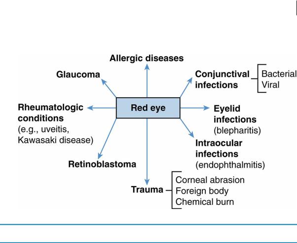
1.Etiology. The differential diagnosis of red eye in infants and children is extensive (Figure 18-1). The most common causes include viral, bacterial, and allergic conjunctivitis, as well as blepharitis (eyelid inflammation).
2.Evaluation
a.History, which often establishes the cause
1.Infectious causes are suggested by history of contact with others who have conjunctivitis.
2.Allergic conjunctivitis is suggested by severe itchiness and is usually seasonal.
3.Conjunctivitis associated with contact lens use may be secondary to allergy to the contact lens solution, to a corneal abrasion, or to a vision-threatening bacterial corneal ulcer.
4.Unilateral conjunctivitis may be associated with a foreign body, corneal ulcer, or herpes simplex keratitis.
b.Ocular examination (I-ARM acronym; see section I.B)
c.Fluorescein staining of the corneal epithelium is performed to evaluate for an abrasion of the corneal tissue. Positive staining is most commonly associated with trauma, but may also be associated with a bacterial corneal ulcer or with herpes simplex keratitis.
3.Specific causes of conjunctivitis. The distinguishing clinical features of the common causes of pediatric red eye are presented in Table 18-3.
a.Bacterial conjunctivitis
1.Etiology. Causes most commonly include nontypeable Haemophilus
644
influenzae, Streptococcus pneumoniae, Moraxella catarrhalis, and Staphylococcus aureus.
2.Clinical features. Purulent discharge, conjunctival erythema, and lid swelling are usually present. Bilateral involvement is common. Some patients have associated otitis media.
3.Diagnosis. History and clinical presentation are the basis of diagnosis.
a.Conjunctival cultures and Gram stain are not performed routinely for mild to moderate infections, and patients are usually treated empirically.
b.However, conjunctival cultures and Gram stain should be obtained in severe cases.
4.Management
a.Topical antibiotics are effective and include bacitracin, sulfacetamide, polymyxin B and trimethoprim sulfate, gentamicin, tobramycin, ofloxacin, and erythromycin.
b.Indications for referral to an ophthalmologist include severe eye involvement, conjunctivitis associated with contact lens use, suspected corneal ulcer, or lack of improvement with topical antibiotics.
b.Viral conjunctivitis
1.Pharyngoconjunctival fever is characterized by an upper respiratory infection that includes pharyngitis, fever, and bilateral conjunctivitis.
a.Etiology. The cause is adenovirus, types 3 and 7.
b.Clinical features
1.Symptoms and signs: severe watery conjunctival discharge, hyperemic conjunctiva, chemosis (conjunctival edema), preauricular lymphadenopathy, and typically a foreign body sensation caused by corneal involvement
2.Highly contagious and lasts for 2–3 weeks
c.Management. Treatment is supportive and includes cool compresses and topical nonsteroidal anti-inflammatory drug (NSAID) drops. Antibiotics may be necessary if bacterial superinfection occurs.
2.Epidemic keratoconjunctivitis is clinically similar to pharyngoconjunctival fever, but symptoms are confined to the eyes.
a.Etiology. The cause is adenovirus, types 8, 19, and 37.
b.Clinical features
1.Symptoms and signs: petechial conjunctival hemorrhage, preauricular lymphadenopathy, and a pseudomembrane along the conjunctiva
2.Photophobia from corneal inflammation (keratitis) caused by a hypersensitivity reaction to the virus (occurs in one-third of patients)
3.Lack of fever or pharyngitis
4.Highly contagious
c.Management. Treatment is supportive, including cool compresses and topical NSAID drops. Children with corneal involvement should be referred to an ophthalmologist.
3.Primary ocular herpes simplex virus
a.Etiology. The cause is herpes simplex virus type 1 (HSV-1) and typically represents the initial exposure to HSV-1 virus.
b.Clinical features
1.Skin eruption with multiple vesicular lesions
645

2.Corneal ulcer (rare)
c.Diagnosis. History, clinical presentation, and positive viral culture or direct fluorescent antibody staining of vesicular fluid are the basis of diagnosis.
d.Management
1.Systemic or topical acyclovir may speed recovery if administered within 1–2 days of onset.
2.Topical antibiotics applied to the skin may prevent secondary bacterial infection.
c.Allergic conjunctivitis
1.Epidemiology. Allergic conjunctivitis is most typically seasonal, often accompanying seasonal allergic rhinitis. It affects 10% of the population.
2.Etiology. The cause is a type 1 hypersensitivity reaction.
3.Clinical features. Marked itching and watery discharge are present.
4.Diagnosis. History and clinical presentation are the basis of diagnosis.
5.Management
a.Removal of environmental allergens
b.Topical mast cell–stabilizing drops, such as cromolyn
c.Topical antihistamines
d.Hemorrhagic conjunctivitis is a dramatic presentation of pediatric red eye in which the child presents with both conjunctivitis and subconjunctival hemorrhage. Causes include infection with H. influenzae, adenovirus, and picornavirus.
e.Blepharitis
1.Definition. Blepharitis is eyelid inflammation.
2.Epidemiology. Blepharitis is one of the most common causes of red eye.
3.Etiology. The usual cause is S. aureus infection.
4.Clinical features. Burning, crusting, and scales at the eyelash base; thickened and hyperemic eyelid margins; broken or absent eyelashes; and a history of awakening in the morning with eyelashes stuck together are characteristic.
5.Diagnosis. History and clinical presentation are the basis of diagnosis.
6.Management. Treatment includes eyelid hygiene, in which eyelids are scrubbed twice daily with baby shampoo. Topical erythromycin ointment may also be applied.
Table 18-2
Etiology, Clinical Features, Diagnosis, and Management of Neonatal Conjunctivitis
Etiology |
Onset and Clinical Features |
Conjunctival Studies |
Management |
|
Chemical |
Within first 24 hours |
Negative Gram stain |
No treatment necessary |
|
Watery discharge |
Few PMNs |
|
|
|
Neisseria |
2–4 days of life |
Gram-negative intracellular |
Intravenous cefotaxime and |
|
gonorrhoeae |
|
diplococci |
topical erythromycin |
|
Purulent |
|
|
|
|
discharge |
|
|
|
|
Eyelid swelling |
Positive gonococcal culture |
Treat parents |
|
|
Can lead to |
|
|
|
|
corneal ulcer |
|
|
|
|
Chlamydia |
4–10 days of life |
Cytoplasmic inclusion |
Oral erythromycin |
|
trachomatis |
|
bodiesPositive DFA or culture |
|
|
Serous or |
Treat parents |
|
|
|
purulent |
|
|
|
|
discharge |
|
|
|
|
Variable lid |
|
|
|
|
swelling |
|
|
|
|
|
|
|
|
|
646
Herpes simplex |
6 days–2 weeks of lifeUsually |
Multinucleated giant cells on |
Intravenous acyclovir and |
virus |
unilateralSerous discharge |
Gram stain |
topical trifluorothymidine |
Positive HSV |
|
|
|
culture |
|
|
|
PMNs = polymorphonuclear neutrophils; DFA = direct fluorescent antibody assay; HSV = herpes simplex virus.
FIGURE 18.1 Differential diagnosis of red eye in older infants and children.
Table 18-3
Distinguishing Clinical Features of Conjunctivitis
Clinical Feature |
Bacterial |
Viral |
Allergic |
Blepharitis |
Discharge |
Purulent |
Watery |
Watery or mucoid |
Minimal |
Itching |
Minimal |
Minimal |
Severe |
Minimal (irritation rather |
|
|
|
|
than itching) |
Preauricular |
Absent |
Common |
Absent |
Absent |
lymphadenopathy |
|
|
|
|
Laboratory findings |
Bacteria and PMNs on |
No bacteria on |
Eosinophils on |
Positive culture for |
|
Gram stain |
Gram stain |
conjunctival scraping |
Staphylococcus aureus |
PMNs = polymorphonuclear neutrophils.
647
IV. Abnormal Tearing
A.Nasolacrimal duct (NLD) obstruction
1.Definition. NLD obstruction is failure of complete canalization of the lacrimal system that results in obstruction to tear outflow. Obstruction typically occurs distally at Hasner valve.
2.Epidemiology. NLD occurs in 1–5% of children.
3.Etiology. The cause of incomplete canalization is unknown.
4.Clinical features
a.Watery eye with increased tear lake (meniscus of tears upon the lower eyelid margin that spill over onto the cheek and eyelid)
b.Matted eyelashes
c.Mucus in the medial canthal area
d.Bilateral involvement in one-third of patients
5.Management
a.Observation only is needed for most children.
1.Over 80% of cases resolve spontaneously within 9–12 months.
2.Nasolacrimal massage may help open the distal obstruction.
3.Topical antibiotics are administered if infection is present.
b.NLD probing, in which a small steel wire is passed through the nasolacrimal system through Hasner valve into the nose, will cure NLD obstruction in most cases. It is typically performed between 6 and 12 months of age.
B.Amniotocele (dacryocele)
1.Definition. Amniotocele is swelling of the nasolacrimal sac.
2.Etiology. The cause is accumulation of fluid as a result of NLD obstruction.
3.Clinical features
a.Bluish swelling in the medial canthal area may be apparent and represents fluid sequestered within the distended nasolacrimal sac.
b.Infection may occur, manifesting as warmth, erythema, tenderness, and increased induration.
4.Management
a.Local massage, if there is no evidence of infection
b.Intravenous antibiotics and urgent NLD probing if infection is present
648
V.Ocular Trauma
A.Retinal hemorrhages
1.Etiology
a.Retinal hemorrhages are highly suggestive of child abuse. (Physical characteristics of child abuse are described further in Chapter 20, section VI.)
b.Nonabuse causes of retinal hemorrhages include birth trauma, leukemia, increased intracranial pressure, malignant hypertension, bacterial endocarditis, immune thrombocytopenic purpura, and rarely, cardiopulmonary resuscitation.
2.Clinical features. Retinal hemorrhages appear as hemorrhagic dots and blots, or hemorrhage within the preretinal vitreous on a dilated funduscopic examination.
B.Corneal abrasion
1.Definition. Corneal abrasion is damage to, and loss of, corneal epithelium.
2.Etiology. The cause is trauma, including injury from contact lens use.
3.Clinical features
a.Severe pain, tearing, and photophobia
b.Foreign body sensation
4.Diagnosis. Identification of the abrasion on fluorescein staining of the cornea is the basis of diagnosis.
5.Management. Complete healing usually occurs within 24–48 hours.
a.Placement of a protective shield or patch for 24–48 hours may be recommended in severe cases.
b.Instillation of a topical antibiotic prevents bacterial superinfection.
c.Ophthalmologic consultation to evaluate for a bacterial corneal ulcer is necessary if the abrasion is associated with contact lens use.
C.Hyphema
1.Definition. Hyphema is blood within the anterior chamber.
2.Etiology
a.Blunt trauma is the most frequent cause. Blunt trauma compresses the globe, and when the globe subsequently re-expands, the iris vasculature tears, resulting in bleeding.
b.Nontraumatic causes include iris neovascularization (associated with diabetes mellitus, intraocular tumors, and retinal vascular diseases), clotting disorders, and iris tumors (e.g., melanoma, juvenile xanthogranuloma).
3.Clinical features
a.Impaired vision. As blood settles, a blood-aqueous fluid level may be seen. A large hyphema may obscure the pupil and iris, thereby impairing vision.
b.Complications
1.Rebleeding 3–5 days after initial injury can occur due to repeat trauma or an underlying bleeding diathesis, or as the clot retracts.
2.Glaucoma
3.Staining of the cornea with blood
4.Optic nerve damage in children with sickle cell disease
4.Management. Treatment includes ophthalmologic consultation, pain control, prevention of vomiting (which may cause a sudden increase in intraocular pressure) and bed rest for at least 5 days.
D.Orbital floor fracture (“blow-out” fracture)
1.Etiology. Blunt trauma to the eye or orbital rim fractures the orbital floor, which is normally thin and easy to fracture.
649
2.Clinical features
a.Orbital fat and the inferior rectus muscle can become entrapped within the fracture, leading to diplopia as a result of restricted vertical eye movement, to strabismus, and to enophthalmos (backward displacement of the globe into the orbit).
b.Numbness of the cheek and upper teeth below the orbital fracture may occur as a result of infraorbital nerve injury.
3.Management
a.Empiric oral antibiotics are administered to prevent infectious contamination of the orbit from organisms from the maxillary sinus.
b.Surgical repair is indicated if diplopia persists 2–4 weeks after injury or if enophthalmos is significant.
650
VI. Congenital Glaucoma
A.Definition. Congenital glaucoma is increased intraocular pressure occurring at or soon after birth.
1.Normal intraocular pressure in infants is 10–15 mm Hg. Infants with congenital glaucoma have intraocular pressures exceeding 30 mm Hg.
2.Congenital glaucoma is very different from adult glaucoma.
a.Adult glaucoma is characterized by increased intraocular pressure that damages the optic nerve but does not change the size of the eye.
b.Congenital glaucoma not only results in optic nerve injury but also expands the size of the eye, because the eye wall is much more elastic during infancy. Congenital glaucoma results in corneal edema, corneal clouding, and amblyopia.
B.Etiology
1.Outflow of aqueous humor is reduced because of maldevelopment of the trabecular meshwork.
2.Most cases are inherited in an autosomal dominant fashion.
3.Other causes include infection (e.g., congenital rubella syndrome), ocular abnormalities (e.g., aniridia [absence of the iris]), or genetic syndromes, (e.g., Sturge–Weber syndrome, neurofibromatosis, Marfan syndrome).
C.Clinical features
1.Ocular enlargement, tearing, photophobia, enlarged cornea, corneal clouding, and a dull red reflex are common.
2.Bilateral involvement is present in 70% of patients.
3.Glaucoma may be initially misdiagnosed as NLD obstruction because of the presence of tearing, but is distinguished by the presence of a normal red reflex in patients with NLD obstruction.
D.Management
1.Surgery to open outflow channels is almost always required.
2.Topical or systemic medications, such as β-adrenergic and carbonic anhydrase inhibitors, may help to lower intraocular pressure.
E.Prognosis. Congenital glaucoma, if not detected and treated surgically early, leads to blindness.
651
VII. Retinopathy of Prematurity (ROP)
A.Definition. ROP is the proliferation of vessels in the immature retina, seen in premature infants exposed to oxygen.
B.Etiology
1.The precise cause of ROP is unknown; however, vascular endothelial growth factors are posited to be involved. High concentrations of oxygen play a major role in the development of ROP.
2.Other risk factors include low birth weight (<1500 g), young gestational age, blood transfusions, respiratory distress syndrome (surfactant deficiency syndrome), and intracranial hemorrhage.
C.Late complications. Myopia, astigmatism, amblyopia, strabismus, and blindness may develop.
D.Management
1.Ophthalmologic examinations are performed every 1–2 weeks in patients with ROP to monitor for normal maturation of retinal vessels.
2.If disease is severe, retinal cryotherapy and laser therapy may be effective to prevent blindness.
E.Screening and prevention
1.Early detection is essential.
2.Minimizing the amount of oxygen delivered and effective treatment of respiratory distress syndrome (see Chapter 4, section VI) are the two most important factors for prevention of ROP.
3.Infants born at a gestational age of 28 weeks or less or with a birth weight of less than 1500 g should have a dilated ophthalmoscopic examination at 4–6 weeks of age.
652
VIII. Leukocoria
Leukocoria is a white pupil and refers to an opacity at or behind the pupil. It may be caused by a cataract, by an opacity within the vitreous, or by retinal disease, such as retinoblastoma.
A.Congenital cataract
1.Definition. Congenital cataract is a crystalline opacity of the lens present at birth.
2.Etiology. The majority of cataracts are idiopathic. Other causes include the following:
a.Genetic syndromes, such as Down, Noonan, Marfan, Alport, and Smith–Lemli– Opitz syndromes
b.Nonsyndromic genetic inheritance
c.Metabolic derangements, such as hypoglycemia, galactosemia, and diabetes mellitus
d.Intrauterine infections, such as cytomegalovirus and rubella
e.Trauma
3.Management. Treatment includes evaluation for underlying disease and early surgery to prevent amblyopia.
4.Prognosis. Congenital cataracts treated within the first weeks of life have a good prognosis, whereas surgery performed after 2–3 months of age is associated with poor visual outcome.
B.Retinoblastoma
1.Definition. Retinoblastoma is a malignant tumor of the retina.
2.Epidemiology
a.Retinoblastoma is the most common ocular malignancy in childhood.
b.The average age at presentation is 13–18 months. More than 90% of cases are diagnosed before 5 years of age, which makes retinoblastoma a tumor of toddlers and preschool children.
3.Etiology
a.Mutation or deletion of a growth suppressor gene on both alleles on the long arm of chromosome 13. Because the development of retinoblastoma requires two deletions or mutations (one on each allele), the cause has been termed the “two-hit” model.
b.Mutations may be sporadic or inherited in an autosomal recessive fashion.
4.Clinical features
a.Leukocoria and strabismus are the two most common presenting signs. Often a careful history will elicit that an observing parent has noticed the leukocoria.
b.Glaucoma, vitreous hemorrhage, retinal detachment, and hyphema are less common presenting signs.
c.Calcification within the tumor, identified on imaging studies of the eye, is a hallmark of retinoblastoma.
5.Diagnosis. Visual inspection with an ophthalmoscope is the basis of diagnosis. Ocular ultrasound or computed tomographic scan of the orbit can further evaluate the tumor and assess for tumor extension.
6.Management
a.Early diagnosis is critical. Retinoblastoma should be suspected in any child with leukocoria.
b.Large tumors involving the macula have a poor prognosis and are generally treated by removal of the entire eye (i.e., enucleation).
c.Smaller tumors may be treated with external beam radiation; however, radiation may induce formation of secondary tumors.
653
d.Very small peripheral tumors may be treated with cryotherapy or laser photocoagulation.
e.Systemic and local intraretinal arterial chemotherapy are additional treatments.
7.Prognosis. Outcome is excellent if retinoblastoma is identified early. The cure rate is 90% if the tumor does not extend beyond the sclera or into the orbit. Retinoblastoma is uniformly lethal if untreated.
654
IX. Strabismus
A.Definition. Strabismus is misalignment of the eyes.
1.Esotropia refers to the eye turned nasally.
2.Exotropia refers to the eye turned laterally.
3.Vertical strabismus refers to the eye turned up or down.
4.Pseudostrabismus is a prominence of the epicanthal folds that results in the false appearance of strabismus, even though the eyes are actually appropriately aligned.
B.Etiology. The cause of most childhood strabismus is usually unknown. However, brain tumors, farsightedness (hypermetropia), or neurologic processes causing paresis of cranial nerves III, IV, or VI may also cause strabismus.
C.Clinical features
1.If strabismus occurs before 5–7 years of age, the child suppresses the image in the deviated eye. If this suppression is prolonged, amblyopia may result.
2.If strabismus occurs later than 5–7 years of age (i.e., acquired strabismus), the mature visual system is unable to suppress the image in the deviated eye, and diplopia results.
3.Acquired strabismus, decreased eye movement, ptosis, decreased vision, and abnormal red reflex are all red flags that suggest a dangerous underlying cause, such as a tumor or neurologic process.
D.Management. Treatment depends on the underlying cause.
1.Ocular patching or use of eye drops to cause blurring to the normal eye to prevent amblyopia is important.
2.Strabismus associated with farsightedness is initially treated with corrective lenses.
3.Surgery is often required to correct any misalignment that does not respond to patching or glasses.
655