

- •Copyright
- •Contents
- •Dedication
- •Preface
- •Acknowledgments
- •Contributors
- •Contributors to the Previous Edition
- •Review Test
- •Answers and Explanations
- •Review Test
- •Answers and Explanations
- •Review Test
- •Answers and Explanations
- •Review Test
- •Answers and Explanations
- •Review Test
- •Answers and Explanations
- •Review Test
- •Answers and Explanations
- •Review Test
- •Answers and Explanations
- •Review Test
- •Answers and Explanations
- •Review Test
- •Answers and Explanations
- •Review Test
- •Answers and Explanations
- •IV. Hypertension
- •VI. Nephrotic Syndrome (NS)
- •VII. Hemolytic Uremic Syndrome (HUS)
- •VIII. Hereditary Renal Diseases
- •IX. Renal Tubular Acidosis (RTA)
- •XI. Chronic Kidney Disease (CKD) and End-Stage Renal Disease (ESRD)
- •XII. Structural and Urologic Abnormalities
- •XIII. Urolithiasis
- •XIV. Urinary Tract Infection (UTI)
- •Review Test
- •Answers and Explanations
- •Review Test
- •Answers and Explanations
- •Review Test
- •Answers and Explanations
- •Review Test
- •Answers and Explanations
- •IV. Food Allergy
- •VI. Urticaria (Hives)
- •VII. Drug Allergy
- •VIII. Asthma
- •IX. Immunology Overview
- •X. Disorders of Lymphocytes (Figure 15-2)
- •XI. Disorders of Granulocytes (Figure 15-3)
- •XII. Disorders of the Complement System
- •Review Test
- •Answers and Explanations
- •Review Test
- •Answers and Explanations
- •Review Test
- •Answers and Explanations
- •Review Test
- •Answers and Explanations
- •Review Test
- •Answers and Explanations
- •Review Test
- •Answers and Explanations
- •Comprehensive Examination
- •Index
Review Test
1.A 1-month-old male infant is brought to the office for a routine health maintenance visit. The infant has been feeding well, and the parents have no concerns. Physical examination is unremarkable except for a lens opacity in the right eye. Which of the following statements regarding this finding is most correct?
A.Because of this infant’s age, he is not susceptible to amblyopia.
B.Immediate referral for cataract surgery is indicated.
C.The opacity was probably not present at birth but has developed after birth.
D.Surgery can be safely delayed until 6 months of age.
E.Red reflex testing is normal and does not identify this finding.
2.A 3-year-old girl with a normal past medical history presents with a 5-day history of bilateral mucoid conjunctival discharge and conjunctival hyperemia. The girl is rubbing her eyes, and her mother believes that her daughter’s eyes are very itchy. Which of the following statements regarding the most likely diagnosis is correct?
A.The girl’s symptoms are secondary to a type 2 hypersensitivity reaction.
B.Topical antihistamines would be effective.
C.Staphylococcus aureus is the likely pathogen.
D.Coexisting rhinitis would be unusual.
E.Topical polymyxin B and trimethoprim sulfate is an effective treatment.
3.A 14-year-old boy who wears contact lenses presents with conjunctival hyperemia and severe photophobia of the right eye. Which of the following statements regarding his diagnosis and management is most correct?
A.Immediate ophthalmologic evaluation is necessary.
B.Viral conjunctivitis is likely and supportive care should be provided.
C.Eosinophils are seen on conjunctival scraping.
D.Fluorescein staining is normal.
E.Topical erythromycin should be prescribed and the patient should follow up if no improvement is seen in 1 week.
4.A 5-year-old boy presents with a history of fever, bilateral watery conjunctival discharge, sore throat, and a foreign body sensation in his eyes. Physical examination is notable for bilateral conjunctival hyperemia with watery discharge and bilateral preauricular lymphadenopathy. Which of the following statements regarding the most likely diagnosis is correct?
A.Topical nonsteroidal anti-inflammatory drugs are contraindicated in the management of this illness.
B.Corneal involvement is unlikely.
C.Cool compresses should be used.
D.Bilateral eye involvement is uncommon.
E.This highly contagious illness is most often caused by Staphylococcus aureus.
5.A 2-year-old child is brought to the office with a 3-month history of “uneven eyes.” Physical examination shows right eye exotropia and leukocoria. Computed tomography of the orbit reveals a large retinoblastoma that involves the macula. Which of the following statements regarding the diagnosis is most correct?
A.This tumor occurs sporadically and is not inherited.
B.Computed tomography reveals calcifications within the tumor.
C.Strabismus is uncommon at presentation.
D.Radiation therapy is effective and has few complications.
E.The age at diagnosis is unusual.
6.A 10-year-old girl is brought to the emergency department after sustaining a baseball injury to
656
the right eye. Which of the following signs or symptoms is most consistent with the suspected diagnosis of a fracture of the orbital floor?
A.Glaucoma
B.Exophthalmos
C.Diplopia
D.Numbness of the lower teeth and chin
E.Hyphema
The response options for statements 7–10 are the same. You will be required to select one answer for each statement in the set.
A.Chemical conjunctivitis
B.Conjunctivitis caused by Neisseria gonorrhoeae
C.Conjunctivitis caused by Chlamydia trachomatis
D.Conjunctivitis caused by herpes simplex virus
For each patient, select the most likely diagnosis.
1.A 7-day-old male infant with purulent conjunctival discharge and mild lid swelling.
2.A 12-hour-old male infant with bilateral watery conjunctival discharge.
3.A 10-day-old female infant with unilateral serous conjunctival discharge and multinucleated giant cells on Gram stain.
4.A 2-day-old female infant with purulent eye discharge and eyelid swelling.
657
Answers and Explanations
1.The answer is B [VIII.A and I.A]. Presentation with a lens opacity is consistent with a congenital cataract, a potentially very serious opacity of the lens. Immediate referral to a pediatric ophthalmologist is warranted. Surgery should occur within the first several weeks of life to ensure a good prognosis. If an opacity is not removed promptly in a young infant, the retina and nervous connections are not appropriately stimulated, and amblyopia (poor vision caused by abnormal retinal stimulation) may develop. Children are most susceptible to amblyopia during the first 3–4 months of life. Congenital cataracts are present at birth. The majority of cataracts are idiopathic, although known causes include trauma, metabolic abnormalities like galactosemia, genetic syndromes, and nonsyndromic genetic inheritance. The Bruckner test, which is used in the assessment of the bilateral red reflex, is usually abnormal in the presence of a cataract.
2.The answer is B [III.B.2.a.(2) and III.B.3.c]. Itching is the hallmark of allergic conjunctivitis, the likely cause of this patient’s symptoms. Symptoms and signs of allergic conjunctivitis also include conjunctival erythema and watery or mucoid discharge. Management includes topical antihistamines, topical mast cell stabilizers, and environmental modification (removing from the environment any known or suspected allergens such as stuffed animals, pets, or carpet). Allergic conjunctivitis is a type 1 hypersensitivity reaction and is commonly seasonal. Uncomplicated allergic conjunctivitis is not associated with bacterial infection. Other signs and symptoms of allergy are commonly found, including allergic rhinitis. Topical antibiotics are ineffective for allergic conjunctivitis.
3.The answer is A [III.B.2.a.(3) and V.B.5.c]. Conjunctivitis associated with contact lens use suggests three major possibilities, including an allergy to the contact lens solution, a corneal abrasion, or a bacterial corneal ulcer, which can be vision-threatening. A possible corneal ulcer requires urgent ophthalmologic consultation. Viral conjunctivitis is unlikely because of the acuity of the presentation, the unilateral findings, and severe photophobia. Eosinophils on conjunctival scraping are found in allergic conjunctivitis. A corneal ulcer usually appears on fluorescein staining. Because a corneal ulcer is a possibility, prescribing antibiotics with follow-up in a week would risk missing the early diagnosis of this serious condition.
4.The answer is C [III.B.3.b.(1)]. This patient’s signs and symptoms are consistent with pharyngoconjunctival fever, a highly contagious illness that lasts for 2–3 weeks and is best managed with supportive care, including topical nonsteroidal anti-inflammatory drugs and cool compresses. Corneal involvement does occur and usually manifests as a foreign body sensation within the eye. Pharyngoconjunctival fever is commonly bilateral. It is a highly contagious illness caused by infection with adenovirus. The presence of preauricular adenopathy and watery discharge is inconsistent with bacterial conjunctivitis.
5.The answer is B [VIII.B]. Retinoblastoma is diagnosed on the basis of visual inspection and imaging studies, such as computed tomography or ocular ultrasound. Calcification within the tumor is a hallmark of retinoblastoma. Retinoblastoma is inherited sporadically or in an autosomal recessive fashion. The two most common presenting features are leukocoria (white light reflex) and strabismus. Management of large tumors often includes enucleation, although small retinoblastomas may be managed with external beam radiation. The side effects of radiation therapy include induction of secondary tumors. The majority of children with retinoblastoma are younger than 5 years.
6.The answer is C [V.D.2]. Orbital floor fracture, or a “blow-out” fracture, is generally secondary to blunt trauma to the orbit and can lead to entrapment of orbital fat and the inferior rectus muscle within the fracture. Glaucoma is typically not a complication of an orbital floor fracture. Entrapment leads to enophthalmos (i.e., retracted globe), restricted
658
vertical movement of the eye, and strabismus with resultant double vision (diplopia). Because the infraorbital nerve may be injured, numbness of the cheek and upper teeth may result. Hyphema, or blood within the anterior chamber, also occurs secondary to blunt trauma. Hyphema should certainly increase your index of suspicion for other injuries associated with blunt trauma, but hyphemas frequently occur without concurrent injuries to the orbital floor.
7.The correct answers are C, A, D, and B, respectively [III.A.1–3 and Table 18-2]. Neonatal conjunctivitis is defined as conjunctivitis occurring within the first month of life. Chlamydia trachomatis causes serous or purulent conjunctivitis with variable lid swelling in infants between 4 and 10 days of age (question 7). Conjunctivitis within the first 24 hours (question 8) is characteristic of chemical irritation from neonatal prophylactic drops or ointment, such as 1% silver nitrate. Herpes simplex virus presents with vesicles, corneal ulcers, and multinucleated giant cells on Gram stain of conjunctival cells (question 9). Neisseria gonorrhoeae causes purulent, rapid onset conjunctivitis with lid swelling in infants between 2 and 4 days of age (question 10).
659

C H A P T E R 1 9
660
Dermatology
Amy E. Gilliam, Lloyd J. Brown
661
I.General Concepts
A.Skin examination should be conducted in good light and should be complete, including evaluation of the scalp, hair, nails, eyes, mouth, palms, and soles. Examination should be both visual and tactile.
B.Description of skin disease requires identification of primary lesions and any secondary characteristics. Configuration and distribution of lesions should also be noted.
1.Primary lesions
a.Macules are <10 mm in size, flat, and nonpalpable, and represent a cutaneous color change. A patch is a large macule (≥10 mm).
b.Papules are epidermal or superficial dermal lesions <10 mm in size that are elevated above the skin surface. A plaque describes large or coalesced papules (≥10 mm).
c.Nodules are dermal lesions that are <10 mm in size and generally below the skin surface, although they may have an epidermal component that rises above the skin surface. A tumor is a large nodule (≥10 mm).
d.Vesicles are fluid-filled papules. A bulla is a large vesicle.
e.Pustules are purulent-filled papules.
f.Cysts are nodules filled with expressible material.
g.Wheals are cutaneous elevations caused by dermal edema.
2.Secondary characteristics include the following:
a.Scale: desquamation of the stratum corneum
b.Crust: dried exudate and debris
c.Pigmentary changes
d.Excoriations: linear erosions into the epidermis caused by fingernail scratches
e.Scars: thickened fibrotic dermis
f.Ulcers: absence of epidermis and some of the dermis
g.Atrophy: thinning of the epidermis or dermis
h.Fissures: linear cracks into the dermis
3.Configuration and distribution
a.Configuration of lesions may be described as linear, annular (i.e., ring shaped), arcuate (i.e., in half-circles), serpiginous (i.e., with a wavy or serpentine border), reticulated (i.e., netlike), grouped, or discrete (i.e., distinct and separate).
b.Distributions include generalized, flexural, acral (hands, feet, buttocks), dermatomal (confined to a dermatome), or other specific locations. Some skin lesions are blaschkoid, meaning they follow the lines of Blaschko, which are the lines of cutaneous development in utero.
C.Diagnostic procedures
1.Woods light can highlight pigmentary changes and some dermatophytes.
2.Scrapings
a.Fungus. Ten percent potassium hydroxide (KOH) can be added to a scraping of a scale or exudate to identify fungal hyphae.
b.Scabies. Examination under a microscope of a scraping of an unscratched lesion or burrow for mites, eggs, or feces may be diagnostic of scabies.
c.Herpes simplex virus (HSV). The base of a vesicle can be scraped for laboratory identification of the herpes virus.
3.Cultures may be obtained for bacteria, virus, fungus, and yeast.
4.Invasive techniques
a.Incision and drainage may be performed for diagnosis, to obtain cultures, or for
662
therapy.
b.Biopsy
1.Shave or tangential biopsy for epidermal and superficial dermal lesions
2.Punch biopsy for epidermal, dermal, and superficial subcutaneous lesions
3.Excisional biopsy for complete lesion removal
5.Immunofluorescent staining of biopsied skin lesions may be useful in diagnosis of autoimmune and/or vasculitic disorders.
D. Management
1.General concepts
a.Absorption of topical agents through the skin of infants and small children is greater than in adults owing to the increased body surface area to weight ratio that children have as compared with adults. This is especially true for premature infants in whom there is also greater absorption through the skin owing to a thinner stratum corneum.
b.Therapeutic efficacy of a topical agent is related to both the active ingredient and the vehicle (e.g., cream, lotion, ointment).
2.Hydration of the skin is critical. Dry, irritated skin is a poor barrier and easily damaged. Moisturizers include the following:
a.Ointments contain little or no water and have maximal water-retaining properties, making them useful for very dry skin.
b.Creams contain 20–50% water and are useful for skin of average dryness.
c.Lotions contain more water than creams and are useful for minimally dry skin or for large surface areas.
d.Solutions and alcohol-based gels are most useful for hair-bearing areas (e.g., scalp).
3.Thickened skin (hyperkeratosis) responds well to treatment with keratolytics, such as salicylic acid, urea, α-hydroxy acids, and retinoic acid.
4.Destructive therapies (e.g., for warts, molluscum contagiosum) include high-dose salicylic acid, cantharidin, podophyllin, 5-fluorouracil, cryotherapy, electrotherapy, and laser therapy.
5.Anti-infective agents include topical antibiotics, antifungals, antivirals, and antiparasitic agents.
6.Anti-inflammatory agents
a.Topical corticosteroids are categorized on the basis of their vehicle and potency. Effectiveness and potential side effects mirror steroid potency. The weakest steroid that will achieve the treatment goal should be used first. In general, only lowpotency corticosteroids should be used on the face or groin because the epidermis is thinner in these areas, resulting in an increased risk of side effects.
1.Systemic side effects. Systemic steroid levels can be achieved if very potent steroids are used on damaged or thin skin for long periods of time. Rare side effects may include adrenal suppression, depressed growth, cataracts, glaucoma, and Cushing syndrome (see also Chapter 6, section IV.E.2).
2.Local side effects are more common and are also more likely to occur after long-term use of topical corticosteroids.
a.Acne (acne rosacea and steroid acne)
b.Hirsutism
c.Folliculitis
d.Striae (especially in the axilla or groin)
e.Hyperor hypopigmentation
f.Skin atrophy
663
g.Ecchymoses and telangiectasias
h.Tachyphylaxis (insensitivity to the medication)
b.Other topical anti-inflammatory agents
1.Tacrolimus ointment and pimecrolimus cream are topical calcineurin inhibitors/immunomodulators used to treat atopic dermatitis, psoriasis, and other inflammatory skin conditions.
2.One to five percent sulfur, formulated with other medications, for acne
3.Tar, used for eczema and psoriasis
4.Antibiotics, used for anti-inflammatory properties to treat acne and perioral dermatitis.
664
II. Newborn Skin Diseases
See Chapter 4, section I.K.
665
III.Inflammatory Disorders
A.Contact dermatitis
1.Definition. Contact dermatitis is inflammation of the epidermis and superficial dermis secondary to direct contact with the skin by a sensitizing substance.
2.Categories
a.Allergic contact dermatitis
1.Etiology. Allergic contact dermatitis occurs as a direct T-cell–mediated response to an exogenous applied allergen. There must be an initial sensitization and then a rechallenge (which may be very small and is not dosedependent) to elicit a reaction. Common causes include poison ivy, oak, or sumac; nickel found in jewelry, belt buckles, or snaps; topical lotions or creams; and perfumes or soaps.
2.Clinical features. Erythematous pruritic papules and vesicles occur in the area that came into contact with the allergen.
3.Management. Treatment involves topical corticosteroids and avoidance of the offending allergen.
b.Primary irritant contact dermatitis
1.Etiology. Primary irritant contact dermatitis is caused by caustic substances that irritate the skin, rather than an allergic reaction. No prior sensitization is required. The reaction is dose-dependent. The most common form is diaper dermatitis, a multifactorial disorder caused by prolonged contact with urine and fecal matter, friction, maceration, and proteases contained in the feces and urine. Secondary infection with Candida albicans may occur.
2.Clinical features of diaper dermatitis include erythema with papules on the skin that is exposed to the diaper surface, including the upper thighs, buttocks, and genitourinary area without involvement of the inguinal creases. Involvement of the inguinal creases, more-intense confluent erythema, and satellite lesions all suggest candidal superinfection.
3.Management of diaper dermatitis includes protecting the skin from urine and stool; application of skin moisturizers, barrier creams, and ointments (e.g., zinc oxide); and frequent diaper changes. Low-potency corticosteroids may be used for severe inflammation. Candidal infection may be treated with topical antifungal medication (e.g., nystatin, clotrimazole).
B.Atopic dermatitis (see Chapter 15, section III)
C.Seborrheic dermatitis
1.Epidemiology. Seborrheic dermatitis predominantly affects two age groups, infants and adolescents.
2.Etiology. The cause is unknown but is thought to be a hypersensitivity reaction to a saprophytic yeast (Pityrosporum ovale) that lives in areas that overproduce sebum.
3.Clinical features. Erythema with overlying “greasy” thick yellow-white scale and sometimes crusting in areas with high numbers of sebaceous glands, such as the scalp, face (including the eyebrows, nose, and beard area), chest, and groin
a.Infants may have dermatitis limited to the scalp, termed seborrheic capitis or cradle cap. Involvement of the face, upper chest, and flexor creases of the extremities may also occur.
b.Adolescents may have dermatitis in the nasolabial folds, pinnae, and scalp.
4.Management
a.Low-potency topical corticosteroids
666
b.Sulfur-, zinc-, or salicylic acid–based shampoos may be used, followed by light scrubbing with a brush to remove crusts. Loose scales may also be removed with mineral oil.
c.Topical antiyeast medications (e.g., clotrimazole) or shampoos (containing zinc pyrithione or ketoconazole) are used to eradicate Pityrosporum ovale.
D.Pityriasis rosea
1.Epidemiology. Pityriasis rosea is uncommon before 5 years of age but is extremely common during late childhood and adolescence.
2.Etiology. The cause is unknown, although it is thought to be an immune response to a virus.
3.Clinical features
a.Papulosquamous skin lesions that begin with a solitary, large 2- to 5-cm scaly, erythematous lesion (herald patch) that is usually located on the trunk or extremities. The herald patch is present for 1–30 days.
b.Approximately 1–2 weeks after the appearance of the herald patch, similar smaller oval pink scaling macules and papules erupt for approximately 3–6 weeks on the trunk and extremities, following skin lines in a “christmas tree” distribution on the trunk.
c.Lesions are pruritic in 50% of cases.
4.Management. Treatment may include topical or systemic antihistamines as well as topical corticosteroids or immunomodulators to help treat pruritus. Exposure to ultraviolet light may shorten the disease course.
E.Psoriasis
1.Epidemiology. Psoriasis occurs in 3% of children in the United States. Although it is more common in adults, 30% of patients develop signs and symptoms during childhood.
2.Etiology. Childhood-onset psoriasis is often a genetic condition. It is caused by immune dysregulation, causing epidermal proliferation and skin inflammation.
3.Clinical features
a.Distribution of skin lesions and severity are variable. If severe, psoriasis may be disfiguring.
b.Lesions are characterized by erythematous scaling papules and plaques often found on the scalp (nongreasy scale without hair loss), ears, elbows, knees, lumbosacral area, and groin. Lesions often have a classic silvery scale.
c.Lesions often demonstrate the Koebner phenomenon in which new lesions develop at sites of skin trauma.
d.Nail involvement is common and may include pits, distal thickening, lifting of the nail bed, and nail destruction.
e.Arthritis during childhood is uncommon.
4.Management. Treatment may include moderateor high-potency topical corticosteroids, topical calcineruin inhibitors, ultraviolet light therapy, vitamin D analogues, topical salicylic acid, tar, retinoids, and anthralin (downregulates epidermal growth factor).
F.Miliaria rubra (heat rash)
1.Etiology. Heat rash is caused by disrupted sweat ducts near the upper dermis (often caused by occlusion or friction) that result in sweat being released onto the skin. The sweat on the skin produces an inflammatory response. Therefore, the more sweat produced and the more occlusion, the more likely heat rash will develop.
2.Clinical features. Small erythematous pruritic papules or vesicles occur in areas of occlusion or in areas that have been rubbed, such as the inguinal region, axilla, chest, and neck.
3.Management. Treatment is avoidance of occlusive clothing to decrease sweating.
667
Medications are unnecessary.
668

IV. Hypersensitivity Disorders
A.Urticaria (see Chapter 15, section VI)
B.Serum sicknesslike reaction may initially mimic urticaria, but it tends to produce skin lesions with a more annular (ring-shaped) appearance with a dusky or bruised-looking central component. It is associated with systemic signs and symptoms including fever, arthralgias, adenopathy, and evidence of organ injury. Viral triggers and medications, such as cephalosporins, are common causes.
C.Erythema multiforme (EM) and Stevens–Johnson syndrome (SJS)
1.Definition. EM is a hypersensitivity reaction to many potential stimuli, including drugs, viruses, bacteria, fungi, protozoa, and systemic disease.
2.Categories. There are two major categories of EM: EM minor and EM major. SJS is now considered to be a separate entity from EM. The classic skin lesion present in both EM and SJS is a target lesion, which is a fixed, dull red, oval macule with a dusky center that may contain a papule or vesicle. Table 19-1 summarizes the causes, clinical features, management, and prognosis of EM and SJS.
D.Toxic epidermal necrolysis is a severe reaction to medications (e.g., anticonvulsants, antibiotics, anti-inflammatory drugs) that results in widespread epidermal necrosis. Clinical features include sloughing of the epidermis (usually >30% skin loss) and severe mucous membrane involvement. Target lesions are usually not seen. Nikolsky sign (skin peels away with lateral pressure) is often present. Mortality is high (10–30%) as a result of the high incidence of complications due to sepsis, dehydration, and electrolyte abnormalities.
Table 19-1
Characteristic Features of the Types of Erythema Multiforme and Stevens–Johnson Syndrome
|
Erythema Multiforme Minor |
Erythema Multiforme Major |
Stevens–Johnson Syndrome |
Major cause |
Herpes simplex virus |
Mycoplasma pneumoniae; |
Medications |
|
|
Medications |
|
Skin findings |
Symmetric target lesions; acral |
Typical symmetric target lesions; |
Widespread atypical, asymmetric |
|
distribution |
acral and truncal distribution |
target lesions, blisters, and necrosis |
Mucous |
Occurs in 25%; only one |
At least two mucosal surfaces |
At least two mucosal surfaces involved |
membrane |
surface involved (often |
involved (often mouth and eyes) |
(often mouth and eyes) |
findings |
mouth) |
|
|
Systemic |
Prodrome of low-grade fever, |
Prodrome of low-grade fever, |
Prodrome of high fever, cough, |
findings |
arthralgias, myalgias |
arthralgias, myalgias |
malaise, headache, arthralgias |
Management |
Supportive care |
Supportive care |
Supportive care |
|
Acyclovir may prevent |
Erythromycin or azithromycin if |
Stop offending drug |
|
recurrence |
M. pneumoniae is suspected |
|
|
|
|
Ophthalmology consultation |
|
|
Stop offending drug |
Consider steroids, IVIG, burn unit |
Prognosis |
Good; possible recurrence |
Good |
High morbidity and mortality (5%) |
IVIG = intravenous immune globulin.
669
V.Infections of the Skin
A.Fungal (dermatophyte) infections can involve hair, skin, and nails.
1.Tinea capitis is a fungal infection of the scalp hair.
a.Etiology. Tinea capitis is most commonly caused by Trichophyton tonsurans (95%), acquired from human-to-human contact, and Microsporum canis (5%), often acquired from animals, such as cats and dogs.
b.Clinical features
1.Patchy hair loss in which the hairs break off at the scalp (black dot ringworm) or in which the broken hairs may be thickened and white
2.Infected areas may have pustules and scale.
3.Kerion (large red boggy nodule/plaque) may be present and represents a hypersensitivity reaction to the dermatophyte.
4.Occipital and posterior cervical lymphadenopathy are suggestive of tinea capitis.
c.Diagnosis. The basis of diagnosis is microscopic evaluation of hairs with 10% KOH to identify fungal hyphae or a positive fungal culture. Hairs fluoresce under Woods light if M. canis is the infecting organism.
d.Management. Treatment includes systemic oral antifungal therapy (e.g., griseofulvin or terbinafine) for 6–8 weeks. Topical antifungal agents are ineffective for infections of the hair. Topical 2.5% or 5% selenium sulfide shampoo may be used as an adjunct to reduce infectivity.
2.Fungal infection of the skin may include tinea corporis (infection on the body), tinea pedis (infection on the foot), tinea cruris (infection in the groin), and tinea faciei (infection on the face).
a.Etiology. Pathogens include M. canis, T. tonsurans, and other Trichophyton species.
b.Clinical features
1.Tinea corporis (“ringworm”) presents as a circular or annular scaly erythematous patch with partial central clearing and an “active” border, which means that the advancing edge or border of the skin lesion is more erythematous and/or scaly than the central portion.
2.Tinea pedis (athlete’s foot) presents most commonly in postpubertal adolescents with scaling, erythema, and maceration between the toes or on the plantar aspect of the foot. Vesicles may also be seen.
3.Tinea cruris usually presents as well-defined scaling and erythema in the groin and inguinal creases.
c.Diagnosis. Clinical features are often the basis of diagnosis. 10% KOH examination of skin scrapings for fungal hyphae or fungal culture can confirm the diagnosis.
d.Management. Treatment includes topical antifungal medications (e.g., terbinafine, ketoconazole, itraconazole).
3.Tinea unguium (onychomycosis) is a fungal infection of the nails characterized by thickening and yellow discoloration of one or several nails (usually toenails). Topical management is not very effective; treatment requires prolonged therapy and is often unsuccessful as onychomycosis tends to recur. Systemic medications, such as griseofulvin, terbinafine, and itraconazole, are therapies of choice and are given for long courses (12+ weeks).
4.Tinea versicolor is a common disorder seen more often in adolescents and young adults. It is caused by a yeast of the Malassezia species (rather than a fungus) that invades the stratum corneum.
670
a.Clinical features vary and include fine, scaly oval macules on the trunk, proximal arms, and face. Macules may be hypoor hyperpigmented and become more prominent with sun exposure. Infection is usually asymptomatic.
b.Diagnosis is usually made by clinical examination but can also be made by identification of fungal hyphae or circular spores on KOH examination of skin scrapings (“spaghetti and meatballs” appearance) or by yellow-green fluorescence on Woods light evaluation. The Malessezia yeast does not grow well on culture.
c.Management. Treatment includes overnight application of 2.5% selenium sulfide weekly for 3–4 weeks, ketoconazole shampoo or cream, or systemic antifungal medications (ketoconazole, itraconazole).
B.Bacterial infections, including impetigo, cellulitis, erysipelas, scarlet fever, toxic shock syndrome, and staphylococcal scalded skin syndrome, are discussed in Chapter 7, section IX.
C.Viral infections
1.Viral exanthem refers to a skin rash associated with a viral infection. Any virus may cause an exanthem, and the rash may take many forms. An enanthem (describing involvement of the oral mucosa) may also be present.
a.Morbilliform: refers to a “measleslike” appearance with diffuse involvement of red macules on the skin
b.Papulovesicular: refers to a rash composed of papules and vesicles
2.Measles and rubella (see Chapter 7, sections XIII.C and D)
3.Erythema infectiosum (fifth disease)
a.Epidemiology. Fifth disease is most common in school-age children, although it can occur at any age.
b.Etiology
1.The cause is parvovirus B19.
2.Fifth disease is transmitted by respiratory secretions.
c.Clinical features
1.Fifth disease begins with upper respiratory symptoms (cough, fever, rhinorrhea) that are followed within 1–2 weeks by a bright red macular rash on the cheeks (“slapped-cheek” appearance) that lasts several days. Patients are generally no longer contagious when the characteristic facial rash appears.
2.A lacy, reticulate rash on the limbs and then the trunk follows the facial rash and generally lasts 3–5 days. In some patients, exercise, heat, or sunlight can induce the lacy rash to recur.
3.Arthralgias may be present, although they are more common in adults.
4.Parvovirus B19 infection may also cause aplastic crisis (especially in patients with hemoglobinopathies), prolonged anemia in immunosuppressed patients, and fetal hydrops or miscarriage in pregnant women.
d.Management. Treatment is supportive. However, intravenous immune globulin may be used treat prolonged anemia in immunosuppressed patients.
4.Roseola infantum (exanthem subitum)
a.Epidemiology. Roseola is most common in children between 6 months and 3 years of age.
b.Etiology. Roseola is most commonly caused by human herpes virus 6 and 7. Other causes include adenovirus, parvovirus B19, and echovirus 16.
c.Clinical features. Roseola begins with 3–5 days of high fever and irritability. Once the fever resolves, a usually asymptomatic pink papular eruption occurs on the trunk that generally fades within 24–48 hours.
d.Management. Treatment is supportive.
671
5.Gianotti–Crosti syndrome (papular acrodermatitis) most often occurs in children between the ages of 6 months and 12 years. Gianotti–Crosti is associated with hepatitis B, Epstein–Barr virus, and enterovirus and echovirus infections. Clinical features include the development of red or flesh-colored flat-topped papules on the extremities with accentuation over the elbows and knees. The cheeks and buttocks can also be involved, but the rash classically spares the trunk. Skin lesions may last for several weeks and may recur. Upper respiratory symptoms may precede the eruption. Treatment is supportive.
6.Varicella (chickenpox)
a.Epidemiology. The incidence of varicella has decreased as a result of routine early childhood immunization in many states. Varicella may occur at any age in unimmunized children and adolescents.
b.Clinical features
1.Intensely pruritic erythematous macules develop acutely after a 7- to 21-day incubation period. The macules develop central vesicles within 1–2 days. The classic lesion is described as a “dew drop on a rose petal” or a vesicle on a red background.
2.Crops of lesions appear during the course of 2–5 days. Hundreds of vesicles may be present, which will crust over.
3.Fever is common.
c.Management. Treatment includes antipyretics, management of bacterial superinfection, antihistamines for itching, and monitoring and treatment of complications. Acyclovir is generally administered intravenously for patients with varicella pneumonia and encephalitis, orally for those at high risk for complications, and topically in the eyes for those with ophthalmic involvement.
d.Complications (Table 19-2)
7.Herpes simplex virus infection (HSV-1 and HSV-2)
a.Pathophysiology
1.Neonatal infection is generally acquired during passage through the birth canal of a mother with primary HSV infection. Two-thirds of neonatal infections are caused by HSV-2 and one-third are caused by HSV-1.
2.Gingivostomatitis is the most common HSV infection during infancy and childhood and is almost always caused by HSV-1.
b.Clinical features
1.Characteristic lesions are grouped vesicles on an erythematous base.
2.Gingivostomatitis presents most often in young infants with grouped vesicles and ulcers on the lips, in the corners of the mouth, and on the tongue. Pain on swallowing, drooling, and fever may be present. Infection lasts 1–2 weeks.
3.Neonatal HSV commonly presents in the first week of life with variable signs and symptoms. Neonates may have only a few vesicles at the site (often the scalp) that was in contact with the infecting maternal lesion, or they may present with signs and symptoms of sepsis, including apnea, lethargy, irritability, and seizures. Serious sequelae include meningoencephalitis, hepatitis, sepsis, shock, and death.
4.Herpetic whitlow describes HSV-1 infection of the thumb or fingers that is usually secondary to thumbor finger-sucking by a child with an oral HSV lesion.
5.HSV resides in the dorsal root ganglion after initial infection, and therefore, recurrent HSV infection may occur. Recurrent HSV lesions are more mild and less symptomatic than primary infection and generally occur on the lip. Fever, illness, ultraviolet light exposure, emotional stress, and trauma may all
672
reactivate the latent virus.
c.Diagnosis. HSV may be diagnosed by identification of epidermal giant cells on microscopic evaluation of a Tzanck preparation, by detection of HSV antigen on direct fluorescent antibody (DFA) testing, or by culture of the base of the lesion.
Polymerase chain reaction (PCR) technology is commonly used to identify HSV in the cerebrospinal fluid.
d.Management
1.Neonatal HSV infection is a medical emergency and requires immediate hospitalization and treatment with intravenous acyclovir.
2.Cutaneous and oral HSV may be treated with oral acyclovir, although treatment must be started promptly to alter the disease course. Oral acyclovir, when given daily, may also prevent recurrent infection.
8.Hand–foot–mouth disease and herpangina
a.Etiology. Hand–foot–mouth disease and herpangina are caused by infection with enterovirus, usually coxsackievirus types A16 (most common), A2, A5, A6, and A10. It is usually seen in children less than 5 years of age.
b.Clinical features include vesicles, papules, or pustules on the palms, soles, or fingertips and shallow ulcers or erosions on the soft palate or tongue. The rash also sometimes affects the buttocks. If only oral lesions are present, the disorder is termed herpangina. Fever may occur with both forms. In some cases, shedding of fingerand toenails, called onychomadesis, is observed a few weeks after the infection.
c.Management. Treatment is supportive.
9.Warts
a.Etiology. The cause is human papillomavirus.
b.Clinical features. Warts may occur on any skin surface and appear as irregularly shaped discrete flesh-colored papules that may be smooth or rough. Warts often increase in size, are contagious, and may spread to adjacent skin. Condylomata acuminata is the term used to describe multiple external warts in the genital area (see Chapter 3, section VII.B.6).
c.Management. Most warts resolve spontaneously within 1–2 years. Treatment to remove or destroy warts may include liquid nitrogen, salicylic acid, cantharidin, podophyllin, laser therapy, and surgical excision. Newer immune therapies are also used to treat warts, which include injections with candida antigen and topical
agents used to stimulate the immune system (e.g., squaric acid, imiquimod). Recurrence after any treatment is high.
10.Molluscum contagiosum
a.Etiology. The cause is a poxvirus.
b.Clinical features
1.Small asymptomatic flesh-colored papules with central umbilication are characteristic.
2.Lesions may be present anywhere on skin with hair follicles, although the proximal extremities and trunk are most commonly involved.
3.Lesions are contagious.
4.Human immunodeficiency virus (HIV) infection may be associated with extensive eruptions of molluscum.
c.Management. Treatment is often observation with expected resolution over months to years without therapy. Removal can be accomplished by curettage or application of cantharidin, podophyllin, trichloroacetic acid, liquid nitrogen, or salicylic acid.
D.Ectoparasites
673

1.Louse infestation may involve the scalp (head lice), body (body lice), or groin (pubic lice).
a.Etiology. Causative organisms include Pediculus humanus, the cause of head and body lice, and Phthirus pubis, the cause of pubic lice. The louse is a small six-legged insect that attaches to the skin and ingests blood.
b.Epidemiology. Louse infestations are associated with crowded living conditions and sharing of hats, clothes, combs, and hairbrushes.
c.Clinical features
1.Head lice are associated with itching. The nits (eggs) may be seen as oval white bodies attached to the hair shaft. The louse may be found on the scalp.
2.Body lice are associated with papules and pustules on the trunk with excoriations.
3.Pubic lice are associated with lice or nits in the groin and black-crusted papules or blue macules (macula cerulea).
d.Management
1.Head lice are treated with 1% permethrin shampoo and a comb to remove the nits. Five percent permethrin, malathion, ivermectin, and therapies meant to suffocate the lice are sometimes used for resistant lice.
2.Body and pubic lice are treated with topical agents that contain malathion, permethrin, or a pyrethrin.
2.Scabies
a.Etiology. Scabies is caused by the mite Sarcoptes scabiei.
b.Clinical features. Pruritic papules or vesicles are most commonly located on the abdomen, dorsum of the hands, groin, axilla, flexor surfaces of the wrists, and interdigital spaces. Infants may have facial and neck involvement. Itching is severe, and S-shaped burrows may be seen.
c.Diagnosis. Microscopic examination of a scraping from an unscratched burrow demonstrating the mite, eggs, or mite feces is diagnostic.
d.Management. Treatment includes an overnight application of 5% permethrin lotion or 1% lindane (adolescents and adults only). Scabies is highly contagious, and therefore, all household contacts should be treated. Treatment should be repeated 7–10 days after the first application of the antiscabetic to catch the newly hatched mites. Itching may persist for up to 30 days after treatment. All bed sheets, pillowcases, and clothing should be washed in hot water.
Table 19-2
Complications of Varicella Infection
Bacterial superinfection (often Staphylococcus aureus)
Necrotizing fasciitis (often Group A Streptococcus)
Scarring
Reye syndrome (associated with simultaneous ingestion of salicylates/aspirin)
Pneumonia
Encephalitis
Acute cerebellar ataxia
Hepatitis
Herpes zoster (reactivation of virus)
Infection during pregnancy:
Teratogenic effects (“congenital varicella syndrome” due to maternal infection during the first 20 weeks of gestation: zigzag scarring of the skin, shortened or malformed extremities, central nervous system damage, and eye abnormalities such as cataracts or chorioretinitis)
“Neonatal varicella” is severe varicella infection in a neonate due to peripartum maternal infection within 1 week of delivery or postnatal exposure. May be fatal.
674
675

VI. Pigmentary Disorders
A.Hypopigmentation. Causes include the following:
1.Postinflammatory hypopigmentation may follow any skin inflammation (e.g., atopic dermatitis) and generally resolves over months to years.
2.Pityriasis alba is thought to be related to atopic dermatitis. It is characterized by hypopigmented, dry, scaly patches, most commonly on the cheeks. Treatment includes moisturizers, sun protection, and low-potency topical corticosteroids or immunomodulators.
3.Vitiligo is a complete loss of skin pigment in patchy areas that is caused by autoimmune melanocyte destruction. Treatment with high-potency topical steroids, topical immunomodulators, and/or ultraviolet light may sometimes produce repigmentation.
4.Oculocutaneous albinism is caused by a genetic defect in melanin synthesis. Clinical features include white skin and hair, blue eyes, and other eye findings, such as photophobia and nystagmus. There is no treatment.
B.Neurocutaneous disorders. Table 19-3 summarizes the clinical features of tuberous sclerosis and neurofibromatosis.
C.Nevocellular nevi are pigmented lesions that may be congenital or acquired.
1.Congenital nevi. These black, brown, tan, or flesh-colored papules or plaques are first detected between birth and 6 months of age. They occur in 1–2% of neonates. All congenital nevi may have an increased risk of malignancy, but giant nevi (>20 cm in diameter) have a 6–7% lifetime risk of development of malignant melanoma. Management of giant nevi often includes excision, if possible, or careful observation.
2.Acquired nevi (moles). Peak ages of development are 2–3 years and 11–18 years of age. Moles are characterized as well-demarcated brown or black papules that increase in size and number during puberty or pregnancy and following sun exposure. Most acquired nevi in childhood are junctional nevi. The risk of malignant transformation is much lower for acquired than that for congenital nevi. Management is careful observation.
Table 19-3
Clinical Features of Two Neurocutaneous Syndromes
|
Tuberous Sclerosis |
Neurofibromatosis Type 1 (NF-1)* |
Inheritance |
Autosomal dominant |
Autosomal dominant |
Skin findings |
Ash-leaf spots (hypopigmented macules seen |
Café-au-lait spots (Figure 19-1) |
|
best under Woods light) |
|
|
Axillary or inguinal freckling |
|
|
Angiofibromas on nose or face (adenoma |
Plexiform neurofibroma or skin |
|
sebaceum) |
neurofibromas (soft flesh-colored |
|
|
nodules) |
Shagreen patch (thickened orange |
|
|
peel appearance) |
|
|
|
Ungual fibromas |
|
CNS findings |
Seizures (95%), including infantile |
Optic glioma (usually present by |
|
spasmsIntracranial calcifications |
3 years of age)Intracranial calcifications |
|
Cortical or subependymal tubers |
CNS neurofibromas |
Systemic findings |
Renal cysts |
Lisch nodules (iris hamartoma) |
Cardiac rhabdomyomas (leading |
Osseous lesions (present by 1 year of age): |
|
cause of neonatal cardiac tumors) |
sphenoid dysplasia or thinning of long bone |
|
|
cortex |
|
Retinal astrocytoma or hamartoma |
Scoliosis |
|
Mental retardation |
Hypertension |
|
|
Learning problems |
|
676
*Neurofibromatosis type 2 accounts for 10% of all cases of neurofibromatosis and is characterized by bilateral acoustic neuromas. Skin findings of café-au-lait spots and neurofibromas are less common findings as compared with neurofibromatosis type 1.
CNS = central nervous system.
Note: Diagnosis of neurofibromatosis type 1 requires two of the following seven clinical findings: six or more café-au-lait spots (≥5 mm in children or 15 mm in adults); two or more neurofibromas or one plexiform neurofibroma; freckling in the axilla or groin; optic glioma; two or more Lisch nodules; characteristic osseous lesion; and first-degree relative with neurofibromatosis type 1.
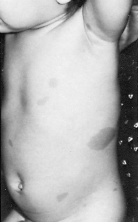
FIGURE 19.1 In pediatric patients with neurofibromatosis type 1, café-au-lait spots usually are larger
than 55 mm in diameter, the edges are well defined, and the intensity of the coloration is uniform.
Reprinted with permission from McMillan JA, Feigin RD, DeAngelis C, Jones MD. Oski’s Pediatrics: Principles and Practice. Philadelphia: Lippincott Williams & Wilkins, 2006.
677
678
VII. Disorders of the Hair
A.Alopecia areata
1.Etiology. The cause is thought to be autoimmune lymphocyte-mediated injury to the hair follicle.
2.Epidemiology. Alopecia areata affects 1 in 1000 persons.
3.Clinical features
a.Complete hair loss occurs as sharply demarcated areas of alopecia without inflammation or scaling. The hair loss is described as occurring suddenly, and the underlying skin is smooth and soft. The hair loss can occur on the scalp or other hair-bearing areas such as the beard, eyebrows, eyelashes, etc.
b.Pitting of the nails occurs in 40% of patients.
c.Subtypes of alopecia areata include alopecia totalis (loss of all scalp hair) and alopecia universalis (loss of all body and scalp hair).
4.Management. Most patients have regrowth of hair within months to years without any treatment. Hair regrowth may be stimluated with the use of topical or injected corticosteroids, topical minoxidil, and topical irritants (anthralin, squaric acid). Wigs and counseling may be necessary owing to the psychological consequences of the significant hair loss.
B.Tinea capitis is a common cause of hair loss [see section V.A.1].
C.Traumatic alopecia is also a common cause of childhood hair loss and may be one of the following two types:
1.Trichotillomania is hair loss that occurs as a result of conscious or unconscious pulling or twisting of hair. Clinical features include irregularly bordered areas of hair loss in which hairs are broken off at different lengths. The scalp may show perifollicular petechiae and excoriations. Eyelashes and eyebrows may also be involved. The cause is unknown, although it may be associated with anxiety. Management includes stress management or counseling and investigation into precipitating events.
2.Traction alopecia is hair loss caused by constant traction or friction and may be the result of tight hair braids, curlers, vigorous scalp massage, or constant rubbing. Clinical features include patchy and jagged, irregularly shaped areas of alopecia especially along the hair line, with thinned, small hairs, and broken hairs. Management is to stop the inciting trauma.
D.Telogen effluvium is the second most common type of alopecia after male pattern baldness. It is caused by any acutely stressful event (e.g., pregnancy, surgery, acute illness, high fever, trauma) that converts hairs from a growing phase (anagen) to a final resting phase (telogen). Clinical features include complaints of generalized excessive hair loss (hair loss > 100 hairs per day compared to normal hair loss of 50–100 hairs per day) 1–3 months after the precipitating event. Hair loss continues for 3–4 months, and then spontaneous regrowth occurs.
E.Other conditions that cause hair loss include hypothyroidism, diabetes mellitus, hypopituitarism, nutritional disorders (e.g., hypervitaminosis A, marasmus, zinc deficiency also known as “acrodermatitis enteropathica”), medications (e.g., warfarin, heparin, chemotherapy, cyclophosphamide, isotretinoin), ectodermal dysplasias, and hair shaft structural defects.
679
680
VIII. Acne Vulgaris
Acne is the most common skin disease, and its sequelae may include scarring and disfigurement with resultant adverse effects on psychosocial development, such as depression and other emotional problems.
A.Pathophysiology. Acne is caused by a combination of the following processes:
1.Excessive shedding and cohesion of cells that line the sebaceous follicles located on the chest, back, and face
2.Increased production of sebum by sebaceous glands under the influence of androgens. Obstruction of sebum outflow from the follicle leads to the formation of comedones.
3.Inflammation as a result of the proliferation of the bacteria, Propionibacterium acnes, and the host immune response to the bacteria
B.Clinical features
1.Acne usually begins 1–2 years before puberty.
2.Noninflammatory (comedonal) acne is characterized by open comedones (blackheads) and closed comedones (whiteheads).
3.Inflammatory acne is characterized by erythematous papules, pustules, nodules, and cysts.
4.Most patients have a combination of comedomal and inflammatory acne.
C.Management. Treatment is individualized based on the disease severity and on the location and type of lesions.
1.Benzoyl peroxide, retinoids (Retin-A) (particularly useful for comedonal acne), sulfur, and salicylic acid are effective topical agents for acne.
2.Antibiotics (oral or topical) are used in combination with the aforementioned medications for more inflammatory acne.
3.Oral contraceptives are helpful in treating acne in teenage girls and young adult women.
4.Systemic isotretinoin (Accutane) is highly effective for all types of acne, including nodular and cystic acne. Females must be tested for pregnancy before and during treatment, and must use effective birth control during treatment to prevent pregnancy because of the risk of teratogenic effects associated with the use of systemic isotretinoin.
681
IX. Vascular Skin Lesions
A.Nevus simplex/flammeus, often called “stork bites,” are congenital capillary malformations present in about 25–50% of newborns that are located on the midline upper face, scalp, and nuchal area. The lesions usually fade over the first 1–2 years of life.
B.Infantile hemangiomas are common vascular skin lesions seen in approximately 5% of infants.
1.Clinical features
a.Hemangiomas often appear shortly after birth and exhibit a growth phase lasting up to 5 months, followed by slow involution (shrinkage and loss of color) over several years.
b.Hemangiomas can present anywhere on the skin (as well as in internal organs such as the liver) as red papules and plaques, deep blue nodules, or both.
2.Management. Most hemangiomas do not require treatment, but treatment can be considered for those that are likely to cause disfigurement, as in the case of facial lesions, or obstruction of vital structures, as can be seen in the periocular location, or when they arise in the airway. Treatments may involve the use of topical and oral β-blockers to prevent growth and speed involution, or laser therapy.
C.Port wine stains (capillary malformations) are present at birth and do not proliferate or involute. They can present anywhere on the skin and are associated with certain syndromes when found in specific locations (e.g., Sturge–Weber Syndrome when located on the face in the distribution of the first or second branches of the trigeminal nerve). Port wine stains can be lightened with laser treatments.
D.Pyogenic granulomas are proliferations of capillaries on the skin, which often arise in response to some sort of trauma. They are most commonly seen during the first 5 years of life and often present as red friable glistening papules or nodules that bleed easily. Surgical removal is the treatment of choice, but laser can be used for small lesions.
682