

Answers and Explanations
1.The answer is D [II.C.5 and Table 7-1]. Fever in an infant younger than 28 days of age must be taken very seriously because the neonate’s immune system is immature. As a result, the current appropriate management for any neonate with fever (temperature >100.4°F [>38°C]) includes a complete workup for serious bacterial infection (SBI) that includes evaluation of the blood, urine, and cerebrospinal fluid for evidence of bacterial infection; administration of empiric intravenous antibiotics; and hospitalization. The risk of SBI in a nontoxic infant younger than 3 months of age is approximately 1–7%. Usual bacteria resulting in infection in this age group include group B streptococcus, Escherichia coli, and Listeria monocytogenes.
2.The answer is B [II.D.3 and Table 7-1]. Because of the patient’s elevated fever, evaluation for a urinary tract infection, including urine culture and urinalysis, is indicated. Because the infant is completely immunized, risk of bacteremia is low enough not to warrant blood tests. If she were incompletely immunized (had not received her 2-, 4-, and 6-month vaccinations), a
complete blood count and blood culture would be indicated and intramuscular ceftriaxone may be given either empirically, or only if the white blood count is ≥15,000 cells/mm3.
Hospitalization is generally not required unless the patient is toxic in appearance, is dehydrated, or has poor ability to return to the physician for follow-up. Neither evaluation of spinal fluid nor a chest radiograph is indicated in this nontoxic patient without respiratory signs or symptoms. Neither intravenous antibiotics nor hospitalization is indicated because the infant is nontoxic and well hydrated.
3.The answer is D [XIII.B]. This patient’s clinical presentation with fever, lymphadenopathy, pharyngitis, and splenomegaly is most consistent with infectious mononucleosis. If a child with infectious mononucleosis is given amoxicillin, a diffuse pruritic rash may develop.
Monospot testing is highly sensitive in older children, but heterophile antibodies do not reliably form in children younger than 4 years of age. Antibody titers are therefore the preferred diagnostic test in such young children. The most common cause of infectious mononucleosis is Epstein–Barr virus. Although supportive care is most appropriate, corticosteroids may be indicated for treatment of infectious mononucleosis complicated by airway compromise, but are not routinely recommended. Splenomegaly is not consistent with the diagnosis of streptococcal pharyngitis.
4.The answer is C [IV.C.3 and Tables 7-3 and 7-4]. This cerebrospinal fluid (CSF) evaluation is most consistent with aseptic meningitis, specifically viral meningitis. Enteroviruses are the most common cause of viral meningitis and most often occur during the summer and fall. Early in viral meningitis, the white blood cell (WBC) count in the CSF may demonstrate a polymorphonuclear cell predominance that shifts to a lymphocyte predominance within 24– 48 hours. The normal protein and glucose and negative Gram stain are also consistent with viral meningitis. Meningitis caused by Neisseria meningitidis or Streptococcus pneumoniae would be reflected by a higher CSF WBC count, lower glucose, and higher protein. Although patients with Lyme meningitis, which is caused by Borrelia burgdorferi, may present with an aseptic CSF profile, the onset is not as acute as in this patient. Patients with Mycobacterium tuberculosis present with a low to very low glucose and elevated protein level in the CSF.
5.The answer is A [IV.B.5]. Empiric therapy of presumed bacterial meningitis should include a
third-generation cephalosporin and the addition of vancomycin until sensitivities are available, because of the high level of pneumococcal antibiotic resistance in many
communities. Ampicillin is not indicated because this child is out of the age range at which Listeria infection occurs. Acyclovir is not indicated because the cerebrospinal fluid profile is most consistent with bacterial meningitis, not viral meningitis. Acyclovir is used to treat neonates with presumed herpes simplex meningitis, or encephalitis or in older children with
291
encephalitis. Corticosteroids are effective in reducing the incidence of hearing loss in Haemophilus influenzae type b meningitis but have not been shown to be effective for other bacterial pathogens.
6.The answer is C [XIII.A.2]. Factors that increase the risk of HIV transmission from mother to infant include high maternal viral load (measured by RNA copy number) at delivery, concomitant chorioamnionitis or other genital tract infections, primary or advanced maternal HIV infection, premature birth, and prolonged rupture of membranes. Transmission may also occur through breast milk. Transmission is decreased through the use of maternal antiretroviral therapy, newborn prophylaxis with antiretroviral agents (e.g., zidovudine), birth by cesarean section, and low maternal viral load.
7.The answer is B [IX.A.5.f]. This patient’s clinical presentation of a sandpaper-like rash associated with pharyngitis and fever is consistent with scarlet fever, caused by erythrogenic toxin–producing strains of group A β–hemolytic streptococcus (GABHS). Although there are multiple complications of GABHS infection, including rheumatic fever, glomerulonephritis, and reactive arthritis, only rheumatic fever will be prevented by treatment with antibiotics.
8.The answer is D [Tables 7-6 and 7-7, XV.B.3]. Infection with the protozoan Giardia lamblia is associated with bulky, foul-smelling stools, weight loss, and day care attendance. Entamoeba histolytica and Clostridium difficile generally cause bloody diarrhea. Escherichia coli infection generally results in short-term watery diarrhea. Day care attendance is also associated with Norwalk virus; however, symptoms of Norwalk virus infection generally last only 48–
72 hours.
9.The answer is A [XIII.C.3, XIII.C.4, XIII.C.5]. This patient’s presentation is most consistent with measles infection. Management includes supportive care, and vitamin A therapy may also be beneficial. Koplik spots are transient, and by the time the rash is present, Koplik spots are no longer appreciated. Bacterial pneumonia is the most common complication of measles
infection and is the most common cause of mortality. Diagnosis is based on confirmation by serologic testing in the presence of typical clinical features. Corticosteroids do not play a role in the therapy of measles.
10.The answers are B, D, G, A, and C, respectively [XV.D.3, XV.A.3, XIV.A.2, XV.C.3, and XV.B.3]. The triad of intracranial calcification, hydrocephalus, and chorioretinitis is consistent with congenital toxoplasmosis, which is caused by Toxoplasma gondii. Entamoeba histolytica may result in asymptomatic infection or colitis. The most common extraintestinal complication is a liver abscess. Aspergillus infection may result in invasive disease or in noninvasive allergic disease characterized by wheezing, eosinophilia, and pulmonary infiltrates. This can occur in patients with severe asthma or with cystic fibrosis. Malaria classically presents with a flulike illness followed by the development of high fevers that cycle in 48to 72-hour paroxysms. Giardia lamblia typically presents with bulky, large-volume, watery stools that eventually lead to weight loss. Outbreaks can occur in day care settings where children are in diapers.
11.The answer is A [XVII.C]. A tuberculin skin test is considered positive depending on a patient’s specific risk factors for acquisition of tuberculosis. A tuberculin skin test ≥10 mm is considered positive if the patient is younger than 4 years of age or if the patient resides or has lived in an area endemic for tuberculosis. Therefore, given that the tuberculin skin test is positive in this patient, a chest radiograph to evaluate for pulmonary tuberculosis is indicated. Children younger than 10 years of age with tuberculosis are unlikely to be contagious because of minimal cough and pulmonary involvement. Medications for tuberculosis disease (e.g., rifampin, isoniazid, pyrazinamide, ethambutol regimen) are indicated if the patient has signs and symptoms of tuberculosis. Gastric aspirates are indicated only if the chest radiograph reveals pulmonary disease.
12.The answers are G, B, A, C, and F, respectively [Table 7-6]. Enterotoxigenic Escherichia coli is the major cause of traveler’s diarrhea and results in nonbloody watery stools. Bloody stools
292
may result from infection with Salmonella, Shigella, Yersinia, Campylobacter, enterohemorrhagic E. coli, and Clostridium difficile. Shigella may be associated with seizures caused by the release of a neurotoxin. Salmonella may be acquired by ingestion of contaminated poultry or by exposure to turtles and lizards that carry the organism. Yersinia may result in mesenteric adenitis that causes pain mimicking acute appendicitis. Infection with Vibrio cholerae generally occurs in developing countries and causes massive fluid loss from the gut. E. coli 0157:H7 causes bloody diarrhea and is the causative agent in hemolytic uremic syndrome.
13.The answers are B, A, E, and D, respectively [IX.A]. Impetigo typically presents with honeycrusted lesions on the face; it is caused by infection with Staphylococcus aureus and group A β- hemolytic streptococcus (GABHS). Buccal cellulitis is characterized by a bluish color to the cheeks of a young child; this condition is typically caused by infection with Haemophilus influenzae type b, which is identified on blood culture. Staphylococcal scalded skin syndrome is manifested by Nikolsky sign or the extension of bullae with lateral pressure applied to the skin. Fever, tender skin, and widespread bullae are present. Erysipelas is characterized by tender, erythematous skin, but the border is well demarcated. Necrotizing fasciitis and toxic shock syndrome are complications of infection with GABHS. Necrotizing fasciitis is a severe skin infection that involves multiple layers of tissue. Toxic shock presents with fever, sunburn rash, and multiorgan failure. Both of these complications are a result of toxin produced by the GABHS.
293

C H A P T E R 8
294

Cardiology
David A. Ferry, Leigh Christopher Reardon
295
I.Congestive Heart Failure (CHF)
A.Definition. Congestive heart failure (CHF) is a clinical syndrome defined as inadequate oxygen delivery by the myocardium to meet the metabolic demands of the body.
B.Pathophysiology. Signs and symptoms of CHF often result from compensatory mechanisms that lead to increased demand on an already compromised myocardium.
1.Hypoperfusion of end organs stimulates the heart to maximize contractility and heart rate in an attempt to increase cardiac output.
2.Hypoperfusion also signals the kidneys to retain salt and water through the renin– angiotensin system in an attempt to increase blood volume.
3.Catecholamines released by the sympathetic nervous system also increase heart rate and myocardial contractility.
C.Etiology. CHF may result from congenital heart disease (CHD), acquired heart disease, and a variety of miscellaneous disorders.
1.CHD may result in CHF.
a.Increased pulmonary blood flow may cause CHF. Examples of congenital lesions that cause increased pulmonary blood flow include a large ventricular septal defect (VSD), a large patent ductus arteriosus (PDA), transposition of the great arteries (TGA), truncus arteriosus, and total anomalous pulmonary venous return (TAPVR).
b.Obstructive lesions may also cause CHF. Examples include severe aortic, pulmonary, and mitral valve stenosis, coarctation of the aorta, interrupted aortic arch, and hypoplastic left heart syndrome.
c.Other causes include arteriovenous malformations and mitral or tricuspid regurgitation, which all lead to volume overload in the heart.
2.Acquired heart disease may also lead to CHF.
a.Viral myocarditis is a common cause of CHF in older children and adolescents.
b.Other cardiac infections (e.g., endocarditis, pericarditis), metabolic diseases (e.g., hyperthyroidism), medications (e.g., doxorubicin, a chemotherapeutic agent), cardiomyopathies, and ischemic diseases (e.g., coronary artery disease)
c.Dysrhythmias, including certain types of tachycardia and bradycardia
3.Miscellaneous causes of CHF include the following:
a.Severe anemia may cause high-output CHF.
b.Rapid infusion of intravenous fluids, especially in premature infants
c.Obstructive processes of the airway, such as enlarged tonsils or adenoids, laryngomalacia, and cystic fibrosis may cause CHF as a result of chronic hypoxemia that results in pulmonary hypertension and right-sided heart failure.
D.Clinical features
1.Tachypnea, cough, wheezing, and rales on examination and pulmonary edema on chest radiograph (CXR) are evidence of pulmonary congestion.
2.Tachycardia, sweating, pale or ashen skin color, diminished urine output, and enlarged cardiac silhouette on CXR are evidence of impaired myocardial performance and therefore poor cardiac output.
3.Hepatomegaly and peripheral edema are evidence of systemic venous congestion.
4.Other signs and symptoms include failure to thrive, poor feeding (common symptom in newborns), and exercise intolerance (common symptom in older children and adolescents).
5.Cyanosis and shock are late manifestations.
E.Management
1.Goals of medical management are to improve myocardial function and relieve
296
pulmonary and systemic congestion.
a.Cardiac glycosides (e.g., digoxin) may increase the efficiency of myocardial contractions and relieve tachycardia.
b.Loop diuretics (e.g., furosemide, ethacrynic acid) reduce intravascular volume by maximizing sodium loss, which in turn leads to diminished ventricular dilation and improved cardiac function.
c.Inotropic medications (e.g., dobutamine, dopamine, epinephrine) are administered intravenously to augment cardiac contractility, thereby increasing the force of the heart’s contraction. They may be used to treat severe CHF.
d.Other medications, such as amrinone and milrinone (phosphodiesterase inhibitors) improve contractility and reduce afterload (the pressure in the aorta that the left ventricle must overcome to eject blood).
2.Interventional catheterization procedures can address some of the underlying causes of CHF (e.g., balloon valvuloplasty for critical aortic and pulmonary valve stenosis).
3.Surgical repair is often the definitive treatment of CHF secondary to CHD.
4.Cardiac transplant is reserved for the most severe cases of CHF that are refractory to medical management.
297

II.Innocent Cardiac Murmurs
A.Definition. Innocent murmurs result from turbulent but normal blood flow, are not caused by structural heart disease, and have no hemodynamic significance.
B.Epidemiology. Approximately 50% of children have an innocent heart murmur at some point during childhood.
C.Clinical features. The most common innocent heart murmurs are presented in Table 8-1.
Table 8-1
Clinical Features of Innocent Heart Murmurs
Murmur |
Age |
Location |
Characteristics |
Still’s murmur (left |
Ages 2–7 years |
Mid-left sternal |
Grade 1–3, systolic |
ventricular outflow tract) |
|
border |
Vibratory, twanging, or buzzing |
|
|
|
Loudest supine |
|
|
|
Louder with exercise and expiration |
Pulmonic systolic murmur |
Any age |
Upper left |
Grade 1–2, peaks early in systole |
(systolic ejection murmur) |
|
sternal border |
Blowing, high-pitched |
|
|
|
Loudest supine |
|
|
|
Louder with exercise and inspiration |
Venous hum |
Any age, |
Neck and |
Continuous murmur |
|
especially school |
below the |
Heard only sitting or standing |
|
age |
clavicles |
Disappears if supine; changes with compression of the |
|
|
|
jugular vein or with neck flexion or extension |
298
III.Acyanotic Congenital Heart Disease
A.Normal cardiac anatomy is depicted in Figure 8-1.
B.Clinical and diagnostic features. Physical examination, CXR, and electrocardiographic (ECG) findings of acyanotic CHD are presented in Table 8-2. Echocardiography confirms the specific anatomic lesions.
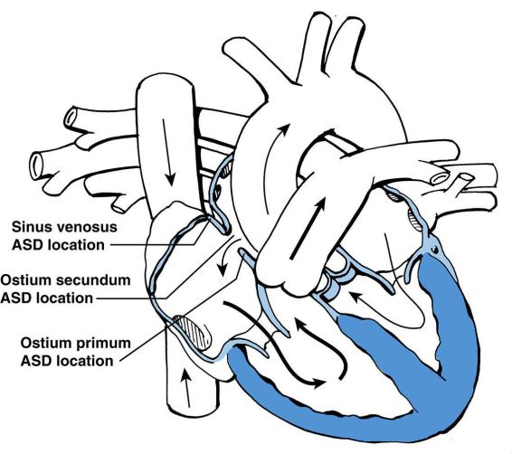
C.Atrial septal defect (ASD) (Figure 8-2)
1.Classification
a.Ostium primum. This type of ASD is a defect in the lower portion of the atrial septum. A cleft, or division, in the anterior mitral valve leaflet is almost always present and may cause mitral regurgitation. Ostium primum ASD is a common congenital heart lesion in Down syndrome.
b.Ostium secundum. This type of ASD is a defect in the middle portion of the atrial septum. Ostium secundum is the most common type of ASD.
c.Sinus venosus. This type of ASD is a defect superiorly and posteriorly in the septum near the junction of the right atrium and superior vena cava (SVC). In sinus venosus ASD, the right pulmonary veins usually drain anomalously into the right atrium or SVC instead of draining into the left atrium.
2.Pathophysiology. Blood flows across the ASD from the left atrium to the right atrium
(i.e., left-to-right shunt). The direction of the blood flow is determined by the relative diastolic compliances of the right and left ventricles (which in turn are determined by systemic and pulmonary vascular resistances [SVR and PVR]). Blood therefore flows from areas of higher resistance to areas of lower resistance. Increased blood flow across the ASD leads to an increase in size of the right atrium and right ventricle and to increased pulmonary blood flow.
3.Clinical features
a.Symptoms are minimal, if any, except in patients with an ostium primum defect who develop mitral regurgitation that results in CHF.
b.Physical examination findings include the following:
1.Increased right ventricular impulse (manifesting as a stronger point of maximal impulse) as a result of right ventricular volume and pressure overload.
2.Systolic ejection murmur (from excessive pulmonary blood flow) best heard at the mid and upper left sternal borders. A mid diastolic filling rumble representing increased blood flow through the tricuspid valve may also be heard.
3.Fixed-split second heart sound. With blood shunting across the ASD from the left atrium to the right atrium to the right ventricle, the second heart sound is widely split because of the increased volume of blood that must be ejected from the right ventricle, leading to later closure of the pulmonic valve compared with the aortic valve (i.e., a wider split of the A2 and P2 components of the second heart sound). The splitting is fixed, and does not vary with respiration, because the blood shunting across the ASD ensures that the right ventricle receives a fixed volume of blood, by balancing the increased volume reaching the right ventricle with inspiration and the decreased volume of blood reaching the right ventricle with expiration (i.e., the normal physiologic variation in timing of aortic and pulmonic valve closure with respiration is absent).
4.Management. Treatment of primum and sinus venosus ASDs is closure by open heart
299
surgery to prevent right-sided heart failure, pulmonary hypertension, atrial dysrhythmias, and paradoxical embolism (a stroke caused by a blood clot traveling from the right atrium to the left atrium via the ASD). Most secundum ASDs can be successfully closed using interventional catheterization procedures with implantation of atrial septal occlusion devices.
D.Ventricular septal defect (VSD)
1.Classification. VSDs are classified by location as inlet, trabecular (muscular), membranous, and outlet (supracristal) (Figure 8-3).
2.Pathophysiology. After birth, as pulmonary vascular pressure decreases, blood flows across the VSD from the left ventricle to the right ventricle, owing to the lower resistance within the pulmonary circulation compared to the resistance within the systemic circulation. However, with time, the pulmonary vessels hypertrophy in response to this increased pulmonary flow. This hypertrophy may lead to increased PVR (pulmonary hypertension). If treated early, the increased PVR is usually reversible. If increased pulmonary blood flow persists, the pulmonary hypertension may become irreversible and even progress to Eisenmenger syndrome [see section III.D.3.d.(2)].
3.Clinical features and course vary greatly depending on the magnitude of the left-to- right shunting across the VSD. The amount of blood flow directed from one side of the heart to the other side (i.e., the shunt) is determined by both the size of the VSD and the degree of PVR. For example, the larger the VSD and the lower the PVR, the greater the blood flow across the VSD and into the pulmonary vessels. The greater the pulmonary blood flow, the more symptomatic the patient. Typical clinical presentations include the following:
a.Small VSDs have little to no shunt across the VSD and may close spontaneously. On examination, small VSDs typically have a high-pitched holosystolic murmur ranging from a grade 3 to grade 4 murmur. A thrill at the lower left sternal border and a grade 4 high-pitched holosystolic murmur is indicative of a very restrictive defect with a high flow velocity across the VSD. Key point: As the size of the VSD decreases, the intensity of the murmur increases.
b.Moderate VSDs may have a large shunt across the VSD that may result in signs and symptoms of CHF. A holosystolic murmur is usually present, and its intensity depends on the size of the shunt. If the left-to-right shunt across the VSD is large (2:1 pulmonary-to-systemic flow—i.e., twice as much blood flows to the lungs as to the systemic circulation), then a diastolic murmur of mitral turbulence may also be heard at the apex (mitral filling rumble representing the excess blood from the lungs now passing through the mitral valve).
c.Large VSDs often cause signs and symptoms of CHF. They have less turbulence across the VSD, so the systolic murmur is shorter and lower in pitch. A mitral filling rumble may be heard at the apex. The S2 tends to be loud and more narrowly split than usual, with P2 (pulmonary closure sound) being accentuated due to high pulmonary artery pressure.
d.PVR may eventually become elevated in moderate or large VSDs in response to chronically high pulmonary flow. When this occurs, clinical features change.
1.When PVR becomes elevated, the right ventricular impulse is noticeably increased and the second heart sound may be single and loud. The mitral filling rumble disappears because of diminished pulmonary blood flow as a result of decreased left-to-right shunting. Symptoms of CHF also diminish as PVR increases, because of the decrease in pulmonary blood flow.
2.If PVR remains elevated, pulmonary hypertension may become irreversible, even if the VSD is surgically closed. In the extreme situation in which PVR
300
increases and exceeds SVR, shunting changes from left-to-right to right-to-left (a condition termed Eisenmenger syndrome). The right-to-left shunt is manifested by cyanosis.
4.Management
a.Medical management of CHF is indicated in a symptomatic child. Large shunts are also associated with a high incidence of pulmonary infections from excessive blood flow.
b.Surgical closure is indicated in the following circumstances:
1.Heart failure refractory to medical management
2.Large VSDs with pulmonary hypertension are usually surgically closed at 3– 6 months of age.
3.Small to moderate VSDs with persistent left ventricular (LV) volume overload are usually surgically closed between 2 and 6 years of age. Some small VSDs may be left alone if there is no LV volume overload in childhood. Sometimes, transcatheter closure of smaller VSDs can be done with excellent results.
E.Patent ductus arteriosus (PDA)
1.Definition. In the fetus, the ductus arteriosus connects the pulmonary artery to the aorta. After birth, as the PaO2 rises, the ductus arteriosus normally fibroses. If it remains open, it is termed a PDA. The incidence of PDA is especially high in preterm infants.
2.Postnatal pathophysiology. In healthy infants the SVR exceeds PVR, and blood therefore flows through the PDA from the aorta to the pulmonary artery (left-to-right shunt). This leads to increased pulmonary blood flow (Figure 8-4).
3.Clinical features. Signs and symptoms depend on the size of the PDA and on the relationship between SVR and PVR.
a.Small PDAs usually produce no symptoms, but moderate or large PDAs almost always result in signs and symptoms of CHF due to increased pulmonary blood flow.
b.Physical examination findings
1.The classic murmur is a “machinery-like” continuous murmur at the upper left sternal border.
2.If the left-to-right shunt is large, there may also be a diastolic rumble of
blood flow across the mitral valve at the apex, a widened pulse pressure
(˃30 mm Hg) and bounding pulses.
c.Risk of pulmonary hypertension caused by excessive pulmonary blood flow through a large PDA over time.
4.Management
a.Indomethacin is often used in premature infants to close a PDA medically.
b.PDAs in the nonneonate are usually closed surgically by transcatheter insertion of a device.
F.Coarctation of the aorta
1.Definition. Coarctation of the aorta is narrowing of the aortic arch, just below the origin of the left subclavian artery and typically at, or just proximal to, the origin of the ductus arteriosus. It may be a discrete narrowing (hourglass) or a long-segment obstruction.
2.Pathophysiology. The narrowed segment obstructs or diminishes flow from the proximal to the distal aorta (Figure 8-5).
3.Clinical features. Signs and symptoms depend on the severity of the obstruction and on the presence of any other associated cardiac abnormalities.
a.Neonates or infants with severe coarctation may depend on a right-to-left shunt through the PDA for perfusion of the lower thoracic and descending aorta. Such
301
infants may be minimally symptomatic initially, but as the PDA closes, symptoms of CHF develop and progress.
1.Blood pressure may be elevated in the upper extremities and low in the lower extremities before the onset of CHF (before the PDA becomes narrower or closes).
2.Once the neonate or infant develops CHF, pulses in all four extremities are poor due to low cardiac output.
b.Older children or adolescents may have no symptoms and may have only hypertension or a heart murmur. Hypertension is typically noted in the right arm, and commonly in the left arm as well, with reduced blood pressure in the lower extremities.
1.Femoral pulse, which normally precedes the radial pulse, is dampened and delayed until after the radial pulse (radiofemoral delay).
2.Blood pressure and pulse findings may be less prominent if collateral vessels (intercostal arteries) develop, which allow the ascending aortic pressure and flow to circumvent the coarctation.
3.Bicuspid aortic valve or aortic stenosis is present in 50% of patients with coarctation of the aorta. If either of these conditions is present, a systolic murmur of aortic stenosis may be heard [see section III.G.3].
4.Bruit of turbulence through the coarctation may be audible at the left upper back near the left scapula.
4.Management
a.Initial management in the symptomatic neonate is directed at improving circulation to the lower body.
1.Intravenous prostaglandin E (PGE) is given urgently to open the ductus arteriosus.
2.Inotropic medications are given to overcome myocardial depression, and low-dose dopamine is used to maximize renal perfusion and function.
b.Corrective repair
1.Surgery is the usual approach in newborns and infants and involves excision of the narrowed segment followed by end-to-end anastomosis. Late recurrence of narrowing may occur in up to 50% of patients.
2.Balloon angioplasty with or without endovascular stent placement is the treatment of choice for short segment coarctations in school-age children and teens who have not undergone surgical correction. It is also the primary intervention for recurrent coarctation after surgical correction.
5.Prognosis is excellent, but long-term concerns include recurrence of coarctation and upper extremity hypertension with exercise.
G.Aortic stenosis
1.Definition. Aortic stenosis is narrowing of the aortic valve. Pathologically, aortic stenosis typically appears as commissural fusion of the three normal leaflets, leading to a bicuspid or unicuspid valve.
2.Pathophysiology. Aortic stenosis results in reduced LV output. At the myocardial level, aortic stenosis results in an imbalance between myocardial oxygen demand (which is higher than usual owing to the increased ventricular work as a result of outflow obstruction) and supply. This may lead to myocardial ischemia. In the neonate, severe aortic stenosis may be associated with hypoplasia of the left ventricle as a result of impaired fetal LV development.
3.Clinical features. Signs and symptoms depend on the severity of the stenosis and age. Physical examination findings are presented in Table 8-2.
302
a.Neonates with severe stenosis (“critical aortic stenosis”) appear normal at birth but develop signs and symptoms of CHF at 12–24 hours of age. Once the PDA closes, all systemic flow must be ejected through the aortic valve. However, in critical aortic stenosis this is not possible and adequate perfusion to the body cannot be maintained.
b.Older children generally have no symptoms until the stenosis becomes severe. Once severe, symptoms include exercise intolerance, chest pain, syncope, and even sudden death.
4.Management. Indications for intervention include CHF, symptoms such as chest pain
or syncope, and documentation of a high resting pressure gradient across the aortic valve (˃50–70 mm Hg).
a.Balloon valvuloplasty is often the initial management approach for aortic stenosis without associated significant regurgitation.
b.Surgery is often required for aortic stenosis with regurgitation and for recurrent stenosis. The aortic valve is replaced with either the patient’s own pulmonary valve (Ross procedure) or a prosthetic valve.
H.Pulmonary stenosis
1.Definition. Pulmonary stenosis is narrowing of the pulmonary valve. Pathologically, fusion of the valve commissures is typically seen.
2.Pathophysiology. Pulmonary stenosis results in increased right ventricular pressure and reduced right ventricular output.
3.Clinical features. Physical examination findings are presented in Table 8-2.
a.Severe pulmonary stenosis in the neonate may be manifested by cyanosis as a result of right-to-left shunting at the atrial level through a patent foramen ovale (PFO).
b.For most children, outflow obstruction is mild to moderate and symptoms are absent.
4.Management. Treatment is balloon valvuloplasty for symptomatic infants with critical
pulmonary stenosis, and for older children with significant gradients across the pulmonary valve (˃35–40 mm Hg) or high right ventricular pressures.
303
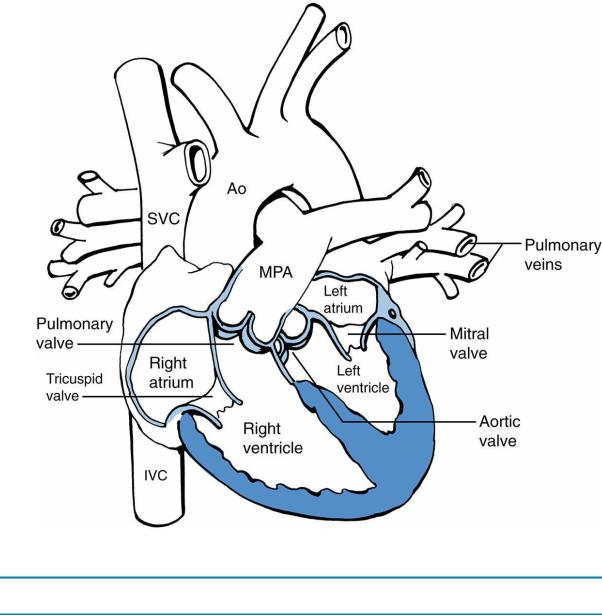
FIGURE 8.1 Anatomy of the normal heart. Ao = aorta; MPA = main pulmonary artery; SVC = superior
vena cava; IVC = inferior vena cava.
Table 8-2
Key Differentiating Clinical Features of Acyanotic Congenital Heart Disease
Heart Lesion |
Physical Examination Findings |
ECG |
Chest Radiograph |
Atrial septal |
Systolic ejection murmur mid-left sternal border |
RAD, RVH, and RAE |
Right atrial and ventricular |
defect |
and ULSB |
|
enlargement |
|
Fixed split S2 |
|
Increased PVM |
|
Diastolic rumble LLSB |
|
|
Ventricular |
High-pitched holosystolic murmur at LLSB, with |
If small: normal or |
If small: normal |
septal defect |
or without a thrillDiastolic rumble at apex if |
mild LVH |
|
|
pulmonary blood flow is high |
If moderate: LVHRVH |
If moderate or large: |
|
|
if pulmonary |
cardiomegaly and increased |
|
|
hypertension present |
PVM |
|
|
|
If elevated PVR: normal or |
|
|
|
decreased PVM |
Patent ductus |
Continuous murmur at ULSB; “machinery-like” |
LVHRVH if |
Cardiomegaly with increased |
arteriosus |
Brisk pulses |
pulmonary |
PVM |
|
|
hypertension present |
|
Coarctation |
Elevated BP in right arm |
Normal or LVH |
Normal heart sizeRib notching |
of the aorta |
Reduced BP in legs |
|
(evidence of collateral flow) |
(older child) |
Dampened and delayed femoral pulse |
|
|
|
Bruit left upper back |
|
|
Aortic |
Ejection click at base and apex (fixed with |
Normal or LVH |
Normal or mild |
304
stenosis |
respiratory cycle) |
|
cardiomegalyProminent |
|
Systolic ejection murmur at base with radiation to |
|
ascending aorta (poststenotic |
|
URSB, apex, suprasternal notch, and carotids |
|
dilation) |
|
Thrill at URSB and suprasternal notch |
|
|
Pulmonary |
Ejection click at apex (variable with respiratory |
RVH |
NormalProminent main |
stenosis |
cycle) |
|
pulmonary artery due to |
|
Systolic ejection murmur at ULSB |
|
poststenotic dilation |
ECG = electrocardiogram; ULSB = upper left sternal border; LLSB = lower left sternal border; URSB = upper right sternal border; S2 = second heart sound; LVH = left ventricular hypertrophy; RVH = right ventricular hypertrophy; BP = blood pressure; RAE = right atrial enlargement; RAD = right axis deviation; PVM = pulmonary vascular markings; PVR = pulmonary vascular resistance.
FIGURE 8.2 Location of atrial septal defects (ASDs). Arrows designate the direction of blood flow. Thick
arrows designate increased blood flow. The right atrium and the right ventricle are enlarged. The location
of the sinus venosus ASD is the posterior–superior aspect of the atrial septum.
305
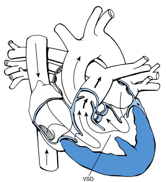
FIGURE 8.3 Trabecular (muscular) ventricular septal defect (VSD). Arrows designate the direction of
blood flow. Thick arrows designate increased blood flow.
306
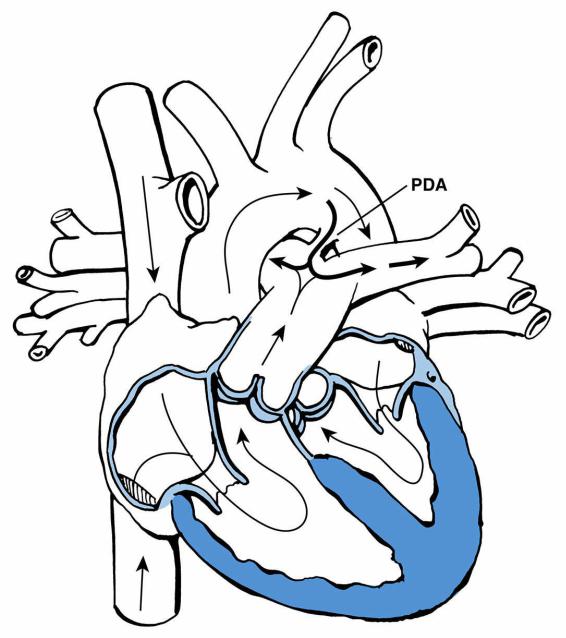
FIGURE 8.4 Patent ductus arteriosus (PDA). Arrows designate the direction of blood flow. Thick arrows
designate increased blood flow.
307
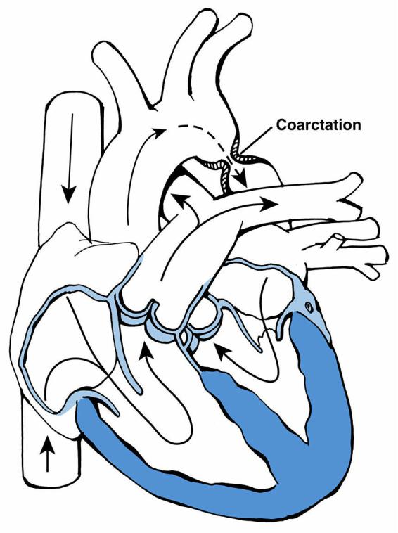
FIGURE 8.5 Coarctation of the aorta. Arrows designate the direction of blood flow.
308
IV. Cyanotic Congenital Heart Disease
A.General concepts
1.Cyanosis may be peripheral, central, or both. Peripheral cyanosis is usually caused by vasomotor instability or vasoconstriction as a result of cold temperature. Central cyanosis, especially apparent in the tongue and inner mucous membranes, may be attributable to both cardiac and noncardiac causes:
a.Noncardiac causes of central cyanosis include pulmonary disease, sepsis, hypoglycemia, polycythemia, and neuromuscular diseases that impair chest wall movement.
b.The most common cardiac causes of central cyanosis may be remembered using the mnemonic “5 T’s”: tetralogy of Fallot, transposition of the great arteries, tricuspid atresia, truncus arteriosus, and total anomalous pulmonary venous return.
2.Evaluation
a.A thorough physical examination is essential.
b.Other initial studies include pulse oximetry (in room air), complete blood count (CBC), arterial blood gas (ABG), ECG, and CXR. The degree of pulmonary blood flow seen on CXR may be useful in diagnosis (Table 8-3).
c.The 100% oxygen challenge test suggests cyanotic CHD when the PaO2 fails to rise above 100–150 mm Hg despite the administration of 100% oxygen.
d.Echocardiogram provides a definitive diagnosis.
3.Diagnosis. The distinguishing features of the five types of cyanotic CHD are presented in
Table 8-4.
B.Tetralogy of Fallot is the most common cause of central cyanosis presenting beyond the newborn period.
1.Definition. Tetralogy of Fallot has four anatomic components:
a.VSD
b.Overriding aorta (aorta overlies a portion of the ventricular septum)
c.Pulmonary stenosis
d.Right ventricular hypertrophy (RVH)
2.Pathophysiology. As a result of right ventricular outflow tract obstruction (RVOT; pulmonary stenosis), blood flows from right to left across the VSD and into the overriding aorta (Figure 8-6), resulting in cyanosis.
3.Clinical features. Signs and symptoms depend on the severity of the RVOT obstruction.
a.Physical examination findings include an increased right ventricular impulse because of RVH, a systolic ejection murmur representing pulmonary stenosis (see Table 8-2), and cyanosis.
b.Cyanosis depends on the interplay between the resistance to flow out of the RVOT and the SVR. It is important to remember that blood always flows from higher resistance to lower resistance.
1.Actions that decrease SVR (e.g., exercise, vasodilation, volume depletion) or increase resistance through the RVOT (e.g., crying, tachycardia) increase right-to-left shunting from the right ventricle through the VSD and to the aorta, resulting in cyanosis.
2.Actions that increase SVR or reduce resistance through the RVOT (e.g., volume infusion, systemic hypertension, Valsalva maneuver, bradycardia) reduce the right-to-left shunt through the VSD and therefore increase systemic arterial saturation.
3.Neonates with severe pulmonary stenosis or atresia present with cyanosis
309
immediately after birth once the PDA closes. These patients are dependent on the PDA for blood flow to the lungs.
c.Tetralogy of Fallot (hypercyanotic or “tet”) spells are characterized by sudden cyanosis and decreased murmur intensity.
1.The trigger may be any condition such as dehydration or crying that decreases arterial oxygen saturation.
2.With desaturation, the child typically becomes irritable and cries, which increases resistance through the RVOT, worsening the cyanosis by increasing the right-to-left shunt, resulting in a cycle of increasing cyanosis.
3.Alterations in consciousness and hyperpnea may occur as a result of severe hypoxia and acidosis.
4.To compensate, a child with tetralogy of Fallot learns to squat. This position (knee-chest position in an infant) increases venous return to the heart and increases SVR, thereby decreasing the right-to-left shunt.
4.Management (Table 8-5 describes management of “tet” spell)
a.Definitive management is complete surgical repair at 2–8 months of age.
b.Some infants with unfavorable anatomy (small pulmonary arteries) or with recurrent tetralogy of Fallot spells may initially undergo a palliative procedure to improve systemic saturation and encourage pulmonary growth. The procedure involves either a modified Blalock–Taussig shunt (Gore-Tex graft interposed between the subclavian and ipsilateral pulmonary artery) or balloon pulmonary valvuloplasty. In the neonate who requires intervention, transcatheter stenting of the PDA is also an option.
C.TGA
1.Definition. TGA occurs when the aorta arises from the right ventricle and the main pulmonary artery from the left ventricle.
2.Pathophysiology (Figure 8-7)
a.Pulmonary and systemic circulations are in parallel, rather than in series.
b.Adequate saturation can only be achieved by shunting blood from one circulation to the other through a PFO, ASD, VSD, or PDA.
3.Clinical features
a.Cyanosis is present at, or shortly after, birth. Cyanosis is more intense if the PFO is small. Cyanosis is less intense if the PFO is large or if there are additional sites of mixing, such as an ASD or VSD.
b.Physical examination findings include an infant who appears healthy but who has central cyanosis, a quiet precordium, and on auscultation, a single S2 (because the aortic valve is anterior and the pulmonary valve is posterior in TGA, the closure of the pulmonary valve is difficult to hear) and no murmur.
4.Management
a.Neonates may require initial management with PGE to improve oxygen saturation by keeping the ductus patent. Emergent balloon atrial septostomy (Rashkind procedure) is an often life-saving procedure that increases the size of an ASD or PFO.
b.Definitive repair is the arterial switch operation, in which the great arteries are incised above their respective valves and implanted in the opposite root. The coronary arteries are attached to the original aorta, so the coronaries must also be incised and reimplanted.
D.Tricuspid atresia
1.Definition. Tricuspid atresia is defined anatomically as a plate of tissue located in the floor of the right atrium in the location of the tricuspid valve. An ASD or PFO is always
310
present to allow for right-to-left shunting.
2.Pathophysiology. Whether a VSD is also present plays a significant role in determining the clinical presentation.
a.If no VSD is present and the ventricular septum is intact, pulmonary atresia is also present. For blood to flow to the lungs in this situation, a PDA must be present. As the PDA constricts after birth, visible cyanosis develops.
b.If a VSD is present, blood flow from the left ventricle through the VSD and into the pulmonary artery (left-to-right shunt) may be adequate, facilitating acceptable systemic oxygen saturations (Figure 8-8).
3.Clinical features. Signs also depend on anatomy.
a.Patients with an intact ventricular septum and pulmonary atresia have no murmur and a single S2.
b.Patients with a VSD have a VSD murmur (see Table 8-2).
c.ECG shows right atrial enlargement, left axis deviation (LAD), and left ventricular hypertrophy (LVH). Tricuspid atresia is the only cause of cyanosis in the newborn period that results in LAD and LVH.
4.Management. Treatment includes staged surgery with eventual Fontan procedure at 3– 6 years of age.
a.A bidirectional Glenn shunt (SVC is anastomosed to the right pulmonary artery) is usually performed at 4–6 months of age.
b.In the Fontan procedure, flow from the inferior vena cava is directed into the pulmonary arteries, usually by means of an extracardiac conduit.
c.The net result of these procedures is systemic venous return directed to the pulmonary artery.
E.Truncus arteriosus
1.Definition. Truncus arteriosus occurs when the aorta and pulmonary artery originate from a common artery, the truncus. The pulmonary arteries usually originate from the proximal truncus, and the truncal valve may be regurgitant or stenotic. A VSD is almost always present.
2.Pathophysiology. Because the aorta and pulmonary arteries are connected and the pulmonary arteries are well formed, excessive blood flows to the lungs and CHF commonly develops. Mixing of desaturated and saturated blood occurs within the truncus, and patients are commonly only mildly desaturated and mildly cyanotic (Figure 8- 9).
3.Clinical features
a.Signs and symptoms of CHF are common.
b.Physical examination findings
1.Systolic ejection murmur at the base from increased flow across the truncal valve and a single S2 caused by the presence of only one atrioventricular (AV) valve
2.Diastolic murmur of flow across the mitral valve at the apex as a result of excessive pulmonary blood flow that returns to the left atrium
3.High-pitched diastolic murmur at the base indicates regurgitation of the truncal valve.
4.Management. Treatment includes medications for CHF and surgical repair early in infancy to close the VSD and to place a valved cadaveric homograft between the right ventricle and pulmonary artery.
F.TAPVR
1.Definition. TAPVR occurs when the pulmonary veins drain into the systemic venous side rather than into the left atrium. Sites of pulmonary venous drainage include
311

supracardiac (into the right SVC or innominate vein), cardiac (into the right atrium or coronary sinus), and infracardiac (into the portal system and eventually into the inferior vena cava).
2.Pathophysiology. Systemic and pulmonary venous blood enters the right atrium and mixes together. As a result, this mixture of systemic and pulmonary venous blood is present in all four cardiac chambers (blood also passes across a PFO or ASD into the left side of the heart), pulmonary arteries, and aorta, resulting in desaturated systemic blood and visible cyanosis on examination.
3.Clinical features
a.Cyanosis is present, and if severe, may indicate obstruction of pulmonary venous return.
b.Pulmonary flow murmur at the mid-left sternal border is caused by increased pulmonary blood flow.
4.Management. Treatment is surgical repair shortly after diagnosis. The pulmonary veins are anastomosed to the back of the left atrium, and the PFO or ASD is closed.
Table 8-3
Cyanotic Congenital Heart Disease Lesions and Pulmonary Blood Flow on Chest Radiograph
Increased Pulmonary Flow |
Decreased Pulmonary Flow |
|
|||
Transposition of the great arteries |
Tetralogy of Fallot |
|
|
||
Total anomalous pulmonary venous return |
Pulmonary atresia |
|
|
||
Truncus arteriosus |
|
Tricuspid atresia |
|
|
|
Single ventricle |
|
|
|
|
|
Table 8-4 |
|
|
|
|
|
Key Differentiating Features of Cyanotic Congenital Heart Disease |
|
|
|||
|
|
|
|
||
|
|
|
|
||
Heart Lesion |
Physical Examination Findings |
ECG |
Chest Radiograph |
||
Tetralogy of Fallot |
Systolic ejection murmur of |
RVH |
Upturned cardiac apex (“boot shaped”) |
||
|
pulmonary stenosis (Table 8-2) |
|
Decreased PVM |
||
|
|
|
|
Right aortic arch (commonly) |
|
Transposition of the |
No murmurSingle S2 |
Normal or |
Small heart with narrow mediastinum (“egg-on-a- |
||
great arteries |
|
|
RVH |
string” appearance) |
|
|
|
|
|
Increased PVM |
|
Tricuspid atresia |
No murmur and single S2 if no |
LAD, |
Small heart |
||
|
VSD |
|
RAE, and |
|
|
|
If VSD is present, systolic |
LVH |
Decreased PVM |
||
|
murmur of VSD (Table 8-2) |
|
|
|
|
Truncus arteriosus |
Single S2 |
|
CVH |
Enlarged heart |
|
|
Systolic ejection murmur along |
|
Increased PVMRight aortic arch (commonly) |
||
|
the left sternal border |
|
|
|
|
|
Diastolic murmur at the apex |
|
|
|
|
Total anomalous |
Pulmonary ejection murmur |
RVH and |
Enlarged heart in older, unrepaired children with |
||
pulmonary venous |
along the left sternal border |
RAE |
supracardiac drainage (“snowman appearance”) |
||
return |
|
|
|
Increased PVM |
|
|
|
|
|
If obstruction is present, small heart and pulmonary |
|
|
|
|
|
edema |
|
RVH = right ventricular hypertrophy; LAD = left axis deviation; RAE = right atrial enlargement; CVH = combined ventricular hypertrophy; PVM = pulmonary vascular markings; LVH = left ventricular hypertrophy; VSD = ventricular septal defect; S2 = second heart sound.
312
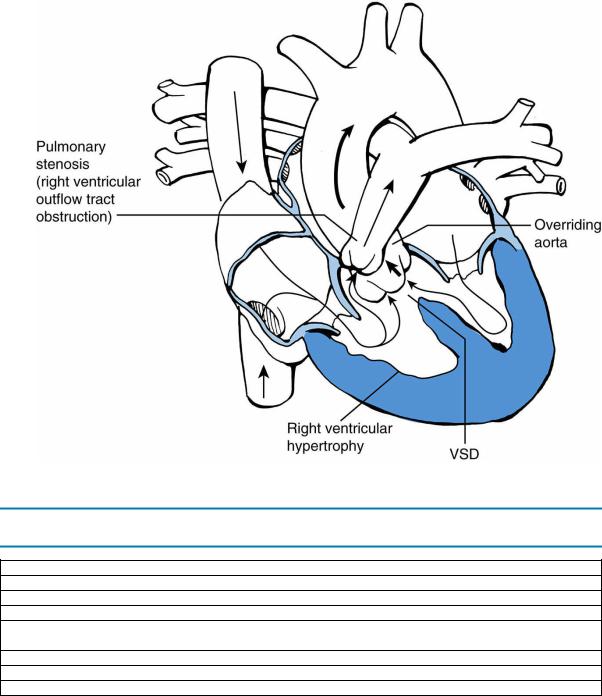
FIGURE 8.6 Tetralogy of Fallot. Arrows designate direction of blood flow. Thick arrows designate increased blood flow. VSD = ventricular septal defect.
Table 8-5
Acute Management of a Tetralogy of Fallot (“Tet” or Hypercyanotic) Spell
Placement in knee-chest position (mimics squatting position)
Intravenous fluid bolus
Oxygen
Sedation (morphine) to decrease agitation, which will then slow down the heart rate
β-Adrenergic blocker (e.g., propranolol) to slow down the heart rate, reduce contractility in right ventricular outflow tract, and augment pulmonary blood flow
Intravenous sodium bicarbonate to correct any acidosis from prolonged hypoxia
Correction of significant anemia with transfusion
Rarely, general anesthesia and surgery
313
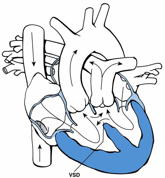
FIGURE 8.7 Transposition of the great arteries. A ventricular septal defect (VSD) is present. Arrows
designate the direction of blood flow.
314

FIGURE 8.8 Tricuspid atresia with atrial septal defect (ASD) and ventricular septal defect (VSD). Arrows designate direction of blood flow. Thick arrows designate increased blood flow.
315
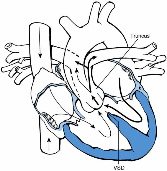
FIGURE 8.9 Truncus arteriosus with ventricular septal defect (VSD). Arrows designate the direction of blood flow. Thick arrows designate increased blood flow. The dashed line represents the other border of the pulmonary artery at the back of the truncus.
316
V.Acquired Heart Disease
A.Kawasaki disease is the most common cause of acquired heart disease in children in the United States and is discussed in Chapter 16, section II.
B.Acute rheumatic fever is the most common cause of acquired heart disease worldwide and is discussed in Chapter 16, section VI.
C.Infective endocarditis
1.Definition. Infective endocarditis is a microbial infection of the endocardium, or internal surface, of the heart.
2.Epidemiology
a.Eighty percent of cases occur in children who have structural abnormalities of the heart. Endocarditis may also occur in anatomically normal hearts, especially in the hearts of neonates and infants.
b.Fifty percent of cases occur soon after cardiac surgery.
3.Etiology
a.Gram-positive cocci, including α-hemolytic streptococcus (Streptococcus viridans) and Staphylococcus species, are the most common bacterial agents.
b.Gram-negative organisms are rare causes of endocarditis.
c.Fungal endocarditis is extremely rare but may occur in a chronically ill child.
4.Pathophysiology
a.Bacteria are introduced into the blood spontaneously or during an invasive procedure. Bacteria then infect injured cardiac endothelium.
b.Fibrin and platelets adhere to the site of injury, creating a growth or vegetation. The vegetation may lead to incompetency of a valve.
c.Distal manifestations of disease may occur, including embolic phenomena and immunologic sequelae (e.g., nephropathy).
5.Clinical features (Table 8-6)
6.Diagnosis. History and physical examination, in addition to several other studies, are the basis of diagnosis.
a.Blood culture is the single most important laboratory test. Three sets of aerobic and anaerobic blood cultures should be drawn to maximize the likelihood of identifying the infecting organism. Because bacteremia is continuous in endocarditis, blood cultures may be drawn even if the patient is afebrile.
b.The erythrocyte sedimentation rate (ESR) is usually elevated, unless polycythemia is present. (Patients with cyanotic CHD may have reactive erythrocytosis.)
c.Other acute-phase reactants (e.g., rheumatoid factor) are found in 50% of patients.
d.Although transthoracic echocardiography may detect vegetations, transesophageal echocardiography is more sensitive than transthoracic echocardiography at identifying vegetations. However, a normal echocardiogram does not exclude endocarditis.
7.Management
a.Intravenous antimicrobial therapy is directed against the identified organism. Treatment for 4–6 weeks is required.
b.Because endocarditis is rarely a medical emergency, therapy may be safely withheld until an adequate number of blood cultures are obtained and the diagnosis is confirmed.
8.Antibiotic prophylaxis for endocarditis is recommended before invasive procedures in certain patients. Such procedures include dental work likely to produce bleeding, surgery (including tonsillectomy), and invasive gastrointestinal or urologic procedures.
317
This applies to a patient with any of the following circumstances:
a.A prosthetic cardiac valve or prosthetic material used for a valve repair
b.A personal history of infective endocarditis
c.CHD
1.Unrepaired cyanotic CHD
2.Completely repaired CHD with prosthetic material or device by surgery or catheter intervention within 6 months after the procedure
3.Repaired CHD with residual defects at or adjacent to a site of a prosthetic patch or device
d.A patient with a history of cardiac transplantation who develops a cardiac valvulopathy (e.g., valvular regurgitation or stenosis)
D.Pericarditis
1.Definition. Pericarditis is inflammation of the pericardial space.
2.Etiology. Causes most commonly include infection, collagen vascular disease (e.g., systemic lupus erythematosus [SLE]), uremia, and inflammatory response after cardiac surgery (postpericardiotomy syndrome).
a.Viral infection is the most common cause of pericarditis in children. Viruses include coxsackievirus, echovirus, adenovirus, influenza, parainfluenza, and Epstein–Barr virus (EBV).
b.Purulent pericarditis is usually caused by bacterial infection, either primary infection or disseminated from pneumonia or meningitis.
1.Staphylococcus aureus and Streptococcus pneumoniae are the most common agents.
2.Patients with purulent pericarditis have a high incidence of constrictive pericarditis owing to the intense inflammatory response.
c.Postpericardiotomy syndrome may occur in as many as one-third of patients whose pericardium has been opened during surgery. Patients typically present 1– 6 weeks after cardiac surgery with fever, pleuritis, and pericarditis with or without a pericardial effusion.
3.Pathophysiology. Inflammation of parietal and visceral pericardial layers leads to exudation or transudation of fluid and impairment of venous return and cardiac filling.
4.Clinical features
a.Symptoms include fever, dyspnea, malaise, and chest pain most intense while supine and relieved when sitting upright.
b.Physical examination findings include a pericardial friction rub, distant heart sounds if the effusion is large, pulsus paradoxus (˃10 mm Hg reduction in systolic
blood pressure on deep inspiration), and hepatomegaly. Cardiac tamponade, or critically impaired LV output due to an acute accumulation of pericardial fluid, may occur and is life-threatening.
5.Diagnosis
a.Pericarditis should be considered in any child with dyspnea and fever, and in any patient who recently underwent cardiac surgery and has hemodynamic instability or nonspecific complaints of dyspnea or malaise.
b.Pericardiocentesis, in which a needle is inserted into the pericardial sac and fluid is withdrawn, is both diagnostic and therapeutic. Fluid should be sent for cell and serologic analysis and for culture.
c.ESR, although not specific for pericarditis, is elevated.
d.Imaging studies
1.ECG may show diffuse ST-segment elevations (Figure 8-10) or low-voltage QRS complexes in patients with large pericardial effusions.
318
2.CXR shows an enlarged heart shadow in patients with large effusions.
3.Echocardiogram demonstrates the size of the pericardial effusion.
6.Management
a.Appropriate antibiotics should be given if bacterial pericarditis is suspected.
b.Anti-inflammatory agents, such as aspirin or steroids, are indicated for viral pericarditis or postpericardiotomy syndrome.
c.Drainage of pericardial effusion by placement of a pericardial catheter or surgical window may be indicated.
E.Myocarditis
1.Definition. Myocarditis is inflammation of the myocardium, characterized by cellular infiltrate and myocardial cell death.
2.Epidemiology. Myocarditis is one of several common causes of sudden death in young athletes. Overall incidence is unknown, but evidence of myocarditis is apparent in 20% of children who die suddenly.
3.Etiology
a.Viruses, such as enteroviruses, especially coxsackievirus
b.Bacteria, such as Corynebacterium diphtheriae, Streptococcus pyogenes, S. aureus, and Mycobacterium tuberculosis
c.Fungi, such as Candida and Cryptococcus
d.Protozoa, such as Trypanosoma cruzi (Chagas disease)
e.Autoimmune diseases, such as SLE, rheumatic fever, and sarcoidosis
f.Kawasaki disease
4.Pathophysiology. Myocarditis may involve infectious infiltration that damages myocardial cells, or activated lymphocytes that are misdirected to attack the myocardium.
5.Clinical features
a.Myocarditis frequently follows a viral or flul-ike illness.
b.Symptoms include dyspnea and malaise.
c.Physical examination shows resting tachycardia, muffled heart sounds, gallop heart rhythm, hepatomegaly, tachypnea, and pulmonary rales.
6.Diagnosis
a.Laboratory studies. Findings include elevated ESR, creatine kinase (CK)–MB fraction, troponin, and C-reactive protein (CRP) in most, but not all, cases. The etiologic organism may be identified by viral serology or polymerase chain reaction (PCR) of endomyocardial biopsy specimens.
b.Accessory tests
1.ECG may show T-wave and ST-segment changes. Atrial or ventricular dysrhythmias may be present.
2.Echocardiogram shows an anatomically normal heart with global ventricular dysfunction. Pericardial effusion and valvular regurgitation may be present.
7.Management. Treatment is largely supportive, with use of inotropic agents, diuretics, and afterload-reducing drugs as needed. Intravenous immune globulin may be helpful in some cases. Cardiac transplantation is an option for patients with CHF refractory to medical management.
8.Prognosis. Outlook is variable. Mortality is 10–20% and is especially high in young infants and in those with ventricular dysrhythmias.
F.Cardiomyopathy
1.Definition. Cardiomyopathy is defined as an abnormality of cardiac muscle manifested by systolic or diastolic dysfunction. There are three types of cardiomyopathy, including dilated cardiomyopathy, hypertrophic cardiomyopathy, and restrictive cardiomyopathy.
319
2.Dilated cardiomyopathy
a.Definition. This primary myocardial disorder (i.e., not attributable to valvular, coronary artery, pericardial, or CHD) is characterized by ventricular dilation and reduced cardiac function.
b.Etiology. Dilated cardiomyopathy is frequently idiopathic, but many causes have been identified.
1.Viral myocarditis
2.Mitochondrial abnormalities
3.Carnitine deficiency
4.Nutritional deficiency, such as selenium and thiamine deficiency
5.Hypocalcemia
6.Chronic tachydysrhythmias
7.Anomalous origin of left coronary artery from the pulmonary artery
(ALCAPA), which results in myocardial ischemia and infarction
8.Medications (e.g., doxorubicin)
c.Clinical features. Signs and symptoms of CHF may be present.
d.Diagnosis. Evaluation should include viral serologies and serum carnitine level. In addition:
1.ECG shows sinus tachycardia, low cardiac voltage, and ST-segment and T- wave changes. With ALCAPA, evidence of infarction is typically noted, with Q waves in leads I and aVL.
2.Echocardiogram shows a dilated left ventricle with poor ventricular function.
e.Management
1.Medical management of CHF
2.Treatment of underlying metabolic or nutritional problem
3.Surgical repair of ALCAPA (implantation of left coronary artery into the aortic sinus)
4.Cardiac transplantation if CHF is unresponsive to medical management (true for all cardiomyopathies, including those listed below).
3.Hypertrophic cardiomyopathy
a.Definition. Hypertrophic cardiomyopathy is LVH in the absence of any systemic or cardiac disease known to cause the hypertrophy. The most typical anatomic finding is asymmetric septal hypertrophy.
b.Etiology. Inheritance is autosomal dominant in 60% of cases. Infants of mothers with diabetes mellitus may have transient septal hypertrophy.
c.Pathophysiology
1.Poor LV filling due to reduced LV compliance
2.Dynamic left ventricular outflow tract (LVOT) obstruction is present and caused by the anterior mitral leaflet being swept into the subaortic region and coming into contact with the ventricular septum during systole.
3.Mismatch between myocardial oxygen demand and supply (owing to hypertrophy) may result in myocardial ischemia.
d.Clinical features. Hypertrophic cardiomyopathy is the most common cause of sudden death in athletes.
1.Symptoms may be absent until syncope or sudden death occurs, or may include chest pain and exercise intolerance.
2.Physical examination shows a classic harsh, systolic ejection murmur at the apex that is accentuated with physiologic maneuvers that reduce LV volume, such as Valsalva or standing (by reducing LV volume, these maneuvers worsen the outflow obstruction, increasing the intensity of the murmur).
320
e.Diagnosis
1.ECG shows LVH, ST-segment and T-wave changes, LAD, and abnormally deep and wide Q waves in the inferior and lateral leads.
2.Echocardiogram shows the hypertrophy.
f.Management. Treatment is generally reserved for patients with symptoms.
1.β-Adrenergic blockers or calcium-channel blockers reduce the LVOT obstruction and improve diastolic compliance.
2.Surgical myomectomy has been recommended for patients with severe obstruction refractory to medical management.
3.Antiarrhythmic medications may be needed, as ventricular dysrhythmias are common.
4.Automatic implantable cardioverter defibrillator, or AICD, is implanted in patients who are felt to be at high risk for ventricular dysrhythmias and sudden death.
5.Participation in competitive athletic sports should be prohibited.
6.β2-agonists, such as albuterol, are contraindicated in hypertrophic cardiomyopathy.
4.Restrictive cardiomyopathy
a.Definition. Restrictive cardiomyopathy is defined as excessively rigid ventricular walls that impair normal diastolic filling.
b.Etiology
1.Amyloidosis
2.Inherited infiltrative disorders (e.g., Fabry disease, Gaucher disease, hemosiderosis, hemochromatosis)
c.Clinical features
1.Symptoms include exercise intolerance (because of limitation of cardiac output), weakness, and dyspnea.
2.Physical examination shows edema, hepatomegaly, and ascites. These findings are caused by elevated central venous pressure (CVP).
d.Management. Treatment is related to reducing CVP with diuretics and improving diastolic compliance with β-adrenergic blockers and calcium-channel blockers.
Table 8-6
Clinical Features of Bacterial Endocarditis
Symptoms
Fever (most common symptom)
Nonspecific complaints, such as malaise, arthralgia, headache, weight loss, night sweats, and anorexia
Signs
New or changing murmur
Splenomegaly
Microscopic or gross hematuria (as a result of embolism or endocarditis-associated glomerulonephritis)
Splinter hemorrhages (linear hemorrhages beneath the nails)
Retinal hemorrhages
Osler nodes (small, raised pink, red, or blue swollen tender lesions on the palms, soles, or pads of the toes or fingers)
Janeway lesions (small, erythematous hemorrhagic lesions on the palms or soles)
Roth spots (round or oval white spots seen in the retina)
321
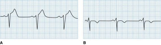
FIGURE 8.10 ST-segment elevation in a patient with acute pericarditis. A. Lead V3 shows the STsegment elevation of acute pericarditis. B. Several days later, the same lead shows that the ST segments have returned to baseline and the T waves have inverted. There are no Q waves.
Reprinted with permission from Kline-Tilford AM, Haut C. Lippincott Certification Review: Pediatric Acute Care Nurse Practitioner. 1st Ed. Philadelphia: Wolters Kluwer, 2016.
322
VI. Dysrhythmias
The most common dysrhythmias of childhood include supraventricular tachycardia (SVT), heart block, and long QT syndrome.
A.SVT
1.Definition. SVT is an abnormally accelerated heart rhythm that originates proximal to the bifurcation of the bundle of His. SVT is the most common dysrhythmia in childhood. It is necessary to distinguish SVT from sinus tachycardia, as detailed in Table 8-7.
2.Pathophysiology. Two types of SVT occur.
a.Atrioventricular reentrant tachycardia (AVRT). Retrograde conduction through an accessory pathway leads to SVT.
b.Atrioventricular node reentrant tachycardia (AVNRT). The conduction abnormality occurs in different pathways within the AV node itself.
c.When anterograde conduction occurs through a bypass tract between the atria and ventricles, Wolff–Parkinson–White (WPW) syndrome is present. WPW is associated with sudden cardiac death.
3.Clinical features. Symptoms include palpitations, chest pain, dyspnea, and sometimes altered level of consciousness. Prolonged SVT may lead to signs and symptoms of CHF, especially in the neonate.
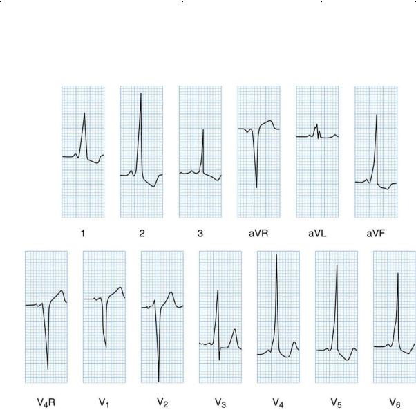
4.Diagnosis. WPW may be identified on ECG by the presence of a delta wave (i.e., slurred upslope of the QRS complex with a short PR interval; Figure 8-11).
5.Management
a.Vagal maneuvers, such as Valsalva, placement of an ice pack to the face, unilateral carotid massage, placing the child upside down, and orbital pressure in an older child may all convert SVT into a sinus rhythm.
b.Intravenous adenosine is the primary medication used for acute conversion to a sinus rhythm. Other medications used acutely include propranolol, digoxin, procainamide, and amiodarone.
c.Synchronized cardioversion may be used in patients who are hemodynamically unstable.
d.Chronic medical management typically includes ß-adrenergic blockers.
e.Patients with WPW should undergo an exercise stress test for sudden cardiac death risk stratification. If the delta wave disappears during exercise, the pathway is considered low risk. If the delta wave persists during exercise, patients should be referred to a cardiac electrophysiologist for possible ablation.
f.Radiofrequency catheter ablation may be used to remove the accessory pathway in chronic SVT or in high-risk WPW.
B.Heart block or AV block
1.Definition and classification
a.Definition. Heart block is delayed or interrupted conduction of sinus or atrial impulses to the ventricles.
b.Classification. Heart block is classified on the basis of ECG findings (e.g., first, second, or third degree) and by the ratio of atrial to ventricular impulses (e.g., 1:1, 3:1, 3:2, and so on).
1.First-degree AV block is prolongation of the PR interval.
2.Second-degree AV block
a.Type I, also known as Wenckebach, is progressive prolongation of the PR interval leading to failed AV conduction.
323

b.Type II is abrupt failure of AV conduction without progressive prolongation of the PR interval.
3.Third-degree AV block is a complete block, with no conduction of atrial impulses to the ventricles.
2.Etiology
a.Congenital third-degree AV block is associated with children born to mothers with SLE.
b.Postsurgical AV block may occur as a result of cardiac surgery, especially after closure of a VSD that lies close to the conduction system.
c.Bacterial endocarditis may be associated with AV block.
3.Clinical features
a.First-degree AV block is typically asymptomatic.
b.Secondand third-degree AV block can have clinical features ranging from being asymptomatic to developing fatigue, dizziness or syncope, and rarely, sudden death.
4.Management
a.No treatment is indicated for first-degree AV block.
b.No treatment is indicated for type I second-degree AV block.
c.Cardiac pacemakers are indicated in type II second-degree AV block and symptomatic third-degree AV block.
C.Long QT syndrome
1.Definition. Long QT syndrome is prolongation of the QT interval. Prolongation increases the risk of lethal ventricular arrhythmias known as torsades de pointes.
2.Etiology
a.In 50% of cases, inheritance is either autosomal recessive (i.e., Jervell–Lange- Nielsen syndrome, associated with congenital deafness) or autosomal dominant (i.e., Romano–Ward syndrome, not associated with deafness).
b.Some cases may result from the use of drugs, which may directly, or in combination, prolong the QT interval (e.g., antifungals, phenothiazines, tricyclic antidepressants, erythromycin, terfenadine).
c.The remainder of cases are likely due to deletions in genes that are normally responsible for repolarization.
3.Clinical features. Presenting signs and symptoms include syncope (most common), seizure, palpitations, or sudden cardiac arrest. Exercise and emotion are often inciting factors.
4.Diagnosis. The corrected QT interval (QTc) is calculated by taking the measurement of the QT interval divided by the square root of the previous RR interval. Diagnosis of long QT is made on the basis of ECG showing a QTc greater than 0.44 seconds (up to
0.49seconds may be normal in the first 6 months of life).
5.Management. Treatment of symptomatic patients includes a β-adrenergic blocker to reduce symptoms, although the QT interval usually remains prolonged. Treatment of asymptomatic individuals is controversial. Other therapeutic modalities include cardiac pacing, left stellate ganglionectomy, and an AICD.
Table 8-7
Features Differentiating Sinus Tachycardia from Supraventricular Tachycardia
Feature |
Sinus Tachycardia |
Supraventricular |
|
Tachycardia |
|||
|
|
||
Rate (beats/minute) |
<230 in newborns |
Frequently >250 |
|
<210 in children |
|
|
|
Heart rate variation |
Present |
Absent |
324
P waves on ECG |
Normal (axis is 0°–90°) |
Absent or abnormal axis |
Predisposing factors |
Fever, infection, anemia, and |
None |
|
hyperthyroidism |
|
Response to intervention (e.g., adenosine, |
Gradual |
Rapid |
antipyretics, fluids) |
|
|
ECG = electrocardiogram. |
|
|
FIGURE 8.11 Electrocardiogram in a patient with Wolff–Parkinson–White syndrome. Note the wide QRS complex, the presence of a delta wave (slurred upstroke of R), and the short PR interval.
Reprinted with permission from Fleisher GR, Ludwig S, Bachur RG. Textbook of Pediatric Emergency Medicine. 6th Ed. Philadelphia: Lippincott Williams & Wilkins, 2010.
325
VII. Chest Pain
Chest pain is a common presenting complaint in children and adolescents. However, this symptom is rarely of cardiac origin. Figure 8-12 schematically presents the differential diagnosis of chest pain.
A.Cardiac chest pain
1.Pericarditis is the most common cause [see section V.D].
2.Chest pain or chest pressure that occurs with exercise or is associated with syncope, shortness of breath, or abnormal heart rhythm should be investigated further by a cardiologist. Appropriate testing (e.g., continuous ECG monitoring, echocardiography, exercise stress test) is necessary.
B.Noncardiac chest pain. Common causes include asthma, esophagitis, and costochondritis (tenderness in one or more costochondral joints).
326

FIGURE 8.12 Differential diagnosis of chest pain in children and adolescents.
327