


Acknowledgments
We would like to recognize the individuals who have made tremendous impacts on our medical education and on our professional growth and development.
I have been so privileged to learn from outstanding teachers and mentors at UCLA School of Medicine, Cedars-Sinai Medical Center, and Stanford School of Medicine. Lee Miller, one of the coeditors of this book, has influenced my career immeasurably as one of the finest teachers I have known. I would not have discovered my love for teaching without his mentorship and encouragement. I am also immensely grateful to my colleagues and friends at the Palo Alto Medical Foundation who, for the past 14 years, have supported me and helped me to grow as a clinician, leader, and educator.
Lloyd J. Brown
In health and education fields, we are constantly striving to be idealized versions of ourselves— better than we truly are. From exposure to those who are genuinely better than me, I have received invaluable education, encouragement, and perpetual inspiration. A partial list of those to whom I am indebted includes Drs. Lee Miller, Kate Perkins, Paul Chung, Alice Kuo, Tom Klitzner, and Ellen Wald. I hope to live up to all of the support that they, and others, have given to me.
Ryan J. Coller
I will forever be grateful for the support, mentorship, and encouragement by the late Dr. David Rimoin, a wonderful friend and leader who touched so many learners and patients. I also cannot thank enough some of my finest teachers and role models during my training, Dr. Leigh Grossman, Dr. Dick Kesler, and Dr. Frank Saulsbury of the University of Virginia School of Medicine, whose impact has been immense. I am so grateful for my incredible partners on this project, Dr. Lloyd Brown and Dr. Ryan Coller, both phenomenal educators and role models who have taught me so much over the years. Finally, my pediatrician colleagues and dear friends at UCLA, Dr. Kate Perkins and Dr. Deb Lehman, whose partnership and friendship I will always cherish.
Lee T. Miller
We also owe so much to our present and past pediatric and medicine–pediatric residents with whom we feel very close, and to the thousands of medical students from all across the country, but most especially to those from Stanford University, from the University of Wisconsin School of Medicine and Public Health, and from the David Geffen School of Medicine at UCLA, who have stimulated us, energized us, and taught us so much along the way.
Lloyd J. Brown
Ryan J. Coller
Lee T. Miller
14

Contributors
Lloyd J. Brown, MD, FAAP, Regional Chair of Pediatrics, Palo Alto Division, Palo Alto Medical Foundation, Palo Alto, California, Adjunct Clinical Associate Professor of Pediatrics, Stanford School of Medicine, Stanford, California
Ryan J. Coller, MD, MPH, FAAP, Chief, Division of Pediatric Hospital Medicine, Assistant Professor of Pediatrics, University of Wisconsin School of Medicine and Public Health, Madison, Wisconsin
Lee Todd Miller, MD, FAAP, Associate Dean for Student Affairs, Professor of Pediatrics, Director, Global Health Education Programs, David Geffen School of Medicine at UCLA, Los Angeles, California
Dorsey Bass, MD, FAAP, Co-Director, Stanford Children’s Inflammatory Bowel Disease Center, Associate Professor of Pediatrics, Stanford University, Stanford, California
Robert M. Bernstein, MD, Medical Director, The Cedars-Sinai Orthopaedic Center, Executive Vice Chair, Department of Orthopaedics, Cedars-Sinai Medical Center, Professor of Orthopaedic Surgery, Los Angeles, California
Carol Diamond, MD, Director of Pediatric Hematology/Coagulation, Associate Professor of Pediatrics, University of Wisconsin School of Medicine and Public Health, Madison, Wisconsin
David A. Ferry, MD, FACC, Managing Partner, Pediatric Cardiology Medical Associates of Southern California, Encino, California, Attending Pediatric Cardiologist, Children’s Hospital Los Angeles, Los Angeles, California
Amy E. Gilliam, MD, FAAD, Pediatric Dermatology, Palo Alto Medical Foundation, Volunteer Assistant Clinical Professor of Pediatrics and Dermatology, UCSF School of Medicine, San Francisco, California
Heather Hindo, MD, FAAP, Pediatrician, Asian Pacific Healthcare Venture, Inc., Los Angeles, California
Rajashri Shuba Iyengar, MD, MPH, FAAP, FAAAI, Division of Allergy and Immunology, Palo Alto Medical Foundation, Palo Alto, California
Elaine S. Kamil, MD, Pediatric Nephrologist at Cedars-Sinai Medical Center and Harbor-UCLA Medical Center, Health Sciences Clinical Professor of Pediatrics, David Geffen School of Medicine at UCLA, Los Angeles, California
James Heaysung Lee, MD, FAAP, Associate Program Director, Pediatrics, Assistant Clinical Professor of Pediatrics, David Geffen School of Medicine at UCLA, Los Angeles, California
Deborah Lehman, MD, FAAP, Professor of Pediatrics, Assistant Dean for Student Affairs, David Geffen School of Medicine at UCLA, Los Angeles, California
Jason T. Lerner, MD, FAES, Associate Chief of Clinical Pediatric Neurology, Associate Professor of Pediatrics, David Geffen School of Medicine at UCLA, Los Angeles, California
Calvin G. Lowe, MD, Attending Physician, Division of Emergency and Transport Medicine, Children’s Hospital Los Angeles, Los Angeles, California, Assistant Professor of Pediatrics, Keck School of Medicine at the University of Southern California, Los Angeles, California
David J. McCulley, MD, Division of Neonatology, Assistant Professor of Pediatrics, University of Wisconsin School of Medicine and Public Health, Madison, Wisconsin
Omondi L. Nyong’o, MD, Associate Principal Investigator, Palo Alto Medical Foundation Research Institute, Associate Clinical Professor, Department of Ophthalmology, UCSF School of Medicine, San Francisco, California
Kathy L. Perkins, MD, PhD, FAAP, Associate Dean for Graduate Medical Education, Professor of Pediatrics, David Geffen School of Medicine at UCLA, Los Angeles, California
Leigh Christopher Reardon, MD, FACC, Director, Pediatric Mechanical Circulatory Support and Transitional Cardiac Care, Assistant Professor of Medicine and Pediatrics, David Geffen School of Medicine at UCLA, Los Angeles, California
Srinath Sanda, MD, Assistant Adjunct Professor of Pediatrics, Division of Endocrinology, Department of Pediatrics, UCSF School of Medicine, San Francisco, California
Monica Sifuentes, MD, Professor of Pediatrics, David Geffen School of Medicine at UCLA, Associate Residency Program
15
Director, Pediatric Clerkship Director, Department of Pediatrics, Harbor-UCLA Medical Center, Torrance, California
Grant D. Syverson, MD, Pediatric Rheumatology, Assistant Clinical Professor of Pediatrics, Sanford Children’s Hospital, University of North Dakota School of Medicine, Fargo, North Dakota
Brian G. Tang, MD, FAAP, Developmental-Behavioral Pediatrics, Palo Alto Medical Foundation, Adjunct Clinical Assistant Professor of Pediatrics, Stanford School of Medicine, Stanford, California
Jessica Tenney, MD, FAAP, FACMG, Division of Medical Genetics, Assistant Professor of Pediatrics, UCSF School of Medicine, San Francisco, California
Rujuta B. Wilson, MD, Associate Program Director, Pediatric Neurology, Assistant Professor of Pediatric Neurology and Psychiatry, David Geffen School of Medicine at UCLA, Los Angeles, California
Lauren J. Witcoff, MD, Pediatric Pulmonologist, Past Clinical Associate Professor of Pediatrics, Stanford School of Medicine, Stanford, California
Derek Wong, MD, FAAP, FACMG, Division of Medical Genetics, Associate Professor of Pediatrics, David Geffen School of Medicine at UCLA, Los Angeles, California
16

Contributors to the Previous Edition
Lloyd J. Brown, MD, FAAP, Regional Chair of Pediatrics, Palo Alto Division, Palo Alto Medical Foundation, Palo Alto, California, Adjunct Clinical Associate Professor of Pediatrics, Stanford School of Medicine, Stanford, California
Lee Todd Miller, MD, FAAP, Associate Dean for Student Affairs, Professor of Pediatrics, Director, Global Health Education Programs, David Geffen School of Medicine at UCLA, Los Angeles, California
Tiffany Merrill Becker, MD, Division of Academic Primary Care Pediatrics, Cedars-Sinai Medical Center, Los Angeles, California
Peter A. Blasco, MD, FAAP, Director, Neurodevelopmental Program, Child Development and Rehabilitation Center, Portland, Oregon, Associate Professor of Pediatrics, Oregon Health and Science University, Portland, Oregon
David Seung Chun, MD, Fellow, Pediatric Cardiology, James Whitcomb Riley Hospital for Children, Indiana University School of Medicine, Indianapolis, Indiana
David A. Ferry, MD, FACC, Managing Partner, Pediatric Cardiology Medical Associates of Southern California, Encino, California, Attending Pediatric Cardiologist, Children’s Hospital Los Angeles, Los Angeles, California
Carole Hurvitz, MD, FAAP, Director, Pediatric Hematology-Oncology, Cedars-Sinai Medical Center, Los Angeles, California, Professor of Pediatrics, David Geffen School of Medicine at UCLA, Los Angeles, California
Elaine S. Kamil, MD, Pediatric Nephrologist at Cedars-Sinai Medical Center and Harbor-UCLA Medical Center, Health Sciences Clinical Professor of Pediatrics, David Geffen School of Medicine at UCLA, Los Angeles, California
Deborah Lehman, MD, FAAP, Professor of Clinical Pediatrics, Assistant Dean for Student Affairs, David Geffen School of Medicine at UCLA, Los Angeles, California
Calvin G. Lowe, MD, Attending Physician, Division of Emergency and Transport Medicine, Children’s Hospital Los Angeles, Los Angeles, California, Assistant Professor of Pediatrics, Keck School of Medicine at the University of Southern California, Los Angeles, California
Elizabeth Mumper, MD, FAAP, President and CEO, Advocates for Children, Lynchburg, Virginia, Associate Professor of Clinical Pediatrics, University of Virginia School of Medicine, Charlottesville, Virginia
Ronald A. Nagel, MD, FAAP, Clinical Assistant Professor of Pediatrics, David Geffen School of Medicine at UCLA, Los Angeles, California
Charles E. Niesen, MD, FRCPC, Division of Pediatric Neurology, Cedars-Sinai Medical Center, Los Angeles, California, Assistant Professor of Pediatrics and Neurology, David Geffen School of Medicine at UCLA, Los Angeles, California
Kathy L. Perkins, MD, PhD, FAAP, Associate Dean for Graduate Medical Education, Professor of Pediatrics, David Geffen School of Medicine at UCLA, Los Angeles, California
Robert K. Rhee, MD, Pediatric Ophthalmologist, Departments of Pediatrics and Surgery, Associate Professor of Ophthalmology, Medical College of Ohio, Toledo, Ohio
Marta R. Rogido, MD, FAAP, Division of Neonatal-Perinatal Medicine, Assistant Professor of Pediatrics, Emory University School of Medicine, Atlanta, Georgia
Annette B. Salinger, MD, FAAP, Palo Alto Medical Foundation, Palo Alto, California, Clinical Instructor, Stanford University School of Medicine, Stanford, California
Srinath Sanda, MD, Assistant Adjunct Professor of Pediatrics, Division of Endocrinology, Department of Pediatrics, UCSF School of Medicine, San Francisco, California
Margaret Sanford, MD, Associate Pediatric Hematologist-Oncologist, Cedars-Sinai Medical Center, Los Angeles, California, Clinical Assistant Professor of Pediatrics, David Geffen School of Medicine at UCLA, Los Angeles, California
Harry W. Saperstein, MD, Director, Pediatric Dermatology, Cedars-Sinai Medical Center, Los Angeles, California, Clinical Associate Professor of Pediatrics and Medicine, David Geffen School of Medicine at UCLA, Los Angeles, California
17
Frank T. Saulsbury, MD, Head, Division of Immunology and Rheumatology, Department of Pediatrics, University of Virginia Health Systems, Charlottesville, Virginia, Professor of Pediatrics, University of Virginia School of Medicine, Charlottesville, Virginia
Nirali P. Singh, MD, FAAP, Fellow, Pediatric Infectious Diseases, Cedars-Sinai Medical Center, Los Angeles, California
Liliana Sloninsky, MD, Associate Pediatric Hematologist-Oncologist, Cedars-Sinai Medical Center, Los Angeles, California, Clinical Assistant Professor of Pediatrics, David Geffen School of Medicine at UCLA, Los Angeles, California
Augusto Sola, MD, FAAP, Director, Neonatal-Perinatal Medicine, Professor of Pediatrics and Obstetrics and Gynecology, Emory University School of Medicine, Atlanta, Georgia
Jerome K. Wang, MD, FAAP, Associate Director, Internal Medicine–Pediatrics Residency Training Program, Cedars-Sinai Medical Center, Los Angeles, California, Assistant Professor of Pediatrics, David Geffen School of Medicine at UCLA, Los Angeles, California
Frederick D. Watanabe, MD, FAAP, Pediatric Liver and Intestinal Transplantation, Children’s Hospital Los Angeles, Los Angeles, California, Associate Professor of Pediatrics, David Geffen School of Medicine at UCLA, Los Angeles, California
Lauren J. Witcoff, MD, Pediatric Pulmonologist, Past Clinical Associate Professor of Pediatrics, Stanford School of Medicine, Stanford, California
Kenneth W. Wright, MD, FAAP, Director, Wright Foundation for Pediatric Ophthalmology, Director, Pediatric Ophthalmology Research and Education, Cedars-Sinai Medical Center, Los Angeles, California, Clinical Professor of Ophthalmology, Keck School of Medicine at the University of Southern California, Los Angeles, California
Sharon L. Young, MD, Associate Medical Director, Children’s Health Clinic, Cedars-Sinai Medical Center, Los Angeles, California, Assistant Professor of Pediatrics, David Geffen School of Medicine at UCLA, Los Angeles, California
18

C H A P T E R 1
19

Pediatric Health Supervision
Kathy L. Perkins, James Heaysung Lee
20
I.Well-Child Care—General Concepts
A.The purpose of routine well-child care is to address the longitudinal health care needs of children from birth through adolescence. This is ideally achieved through a family-centered medical home model where the pediatric care team works in partnership with the family to ensure that all health-related needs are met. Features of well-child care include the following:
1.Age-specific health supervision according to recommended periodicity schedules
2.Disease detection through surveillance and screening:
a.Assessment of growth and development
b.Screening tests to detect asymptomatic diseases (e.g., vision, hearing, newborn metabolic screening, anemia, and lead screening)
c.Developmental screening
3.Disease prevention
a.Primary prevention
1.Sudden infant death syndrome, or SIDS, prevention (“back to sleep”), for example
2.Immunizations
b.Secondary prevention (e.g., care coordination for children with special health care needs)
B.The content of the well-child visit includes
1.Eliciting parental and patient concerns
2.History provides an opportunity to obtain diagnostic information and to form a doctor– patient–family alliance. The interview is shaped by family and patient concerns and by age-specific trigger questions about common problems (e.g., sleep, nutrition, behavior).
3.Developmental surveillance is gathered through age-specific questioning, developmental questionnaires, observations during the visit, screening tests, and a review of academic school performance.
4.Observation of parent–child interaction
5.Physical examination should be comprehensive and also should focus on growth (i.e., length/height, weight, head circumference, and body mass index).
6.Additional screening tests depend on the age of the child and may include lead level, hemoglobin, lipid levels, blood pressure, and hearing and vision assessment.
7.Immunizations
8.Anticipatory guidance includes a discussion of safety issues and upcoming developmental issues.
21
II.Growth
A.Normal growth
1.Weight, height, head circumference (until 2 years of age), body mass index (starting at 2 years of age), and sexual maturity are monitored routinely during well-child care to assess for adequacy of growth and development.
2.Standardized growth curves, produced by the World Health Organization (WHO) and Centers for Disease Control and Prevention (CDC), reflect average values for age for 95% of children and are used to plot weight, height, body mass index, and head circumference over time.
3.Tables 1-1, 1-2, and 1-3 detail general “rules of thumb” for expected gains in weight, height, and head circumference. Sexual maturity rating scales are found in Chapter 3,
Figures 3-1, 3-2, and 3-3.
B.Growth disturbances are defined as growth outside of the usual pattern. Three common types of growth disturbance include failure to thrive (FTT), short stature, and head growth abnormalities. Short stature is discussed in Chapter 6, section I.
1.FTT
a.Definition. Although there are multiple definitions, FTT is a term most commonly used to describe a growth rate less than expected for a child, and is of particular concern when a child’s weight crosses two major percentile isobars on a growth chart.
b.FTT may involve all growth parameters, although weight gain is generally the most affected. FTT should be distinguished from isolated short stature, in which height is the most abnormal growth parameter (see Chapter 6, section I).
c.In children with FTT, weight is typically affected before linear growth, which is usually affected before head circumference growth. Head circumference is initially spared in FTT.
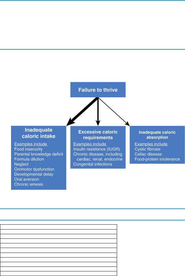
d.Classification. FTT was previously categorized as either nonorganic (due to psychosocial factors) or organic (due to underlying organ system pathology). This classification is no longer recommended because these categories coexist in many cases. A more appropriate categorization of FTT describes whether there is inadequate caloric intake, inadequate caloric absorption, inadequate utilization of nutrients, or excessive metabolic demand. The most common etiology of FTT is inadequate intake of calories, which may result from a disturbed parent–child bond. The differential diagnosis of FTT is outlined in Figure 1-1.
e.Evaluation of FTT requires a careful history and physical examination, a complete dietary history, and observation of the parent–child interaction. No laboratory tests are routinely indicated in the initial evaluation of FTT. Laboratory tests should be reserved for cases where the history or physical examination suggests a specific etiology for which laboratory testing will be helpful or for the child who fails to respond to nutritional interventions.
2.Head growth abnormalities include abnormalities in size (microcephaly/macrocephaly) or shape (deformational plagiocephaly/craniosynostosis).
a.General concepts. Almost all head growth occurs prenatally and during the first 2 years of life.
1.Head circumference at birth is 25% of the normal adult head size, and it increases to 75% of the normal adult head size by 1 year of age.
2.Note that caput succedaneum (scalp edema) and cephalohematoma
(subperiosteal hemorrhage of the newborn cranium after a traumatic delivery)
22
may interfere with accurate head circumference measurement in newborns.
b.Microcephaly
1.Definition. Head circumference is 2–3 standard deviations below the mean for age.
2.Incidence is 1–2/1000 children.
3.Etiologies are classified as either congenital or acquired (Table 1-4).
a.Congenital microcephaly is associated with abnormal induction and migration of the brain tissue.
b.Acquired microcephaly is caused by a cerebral insult in the late third trimester, perinatal period, or first year of life. Affected children are born with a normal head circumference that does not grow after the cerebral insult.
4.Clinical features
a.Because head size generally reflects brain size, microcephaly is always associated with a small brain.
b.Microcephaly is usually associated with developmental delay and intellectual impairment.
c.Microcephaly may be associated with cerebral palsy or seizures.
c.Macrocephaly
1.Definition. Head circumference > 95% for age.
2.Unlike in microcephaly, the size of the head in a patient with macrocephaly does not necessarily reflect brain size.
3.Etiologies
a.Benign familial macrocephaly, associated with an otherwise normal physical examination and a family history of large heads
b.Overgrowth syndromes (e.g., Sotos syndrome), in which all growth parameters are enlarged
c.Metabolic storage disorders (e.g., Canavan syndrome, gangliosidoses)
d.Neurofibromatosis (see Chapter 19, Table 19-3)
e.Achondroplasia (see Chapter 5, section IV.H.1)
f.Hydrocephalus (see Chapter 12, section II)
g.Space-occupying lesions (e.g., cysts, tumors)
4.Evaluation includes measurement of parental head circumferences, and a careful physical examination that includes observation for split cranial sutures, bulging anterior fontanelle, irritability, or vomiting, all of which may suggest elevated intracranial pressure. Head ultrasound or computed tomography (CT) scan is performed to rule out hydrocephalus, if suggested by physical examination. Genetic evaluation may be useful if a genetic syndrome is suspected.
d.Craniosynostosis
1.Definition. Premature closure of one or more of the cranial sutures (Figure 1- 2).
2.Etiology is often unknown. 80–90% of cases are sporadic and 10–20% are familial or part of a genetic syndrome (e.g., Crouzon and Apert syndromes). Known risk factors include intrauterine constraint or crowding, and metabolic abnormalities including hyperthyroidism and hypercalcemia.
3.Clinical features. Head shape in craniosynostosis is determined by which suture closes prematurely, because bone growth perpendicular to the affected suture is arrested.
a.Premature closure of the sagittal suture results in an elongated skull
23

(termed dolichocephaly or scaphocephaly) and is the most common form of craniosynostosis (Figure 1-3).
b.Premature closure of the coronal suture results in a shortened skull (termed brachycephaly). This is more common in boys and may be associated with neurologic complications such as optic nerve atrophy.
c.Premature closure of the metopic suture leads to a triangular-shaped head and prominent longitudinal central ridge on the forehead (termed trigonocephaly).
d.Premature closure of multiple sutures is rare, and is associated with severe neurologic compromise.
4.Diagnosis is made by physical examination of the head. Craniosynostosis is usually noted by 6 months of age. The diagnosis may be confirmed by skull radiographs and head CT scan.
5.Management is surgical repair, most often indicated when cosmetic concerns are significant.
e.Plagiocephaly
1.Definition. Asymmetry of the infant head shape not associated with premature suture closure
2.Clinical features. The most common type of plagiocephaly is positional plagiocephaly (Figure 1-4), associated with flattening of the occiput and prominence of the ipsilateral frontal area. Viewed from above, the skull has a parallelogram shape with the back of the head, ear, and forehead of one side displaced anteriorly.
a.May be associated with congenital muscular torticollis (see Chapter 17, section II.A.1), in which fetal head constraint during the third trimester can result in both trauma to the sternocleidomastoid and deformations of the skull.
b.Incidence has increased as a result of recommendations that infants sleep on their backs to decrease the risk of SIDS.
c.Management may include range of motion exercises for associated torticollis, repositioning the head during sleep, and increased time in the prone position when awake (“tummy time”). Cranial remodeling helmet therapy was a common treatment, but the American Academy of Pediatrics (AAP) updated its position in 2011 stating that helmet therapy was unnecessary for most babies with plagiocephaly.
Table 1-1
Rules of Thumb for Expected Increase in Weight
Age |
Expected Weight Increase |
Birth–2 weeks (average birth weight is 3.5 kg) |
Lose up to 10% of birth weight, usually in the first week of life |
|
Regain birth weight by 2 weeks of age |
2 weeks–3 months |
30 g/day |
3–6 months |
20 g/day |
|
Double birth weight by 4–6 months of age |
6–12 months |
10 g/day |
|
Triple birth weight by 12 months of age |
1–2 years |
250 g/month or 3 kg/year |
|
Quadruple birth weight by 2 years of age |
2 years–puberty |
2 kg/year |
30 g = 1 oz body weight.
Table 1-2
Rules of Thumb for Expected Increase in Height
24
Age |
Expected Height Increase |
|
0–12 months (average birth length is 50 cm) |
25 cm/year |
|
|
Birth length increases by 50% at 12 months of age |
|
13–24 months |
12.5 cm/year |
|
2 years–adolescence |
6.25 cm/year |
|
|
Birth length doubles by age 4 years |
|
|
Birth length triples by age 13 years |
|
Table 1-3
Rules of Thumb for Expected Increase in Head Circumference
Age |
Expected Head Circumference Increase |
0–2 months (average birth head circumference is 35 cm) |
0.5 cm/week |
2–6 months |
0.25 cm/week |
By 12 months |
Total increase = 12 cm since birth |
FIGURE 1.1 Differential diagnosis of failure to thrive (FTT). Inadequate caloric intake is the most common cause of FTT, followed by excessive caloric requirements. Inadequate caloric absorption is the least common cause of FTT. IUGR = intrauterine growth restriction.
Table 1-4
Causes of Microcephaly
Congenital
Early prenatal infection (e.g., HIV infection, TORCH)
Maternal exposure to drugs and toxins (e.g., fetal alcohol syndrome)
Chromosomal abnormality (e.g., trisomy 13, 18, or 21)
Familial microcephaly (autosomal dominant or autosomal recessive inheritance)
Maternal phenylketonuria
Acquired
Late third trimester or perinatal infections
Meningitis or meningoencephalitis during first year of life
Hypoxic or ischemic cerebral insult
Metabolic derangements (e.g., hypothyroidism, inborn errors of metabolism)
TORCH = toxoplasmosis, other (syphilis), rubella, cytomegalovirus, herpes simplex virus; HIV = human immunodeficiency virus.
25
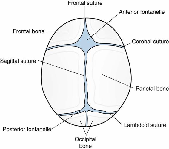
FIGURE 1.2 Cranial sutures in the neonatal skull.
Reprinted with permission from Pillitteri A. Maternal and Child Nursing. 7th Ed. Philadelphia: Lippincott Williams & Wilkins, 2013.
26

FIGURE 1.3 Types of craniosynostosis.
Reprinted with permission from Mongan P, Soriano SG, Sloan TB, Gravlee GP. Practical Approach to Neuroanesthesia. Philadelphia: Lippincott Williams & Wilkins, 2013.
27

FIGURE 1.4 Infant with positional plagiocephaly.
Reprinted with permission from Kyle T, Carman S. Essentials of Pediatric Nursing. 2nd Ed. Philadelphia: Lippincott Williams & Wilkins, 2013.
28
III.Immunizations
A.Immunizations are among the most important components of well-child care and disease prevention. Recommendations for routine immunizations are updated annually. For the most up-to- date recommendations on the childhood immunization schedule, please visit www.cdc.gov.
B.Types of immunizations
1.Active immunization involves induction of long-term immunity through exposure to live attenuated or killed (inactivated) infectious agents.
a.Live vaccines are more likely to induce long-lasting immunity, but carry the risk of vaccine-associated disease in the recipient, or in a secondary host. As a result, live vaccines should generally be avoided in patients with compromised immunity
(e.g., cancer, congenital or drug-induced immunodeficiencies, acquired immune deficiency syndrome). Examples of live vaccines include intranasal influenza; varicella; and measles, mumps, and rubella (MMR) vaccines.
b.Nonlive vaccines are not infectious and tend to induce immunity for shorter periods, thus requiring booster immunizations. Examples include diphtheria, tetanus, and acellular pertussis (DTaP); hepatitis A (Hep A) vaccine and hepatitis B vaccine (HBV); inactivated polio vaccine (IPV); Haemophilus influenzae type b (HIB); inactivated influenza; pneumococcal and meningococcal vaccines.
2.Passive immunization involves delivery of preformed antibodies to individuals who have no active immunity against a particular disease, but have either been exposed to or are at high risk for exposure to the infectious agent. Examples include the following:
a.Varicella zoster immune globulin (VZIG) for immunocompromised patients who have been exposed to varicella and are at high risk for severe varicella infection, including newborns born to mothers who develop chicken pox between 5 days before and 2 days after delivery.
b.Hepatitis B immune globulin, given to newborns born to hepatitis B–positive women
c.Hepatitis A immune globulin, given as “predeparture” passive immunization to some visitors to high risk areas
C.Specific immunizations
1.Hepatitis B vaccine (HBV)
a.Rationale for vaccine: 90% of affected infants will develop chronic infection.
240 million people are chronically infected worldwide, with up to 30 million new cases every year. More than 750,000 people die annually from hepatitis B and its complications.
b.Type of vaccine: inactivated
c.Timing of vaccination: HBV is given as a three-shot series within the first year of life.
2.DTaP
a.Rationale for vaccine: Diphtheria, tetanus, and pertussis all may cause serious disease, especially in young infants. Outbreaks of pertussis with significant mortality continue to occur in the United States.
b.Type of vaccine
1.Vaccine is inactivated.
2.DTP, which contained whole-cell, killed Bordetella pertussis, had a high rate of side effects and was replaced with DTaP (1996), a vaccine that contains purified components (acellular) of B. pertussis, with lower rates of vaccineassociated fever, seizures, and local reactions. DTaP is safer but has a waning
29
immunity to pertussis over time.
c.Timing of vaccination
1.DTaP is recommended at 2, 4, and 6 months of age with boosters at 12– 18 months and 4–6 years of age.
2.Tdap (has less diphtheria component compared with DTaP) is a booster immunization, offering continued protection from these diseases in the adolescent and adult age group. Tdap is recommended at age 11–12. After receiving at least one Tdap, the vaccine should be given every 10 years thereafter for continued protection. Note that Tdap rather than DTaP is given to children 7–10 years of age if they did not complete their series of DTaP by their seventh birthday.
3.Oral polio vaccine (OPV) and IPV
a.Rationale for vaccine: Poliovirus is an enterovirus with propensity for the central nervous system, which may cause transient or permanent paresis of the extremities and meningoencephalitis. Polio has been eradicated from the Western hemisphere and South Pacific, but remains in isolated pockets throughout the world.
b.There are two types of vaccines.
1.Live attenuated (OPV), administered orally
a.Advantages include ease of administration and induction of both host immunity and secondary immunity, because it is excreted in the stool of the recipient and may infect, and thus immunize, close contacts.
b.Disadvantages include the induction of vaccine-related polio, a rare complication. Because of the risk of vaccine-related polio, OPV is no longer used in the United States.
2.Nonlive or inactivated (IPV), administered subcutaneously or intramuscularly, does not cause vaccine-related polio, but also does not confer secondary immunity.
c.Timing of vaccination: In the United States, only IPV is recommended and is given at 2 and 4 months of age, with boosters at 6–18 months and 4–6 years of age.
4.Rotavirus vaccines
a.Rationale for vaccine: Rotavirus infections are the most common cause of severe acute gastroenteritis worldwide. Worldwide, each year approximately 2 million children are hospitalized and 450,000 children die from disease caused by rotavirus. Rotavirus is highly contagious and nearly every child is at risk of getting infected.
b.Type of vaccine: oral, live vaccine
c.Timing of vaccination: recommended at 2, 4, and 6 months of age
5.HIB vaccine
a.Rationale for vaccine: HIB was a serious cause of invasive bacterial infection, including meningitis, epiglottitis, pneumonia, and sepsis, before vaccine licensure in 1985. Since licensure, HIB has become a rare cause of such infections.
b.Type of vaccine: HIB vaccine is a conjugate vaccine (inactivated) with H. influenzae polysaccharide linked to various protein antigens, including diphtheria or tetanus toxoids, to augment immunogenicity.
c.Timing of vaccination: HIB vaccine is recommended at ages 2, 4, and 6 months with a booster at ages 12–15 months. After 15 months of age, the vaccine confers lasting immunity. Therefore, children who receive the HIB vaccine at 15 months of age or older do not require additional doses, no matter how many doses were given before 15 months of age.
6.MMR vaccine
30
a.Rationale for vaccine: MMR immunizes against three viral diseases. Especially important in areas where misinformation regarding the vaccine has led to decreased vaccination rates and unfortunate outbreaks of the following:
1.Measles is a severe illness with complications that include pneumonia associated with significant mortality. Classic signs include cough, coryza, conjunctivitis, and Koplik spots (an enanthem with white lesions on a reddened base on the inner cheek), followed by a classic rash (an exanthem described as morbilliform, starting on the face and moving caudally).
2.Mumps is most commonly associated with parotitis but may also cause meningoencephalitis and orchitis.
3.Rubella causes a mild viral syndrome in children, but may cause severe birth defects (commonly hearing loss and heart defects) in offspring of susceptible women who are infected during pregnancy.
b.Type of vaccine: live attenuated vaccine
c.Timing of vaccination: MMR vaccine is recommended at 12–15 months of age with a booster at 4–6 years of age. A special indication exists for infants 6–12 months of age traveling to countries where MMR is endemic. Because this dose is given before the recommended time of 12–15 months, it does not count toward the two-shot series and the child will subsequently need two additional doses at the appropriate times.
7.Varicella vaccine
a.Rationale for vaccine: Varicella is the virus responsible for chicken pox and zoster. Varicella often causes uncomplicated illness, but may cause severe disease in very young and in older or immunosuppressed patients.
b.Type of vaccine: live attenuated vaccine
c.Timing of vaccination: Vaccine is recommended at 12–18 months of age with a booster at 4–6 years of age. It is commonly given in conjunction with MMR vaccine.
8.Hep A vaccine
a.Rationale for vaccine: Hep A is the most common viral cause of hepatitis worldwide, although it is asymptomatic in up to 70% of infected children younger than 6 years. More severe disease is seen in older children and adults, although it is rarely associated with fulminant hepatitis.
b.Type of vaccine: inactivated
c.Timing of vaccination: Hep A vaccine is a two-dose vaccination series recommended for all children starting at 1 year of age. The booster should given at least 6 months later but ideally no later than 2 years of age. Children not fully vaccinated by 2 years of age should be vaccinated at subsequent visits.
9.Pneumococcal vaccines (PCV-13 and PPSV-23)
a.Rationale for vaccine: Pneumococcal (Streptococcus pneumoniae) disease causes more deaths in the United States each year than all other vaccine-preventable illnesses combined. It is a common cause of acute otitis media, pneumonia, and invasive bacterial infections (bacteremia and meningitis) in the pediatric population.
b.There are two types of vaccines. Both are inactivated.
1.PPSV23 is composed of polysaccharide capsular antigens from 23 pneumococcal serotypes.
a.Major advantage is that the vaccine contains antigens from pneumococcal strains causing almost all cases of bacteremia and meningitis during childhood.
b.Major disadvantage is that the vaccine has little immunogenicity in children younger than 2 years.
31
c.Indications. Vaccine is indicated in children with an immunocompromising condition and and for those with functional or anatomic asplenia. It is also indicated immunocompetent children with chronic heart disease, chronic lung disease, diabetes mellitus, cerebrospinal fluid (CSF) leak or cochlear implants.
d.Timing of vaccination: One dose should be given at 2 years of age and at least 8 weeks after the final dose of PCV13 (see below). Children with an immunocompromising condition or functional/anatomic asplenia should get a second dose 5 years after the first PPSV23.
2.PCV13 is composed of 13 pneumococcal serotypes. PCV13 includes the 13 serotypes that account for most invasive pneumococcal diseases.
a.Major advantages include immunogenicity and efficacy in preventing meningitis, pneumonia, bacteremia, and otitis media from the most common pneumococcal strains in children younger than 2 years.
b.Major disadvantage is that it does not confer as broad coverage against as many pneumococcal strains as PPSV-23.
c.Timing of vaccination: PCV-13 is recommended for all children at 2, 4, and 6 months of age, with a booster at 12–15 months of age. Healthy children who fall behind schedule should be caught up before their fifth birthday.
10.Influenza vaccines
a.Rationale for vaccine: Influenza can be a very serious illness that may result in hospitalizations and even death, regardless of a child’s medical history. About 20,000 children are hospitalized annually in the United States with influenza, and around 40% of children who die from influenza have no significant past medical history. Children under 5 years of age and those with conditions that predispose to complications of influenza are considered to be at high risk.
b.There are two types of vaccines, with recommendations for administration updated annually.
1.Live attenuated influenza vaccine (LAIV), administered intranasally
a.Advantages: no need for an injection.
b.Disadvantages: it is only available for children 2 years of age or older without contraindications to the vaccine. It can cause nasal congestion and rhinorrhea for several days.
2.Inactivated influenza vaccine (IIV), administered intramuscularly
a.Advantages: may be administered as early as at 6 months of age. There are no contraindications owing to underlying medical conditions, unless the child has a history of anaphylaxis to a previous influenza vaccination.
b.Disadvantages: requires injection
c.Timing of the vaccination: Every child aged 6 months and older should receive the influenza vaccination annually. Between 6 months and
8 years of age, first-time vaccine recipients need two doses of vaccine, spaced at least one month apart.
11.Human Papilloma Virus (HPV) vaccines
a.Rationale for vaccine: HPV is the most common sexually transmitted disease in the United States, affecting the majority of sexually active adults. There are about 150 different types of HPV, some of which cause significant health problems such as genital warts and certain cancers (e.g., cervical, vaginal, anal, penile, and oropharyngeal). Thus, HPV vaccine is one of the two cancer-preventive vaccinations, the other being Hepatitis B vaccine.
32
b.Type of vaccine: inactivated vaccine
c.Timing of vaccine: All children aged 11–12 years should receive the two-dose series of HPV, with the second dose administered 6 months after the first. If the first dose is not started until age 15 or older, three doses of vaccine are required. Young men may be vaccinated up to 21 years of age, and women may be vaccinated up to
26 years of age. Ideally, the series is initiated during the preteen years, to facilitate development of an immune response before the initiation of sexual activity.
12.Meningococcal vaccine (MCV)
a.Rationale for vaccine: Infection with Neisseria meningitidis can cause meningitis and sepsis, which is fatal in up to 15% of children. Up to 20% of survivors will develop permanent disabilities (e.g., loss of limbs, deafness, central nervous system damage). Rates of disease are highest in infants < 1 year of age, and in teens and young adults ages 16–23 years. Rates of meningococcal disease have been declining since 1990, which is thought to be a consequence of increased vaccination use.
b.Type of vaccine: inactivated vaccine
c.Timing of vaccine: All children aged 11–12 years should receive the quadrivalent vaccine to protect against serotypes A, C, W, and Y. A booster vaccination is given at 16–18 years of age. Children as young as 2 months old may receive the first dose of a four-dose meningococcal vaccine series if they are at high risk (e.g., anatomic or functional asplenia, complement deficiency, travel to hyperendemic or epidemic regions), with administration of doses recommended at 2, 4, 6, and 12–15 months of age. Vaccine for serotype B is recommended for children ≥ 10 years of age at increased risk of serotype B infection (e.g., community outbreak, functional/anatomic asplenia, and complement pathway abnormalities).
D.Adverse effects of immunization
1.Most vaccine side effects are mild to moderate in severity and occur within the first 24– 48 hours after administration (e.g., local inflammation and low-grade fever).
2.Because MMR and varicella vaccines are live attenuated vaccines, fever and rash may occur 1–2 weeks after immunization (i.e., after the incubation period of the virus).
3.Serious side effects, including those that are life-threatening or result in permanent disability, are rare (e.g., vaccine-related polio after OPV).
4.While there is no evidence that neither vaccines nor preservatives such as thimerosal are linked to developmental disabilities, as a precautionary measure the Food and Drug Administration (FDA) and the Centers for Disease Control and Prevention (CDC) eliminated thimerosal from vaccines in 2001. Of the vaccines currently recommended for children younger than 6 years, only some forms of influenza vaccine contain small amounts of thimerosal.
5.There is no evidence of a link between vaccines and autism. E. Contraindications and precautions to immunization
1.Contraindications to immunization include the following:
a.Anaphylaxis to a vaccine or its constituents
b.Encephalopathy within 7 days after DTaP/Tdap vaccine
c.Immunodeficient patients should not receive live vaccines including OPV, MMR, rotavirus, and varicella vaccines. Household contacts of immunodeficient patients should not receive OPV vaccine, as it is shed in the stool.
d.Patients with a history of intussusception should not receive rotavirus vaccine.
e.Pregnant patients should not receive live vaccines, including OPV, MMR, and varicella.
2.Precautions (i.e., caution should be exercised) to immunization include the following:
a.For all vaccines, moderate to severe illness (with or without fever). Note that mild
33
illnesses, including febrile illnesses, are not contraindications to immunization.
b.DTaP vaccine
1.Temperature of 40.5°C within 48 hours after prior vaccination
2.Collapse or shocklike state within 48 hours after prior vaccination
3.Seizures within 3 days after prior vaccination
4.Persistent, inconsolable crying lasting ≥3 hours occurring within 48 hours after prior vaccination
c.MMR and varicella vaccines: Intravenous Immunoglobulin (IVIG) administration within the preceding 11 months, which might interfere with the patient’s immune response to these live vaccines
34
IV. Well-Child Screening
A.The focus of each well-child visit is to identify undetected problems or the risks for such problems.
B.Screening assessments include a complete history and physical examination, growth measurements, blood pressure measurements, strabismus and vision screening, hearing screening, tuberculosis risk assessment, and laboratory screening.
C.Vision screening for ophthalmologic disorders begins at birth and is detailed in Chapter 18.
D.Hearing screening
1.Universal newborn hearing screening is recommended by 1 month of age, with confirmation of an abnormal screen by 3 months of age. Early intervention for hearing impairment is associated with better developmental language outcomes, and should be initiated no later than 6 months of age.
2.Two types of audiometric tests are used for the newborn.
a.Otoacoustic emission (OAE) measures sounds generated by normal cochlear hair cells that are detected by a microphone placed into the external auditory canal. This screening test is routinely performed in newborn nurseries before infant discharge. Administering the test takes only a few minutes and is painless, and the expenses for equipment and training are lower than for brainstem auditory-evoked response/auditory brainstem response (BAER/ABR) as below. OAE accuracy may be affected by debris or fluid within the external or middle ear.
b.BAER/ABR measures electroencephalographic (EEG) waves generated in response to clicks via electrodes placed on the infant’s scalp. Compared with OAE, BAER is a more accurate test, but it requires costly equipment, trained operators, and sometimes sedation. BAER is typically performed to confirm abnormalities found on OAE.
E.Neonatal metabolic (state) screening
1.Many metabolic diseases can be diagnosed and treated in the newborn period.
2.Although there is some variability among states as to which conditions are screened, all states screen for congenital hypothyroidism, phenylketonuria (PKU), and galactosemia. Each of these conditions is treatable, and if not detected early, each leads to irreversible brain injury.
3.Most states screen for sickle cell anemia and other hemoglobinopathies, because early intervention in a comprehensive treatment program significantly decreases morbidity and mortality.
4.Newborn screening tests for additional metabolic disorders (e.g., congenital adrenal hyperplasia) are performed in some states. The most extensive state screening panels screen for 50 disorders or more.
F.Cholesterol and lipid screening
1.Universal screening for hypercholesterolemia, regardless of body mass index (BMI) or risk factors, has been recommended by the National Heart, Lung and, Blood Institute (NHLBI) and endorsed by the American Academy of Pediatrics (AAP). Obtaining a nonfasting non-HDL cholesterol level is recommended for all children between 9 and
11 years of age, and again between 17 and 21 years of age. For children with an abnormal result on the screen, a fasting lipid profile should be obtained.
2.Targeted screening for hypercholesterolemia (consisting of fasting lipid profile performed twice within 2–12 weeks) is recommended for children between 2–8 years of age and 12–16 years of age with one of the following risk factors:
a.History of diabetes, hypertension, BMI > 95th percentile (if 2–8 years old) or
35
BMI > 85th percentile (if 12–16 years old), or smoking
b.History of one of the following moderateor high-risk medical conditions: nephrotic syndrome, human immunodeficiency virus (HIV), Kawasaki disease with history of coronary aneurysms, and end-stage renal disease
c.Parent with total cholesterol > 240 mg/dL or known dyslipidemia
d.Parent, grandparent, aunt/uncle, or sibling with myocardial infarction, angina, stroke, or coronary artery bypass graft/stent/angioplasty at <55 years in males and <65 years in females
3.Interventions for elevated cholesterol should include dietary and behavioral modifications, and may include referral to a registered dietician for nutrition counseling for the family.
G.Iron-deficiency anemia screening
1.Iron-deficiency anemia occurs most commonly in children < 6 years of age, peaking between 9 and 15 months of age, but may occur throughout the entire pediatric age range. More than 10% of 1 year olds are iron deficient.
2.Risk factors for iron-deficiency anemia include:
a.Prematurity
b.Low birth weight
c.Early introduction of cow’s milk (before 12 months of age) or consumption of 24 oz or more of milk a day
d.Insufficient dietary intake of iron
e.Low socioeconomic status
3.Universal screening of hemoglobin levels is recommended at 12 months of age. Subsequently, selective screening should be based on annual risk assessment.
H.Routine urinalysis screening is not recommended. Analysis of urine samples are recommended only when clinically indicated.
I.Tuberculosis screening
1.Screening for latent tuberculosis infections is recommended for children at risk for tuberculosis. Tuberculosis screening may be performed through the use of tuberculin skin testing with intradermal injection of purified protein derivative (PPD, Mantoux skin test). Alternatively, interferon-gamma release assays (IGRAs), which are in vitro tests of cell-mediated immune response, may be used in children older than 5 years to screen for latent tuberculosis. They are useful in the evaluation of children who have received the Bacillus Calmette–Guérin (BCG) vaccine. Screening is indicated for children with one or more of the following risk factors:
a.Birth place in a high-risk country other than United States, Canada, Australia, New Zealand, or Western and Northern European countries
b.Travel to an endemic region (e.g., Africa, South America, Asia, Eastern Europe) for a cumulative total of 1 week or more
c.Exposure to anyone with tuberculosis
d.Close contact with a person who has had a positive screen for latent tuberculosis
J.Lead screening
1.Lead intoxication (plumbism): No safe blood lead level (BLL) in children has been identified, and lead intoxication remains a public health risk. While the incidence and severity of lead poisoning in the United States are decreasing, in large part owing to effective screening programs and increased public awareness, there is still a high number of affected children living in deteriorating housing built before the 1970s.
2.Risk factors for lead intoxication include the following:
a.Ingestion of lead-containing paint from homes built before 1978
b.Drinking water from lead-containing pipes
36
c.Exposure to lead smelters, industrial manufacturing sites, or to adults with occupational exposures leading to contamination of clothes and hands
d.Use of lead-glazed pottery, imported toys, and imported candies (Mexico, Malaysia, China, and India) that contain tamarind, chili powder, or salt
e.Use of lead-containing folk remedies
f.Exposure to soil with high lead levels due to decades of burning leaded gasoline
3.Clinical features of lead intoxication
a.Children < age 6 are most susceptible to the effects of lead owing to a variety of mechanisms, including increased permeability of the blood–brain barrier to lead, higher rates of iron-deficiency anemia leading to increased lead absorption, and increased hand-to-mouth behaviors amongst infants and toddlers.
b.Acute lead intoxication may lead to anorexia, apathy, lethargy, anemia, irritability, and vomiting. These symptoms may progress to encephalopathy (see also Chapter 20, section VIII.D.4).
c.Chronic lead intoxication is most commonly asymptomatic; however, even patients with very low levels of lead (≥5 µg/dL) may suffer neurologic sequelae, including developmental delay, learning problems, and intellectual disability.
4.As the prevalence of childhood lead poisoning declines, there has been a movement to focus on primary prevention, and to transition from universal screening to targeted screening. AAP recommends that screening be performed in accordance with state laws when applicable. If no state screening program is in place, then universal screening at 1 and 2 years of age is recommended if the community has any of the following risk factors:
a.≥27% of the housing was built before 1950.
b.≥12% of children 12–36 months of age have BLLs ≥ 10 µg/dL.
c.Information on the prevalence of elevated BLLs in the community is unavailable or inadequate.
5.Intervention and management of lead poisoning is based on serum lead levels after repeat testing, and generally includes education to decrease exposure and chelation for very high lead levels.
37
V.Circumcision
A.Approximately 60% of male infants in the United States are circumcised.
B.Though the existing evidence is not sufficient to recommend routine circumcision, the AAP states that the health benefits of circumcision outweigh the risks, and these benefits justify access for families interested in circumcision.
1.Circumcision is associated with a significant reduction in risk of acquiring HIV and other sexually transmitted infections.
2.Circumcised patients have a lower risk of penile cancer, although this type of cancer is very rare in all males.
3.Urinary tract infections (UTIs) are 10 times more common in uncircumcised male infants, particularly in the first 3 months of life.
4.Uncircumcised males may ultimately require circumcision for the following conditions:
a.Phimosis is an inability to retract the foreskin. Physiologic phimosis usually resolves around 5–7 years of age but can be seen in older boys. Ballooning of the foreskin can be normal as long as the ballooning does not require manual compression by the patient. Signs of pathologic phimosis include dysuria, painful erection, bleeding from the preputial orifice, and recurrent balanoposthitis (inflammation of the glans penis and foreskin) and necessitate a referral for urologic evaluation.
b.Paraphimosis occurs when the retracted foreskin cannot be returned to its normal position and acts as a tourniquet, leading to edema from obstruction of venous and lymphatic flow. This is a urologic emergency because ischemia distal to the obstruction may occur. Surgery is required if attempts at manual reduction fail.
c.Balanitis is inflammation of the glans of the penis. It may be associated with Candida spp. or Gram-negative infections in infants, and with sexually transmitted infections.
C.Pharmacologic analgesia is strongly recommended during circumcision.
D.Complications from circumcision include bleeding, infection, poor cosmesis, phimosis (secondary to insufficient foreskin removal), urinary retention, and injury to the glans or urethra.
E.Contraindications to circumcision include anatomic penile abnormalities (e.g., hypospadias, penile torsion), because the foreskin may be required for the reconstruction, as well as medical concerns (e.g., prematurity, bleeding diatheses) associated with higher complication rates.
38
VI. Pediatric Dental Care
A.Counseling about dental care is an important component of anticipatory guidance.
B.Tooth eruption
1.Range for initial tooth eruption is between 3 and 16 months of age, with an average age of 6 months.
2.The first tooth is generally a lower central incisor.
3.Primary teeth (20 teeth in total) are generally established by 2–3 years of age.
4.Secondary (permanent) tooth eruption also begins with the lower central incisor, between 5 and 7 years of age, and generally finishes by 13–14 years of age. There are 32 secondary teeth.
5.Tooth eruption may be early or delayed.
a.Early dental eruption is defined as primary eruption before 3 months of age.
Causes include familial causes, hyperthyroidism, precocious puberty, and growth hormone excess.
b.Delayed dental eruption is defined as primary eruption that has not occurred by
18 months of age. Causes include familial delayed eruption, hypothyroidism, hypopituitarism, vitamin D deficiency, and genetic syndromes such as Down syndrome and ectodermal dysplasia (associated with conical-shaped teeth, dysmorphic facial features, decreased numbers of sweat glands, and alopecia).
C.Dental hygiene
1.Tooth brushing should begin as soon as teeth erupt. A moist washcloth or gauze pad may be used initially, and a soft toothbrush may be used as early as tolerated. Parents should initially do the brushing to ensure that the teeth are actually being cleaned. A rice-sized amount of fluoride toothpaste should be used after tooth eruption. At age
3 years, a pea-sized amount of fluoride toothpaste should be used.
2.Dental floss to remove plaque from between teeth should be initiated when tight contact exists between teeth.
3.Fluoride
a.Children who consume optimal amounts of fluoride from birth until adolescence have 50–75% less dental decay than expected. Fluoride protects against demineralization, promotes remineralization, and decreases the negative effects of cariogenic bacteria in the mouth.
b.Sources of fluoride include food, tap water, some bottled waters, fluoride varnish, and fluoride toothpaste. Fluoride varnish, applied every 3–6 months starting at tooth emergence through age 5, has been shown to prevent early childhood cavities.
c.Excess fluoride can lead to fluorosis.
1.Fluorosis affects permanent teeth and leads to abnormalities in dental enamel and dentin. Most cases of fluorosis are mild; moderate and severe forms are uncommon in the United States.
2.Effects are usually cosmetic and include white streaks, pitting, or brown-gray staining. Structural abnormalities associated with fluorosis are extremely rare.
3.Children are most vulnerable to fluorosis between 15 and 30 months of age.
After 8 years of age, children are no longer at risk for fluorosis because permanent tooth enamel is fully mineralized.
d.Although care must be taken to prevent fluorosis, oral fluoride supplementation remains important starting at 6 months of age for children whose water supply is deficient in fluoride (<0.3 ppm).
D.Dental abnormalities
39
1.Early dental eruption. Occasionally infants are born with teeth or have teeth that erupt within the first month of life.
a.Definitions
1.Natal teeth are those that are present at birth.
2.Neonatal teeth are those that emerge during the first month of life.
b.Among early erupting teeth, mandibular central incisors are the most common. More than 90% of early erupting teeth are primary teeth and 10% are supernumerary teeth (teeth in excess of usual number).
c.Management. Extraction is indicated if the teeth are hypermobile, are at risk for aspiration, cause breastfeeding difficulty, or cause trauma to the infant’s lip or tongue.
2.Dental caries
a.Epidemiology. Early childhood caries is the most common chronic infection in children <6 years of age. Up to 60% of 12–19 year olds have at least one documented cavity. Caries disproportionately affect poor and minority populations.
b.Etiology. Development of dental caries requires four components: teeth, carbohydrate exposure, bacteria, and time.
1.Once teeth have erupted, they can be colonized by cariogenic bacteria. Children can acquire these bacteria from colonized parents and siblings. Streptococcus mutans and Lactobacillus species are the most common bacterial agents.
2.Carbohydrates that are retained around the teeth may serve as a substrate for bacteria capable of causing caries. Severe tooth decay among infants with excessive exposure to sugar from milk, formula, or juice are sometimes called “baby bottle” or “early childhood” caries.
3.The bacteria metabolize the carbohydrates and create acidic by-products. Over time, the acid leads to demineralization of the enamel. Multiple insults can cause collapse of the enamel’s surface, leading to cavities.
c.Clinical features. Caries commonly involve the maxillary incisors, canines, and grooved surfaces of the molars. Cavities are initially chalky white or yellow, signifying loss of enamel. After further demineralization, the lesion will become brown and irreversible.
d.Management of dental caries involves a combination of restorative therapy (e.g., fillings) and preventive measures (e.g., limited sugar intake, fluoridated water, toothpaste, varnishes, etc.). Severe cases may require surgical interventions, such as placement of dental crowns or extraction.
E.Dental trauma
1.A permanent tooth that has been traumatically avulsed may be reimplanted if the avulsed tooth is placed into the socket rapidly.
a.Extraoral time is the most important factor affecting the prognosis for successfully reimplanting a tooth. The tooth should be held by the crown and washed in water for 10 seconds and then reimplanted.
b.If the tooth cannot be reimplanted at home, prognosis is highest if the avulsed tooth is stored in liquids such as cold milk, which extends viability time for the tooth when compared with that for a tooth stored in water.
c.After 30 minutes or longer, a dry stored tooth is unlikely to be successfully reimplanted.
2.Management. After referral to a dentist, radiographs should be obtained to rule out fractures if there is a history of trauma. Even if the avulsed tooth is a primary tooth,
40
radiographs are indicated to assess the status of other unerupted permanent teeth. 3. Avulsed primary teeth do not require reimplantation.
41
VII. Developmental Screening
One of the most important surveillance measures of well-child care is assessment of developmental milestones, which occurs during each visit from infancy through school age. School performance substitutes for formal developmental assessment in typically developing children older than 5–
6 years. Development is discussed in depth in Chapter 2.
42

VIII. Anticipatory Guidance
A.Anticipatory guidance includes both patient and parent education, and is provided during each well-child visit. Guidance is tailored to the child’s developmental level and to anticipated changes in the child’s development before the next scheduled well-child visit. It is one of the most important aspects of the well-child visit.
B.Topics covered by anticipatory guidance include
1.Family support
2.Child development
3.Mental health
4.Healthy weight and nutrition
5.Physical activity
6.Oral health
7.Healthy sexual development and sexuality
8.Safety and injury prevention
9.Community relationships and resources
C.Age-appropriate anticipatory guidance topics for children from birth to 5 years of age are detailed in Table 1-5.
Table 1-5
Age-Appropriate Anticipatory Guidance Topics for Discussion for Children From Birth to Age 5
|
Child |
Safety and Illness |
|
|
|
Age |
Development/Healthy |
Healthy Weight and Nutrition |
Family Support |
||
Prevention |
|||||
|
Habits |
|
|
||
|
|
|
|
||
Newborn |
Cord care |
Rear-facing car seat |
Breastfeeding |
Talk, read, |
|
|
|||||
|
Circumcision |
until 2 years of age |
Feeding schedule for |
and sing to |
|
|
care |
Sleep on back |
breastfeeding or formula |
baby |
|
|
Skin and nail |
Hot water heater |
feeding on demand (8– |
Approach to |
|
|
care |
<120°F |
12 times/day) for 4–6 weeks |
crying |
|
|
Normal vaginal |
Never leave alone |
of age |
Burping and |
|
|
discharge and |
Early signs of illness: |
Supplement with vitamin D |
spitting up |
|
|
bleeding |
fever, failure to eat, |
(400 IU) in the first year of |
Thumb |
|
|
Normal |
vomiting, diarrhea, |
life to prevent rickets for |
sucking and |
|
|
sneezing and |
dehydration, |
both breastfeeders and |
pacifiers |
|
|
hiccups |
irritability or |
infants taking in less than |
Normal sleep |
|
|
Stools change |
lethargy, jaundice, |
1 L/day of formula |
patterns and |
|
|
from |
or rash |
If formula feeding, ensure |
sleeping |
|
|
meconium to |
Use of thermometer |
proper preparation of |
arrangements |
|
|
transitional |
|
formula |
Never shake |
|
|
Amount of |
|
|
a baby |
|
|
clothing needed |
|
|
Screen for |
|
|
and |
|
|
maternal |
|
|
temperature |
|
|
depression |
|
|
regulation |
|
|
|
|
|
|
|
|
|
|
1 month |
Sleeping 18– |
Same as newborn |
Same as newborn |
Colic may begin at |
|
|
3–4 weeks of age |
||||
|
20 hours/day, |
|
No cereal |
||
|
|
and last till 3– |
|||
|
3–4 hours at a |
|
No solids |
||
|
|
4 months of age |
|||
|
stretch |
|
No honey until age 1 |
||
|
|
|
|||
|
Stooling |
|
Relief bottle for |
|
|
|
decreasing in |
|
breastfeeding moms |
|
|
|
frequency and |
|
|
|
|
|
changes to |
|
|
|
|
|
brown and |
|
|
|
|
|
more formed |
|
|
|
|
|
|
|
|
|
43
|
Some infants |
|
|
|
|
strain with |
|
|
|
|
stool |
|
|
|
|
|
|
|
|
2 months |
Sleep 4–8 hours |
Risk of aspiration of |
Same as 1 month old |
Establish |
|
||||
|
at a stretch |
small objects |
|
bedtime |
|
Feed every 3– |
|
|
routine |
|
4 hours |
|
|
Encourage |
|
Stooling qod to |
|
|
vocalizations |
|
3–4 times/day |
|
|
Most |
|
|
|
|
working |
|
|
|
|
mothers |
|
|
|
|
return to |
|
|
|
|
work by 2– |
|
|
|
|
4 months |
|
|
|
|
|
4 months |
Feeding every |
Aspiration risk |
Breastmilk/formula |
Continue |
|
||||
|
4–5 hours |
No infant walkers |
continues to be primary |
encouraging |
|
Sleeping 6– |
|
source of nutrition through |
vocalizations |
|
8 hours at a |
|
1 year of age. |
—talking, |
|
stretch |
|
Maintenance amounts of iron |
singing, and |
|
|
|
should be added to the |
reading to |
|
|
|
vitamin D supplementation. |
infant |
|
|
|
Iron-fortified infant cereal |
Introduce |
|
|
|
and solids can be introduced |
transitional |
|
|
|
|
object (e.g., |
|
|
|
|
toy, stuffed |
|
|
|
|
animal, |
|
|
|
|
blanket) |
|
|
|
|
|
6 months |
Feedings |
Baby-proof home |
More solids introduced |
Provide |
|
||||
|
spaced out |
Pool and water |
Continue breastor bottle- |
opportunities |
|
Sleeping |
safety |
feeds plus solids |
for |
|
through night |
Weapons and pet |
Avoid foods with aspiration |
exploration |
|
for many |
safety |
risk or choking risk (e.g., |
Establish |
|
|
Child gates on stairs |
peanuts, popcorn, hot dogs, |
nighttime |
|
|
Aspiration risk |
carrots, celery sticks, whole |
routine |
|
|
Sunscreen use |
grapes, raisins, corn) |
Transitional |
|
|
No infant walker |
|
object |
|
|
|
|
To discipline, |
|
|
|
|
use |
|
|
|
|
distraction |
|
|
|
|
and routines |
|
|
|
|
Discuss |
|
|
|
|
separation |
|
|
|
|
anxiety |
|
|
|
|
Encourage |
|
|
|
|
reading, and |
|
|
|
|
give infant |
|
|
|
|
the first book |
|
|
|
|
|
9 months |
Drinks from |
Same as 6 months |
Same as 6 months |
Observe |
|
||||
|
cup |
|
|
increasing |
|
Eats |
|
|
independence |
|
appropriate |
|
|
and |
|
finger foods |
|
|
autonomy |
|
|
|
|
Anticipate |
|
|
|
|
separation |
|
|
|
|
anxiety and |
|
|
|
|
sleep |
|
|
|
|
disturbances |
|
|
|
|
|
|
|
|
|
|
44
12 months |
Feeding |
Child-proof house |
Switch to whole milk |
Praise good |
|
3 times/day |
|
Drink from cup |
behavior |
|
plus 2–3 snacks |
|
Table food—watch |
Encourage |
|
Establish sleep |
|
aspiration risk |
language |
|
hygiene— |
|
Weight gain slows down, |
development |
|
transitional |
|
intake decreases, do not force |
—read to |
|
objects, bedtime |
|
foods |
toddler |
|
routine, |
|
|
Encourage |
|
sleeping |
|
|
exploration |
|
through night |
|
|
and initiative |
|
Begin weaning |
|
|
Discipline |
|
from bottle |
|
|
with |
|
|
|
|
distraction, |
|
|
|
|
gentle |
|
|
|
|
restraint, |
|
|
|
|
“time out” |
|
|
|
|
No “screen |
|
|
|
|
time” |
|
|
|
|
(television, |
|
|
|
|
phones, |
|
|
|
|
tablets) |
|
|
|
|
|
15 months |
Feeds self |
Same as 12 months |
Same as 12 months |
Begin |
|
||||
|
Naps 1– |
|
|
discussion of |
|
2 times/day |
|
|
toilet training |
|
|
|
|
readiness; do |
|
|
|
|
not push; be |
|
|
|
|
patient |
|
|
|
|
|
18 months |
Feeds self |
Same as 12 months |
Avoid cookies and sweets as |
View |
|
||||
|
May begin |
|
bribe to eat |
negativism as |
|
toilet training |
|
Whole milk < 24 oz/day |
budding |
|
Naps 1– |
|
|
independence |
|
2 times/day |
|
|
Encourage |
|
|
|
|
language |
|
|
|
|
development |
|
|
|
|
by reading, |
|
|
|
|
singing, and |
|
|
|
|
talking |
|
|
|
|
Make |
|
|
|
|
discipline |
|
|
|
|
brief and |
|
|
|
|
specific |
|
|
|
|
Anticipate |
|
|
|
|
night terrors, |
|
|
|
|
nightmares, |
|
|
|
|
night fears |
|
|
|
|
Do not expect |
|
|
|
|
toddler to |
|
|
|
|
share |
|
|
|
|
|
2 years |
Change to bed |
Same as above, |
Food struggles are common |
Anticipate |
|
||||
|
from crib |
plus the following: |
Change to 2% milk |
new fears, |
|
Initiate toilet |
Can make car seat |
Never force child to eat |
indecision— |
|
training if not |
forward facing |
Offer nutritious foods |
provide |
|
prior |
Will not transition to |
|
reassurance |
|
|
booster seat until at |
|
Anticipate |
|
|
least 4′9″ (8–12 years |
|
parallel play, |
|
|
old). |
|
sibling rivalry |
|
|
|
|
if new baby |
|
|
|
|
expected |
|
|
|
|
Acknowledge |
|
|
|
|
conflict but |
|
|
|
|
|
45
|
|
|
|
do not allow |
|
|
|
|
behavior; |
|
|
|
|
biting and |
|
|
|
|
hitting are |
|
|
|
|
common |
|
|
|
|
No more than |
|
|
|
|
2 hours of |
|
|
|
|
“screen time” |
|
|
|
|
|
3 years |
90% bowel |
Bicycle helmets |
Continue healthy food |
Use correct |
|
||||
|
trained |
Street safety |
choices |
terms for |
|
85% bladder |
Stranger danger |
|
genitalia |
|
trained (day) |
Firearm safety |
|
Introduce |
|
65% bladder |
Tricycle safety |
|
notion that |
|
trained (night) |
Pet and water safety |
|
some areas of |
|
|
|
|
the body are |
|
|
|
|
private |
|
|
|
|
Anticipate |
|
|
|
|
preschool |
|
|
|
|
Observe |
|
|
|
|
beginning of |
|
|
|
|
sharing |
|
|
|
|
during play |
|
|
|
|
|
4 years |
95% bowel |
Swimming lessons |
Continue healthy food |
Provide |
|
||||
|
trained |
Scissor and pencil |
choices |
opportunities |
|
90% bladder |
use |
Anticipate imitating peers in |
for |
|
trained (day) |
Firearm safety |
eating choices |
socialization |
|
75% bladder |
Bicycle helmets |
|
with other |
|
trained (night) |
|
|
children |
|
|
|
|
Establish and |
|
|
|
|
enforce |
|
|
|
|
consistent, |
|
|
|
|
explicit rules |
|
|
|
|
for safe |
|
|
|
|
behavior |
|
|
|
|
Observe |
|
|
|
|
beginning of |
|
|
|
|
imaginative |
|
|
|
|
play |
|
|
|
|
Observe |
|
|
|
|
discovery of |
|
|
|
|
sexual |
|
|
|
|
identity |
|
|
|
|
|
5 years |
Sleeps 10– |
Bicycle helmets |
Continue healthy food |
Library cards, |
|
||||
|
12 hours/night |
Stranger danger |
choices, including school |
learning to |
|
Teach personal |
Firearm safety |
lunch |
read |
|
care and |
Phone number |
|
Rules for |
|
hygiene |
memorized |
|
bedtime, TV |
|
|
|
|
watching |
|
|
|
|
Age- |
|
|
|
|
appropriate |
|
|
|
|
chores |
|
|
|
|
School |
|
|
|
|
participation |
|
|
|
|
encouraged |
|
|
|
|
|
46
47