

- •Copyright
- •Contents
- •Dedication
- •Preface
- •Acknowledgments
- •Contributors
- •Contributors to the Previous Edition
- •Review Test
- •Answers and Explanations
- •Review Test
- •Answers and Explanations
- •Review Test
- •Answers and Explanations
- •Review Test
- •Answers and Explanations
- •Review Test
- •Answers and Explanations
- •Review Test
- •Answers and Explanations
- •Review Test
- •Answers and Explanations
- •Review Test
- •Answers and Explanations
- •Review Test
- •Answers and Explanations
- •Review Test
- •Answers and Explanations
- •IV. Hypertension
- •VI. Nephrotic Syndrome (NS)
- •VII. Hemolytic Uremic Syndrome (HUS)
- •VIII. Hereditary Renal Diseases
- •IX. Renal Tubular Acidosis (RTA)
- •XI. Chronic Kidney Disease (CKD) and End-Stage Renal Disease (ESRD)
- •XII. Structural and Urologic Abnormalities
- •XIII. Urolithiasis
- •XIV. Urinary Tract Infection (UTI)
- •Review Test
- •Answers and Explanations
- •Review Test
- •Answers and Explanations
- •Review Test
- •Answers and Explanations
- •Review Test
- •Answers and Explanations
- •IV. Food Allergy
- •VI. Urticaria (Hives)
- •VII. Drug Allergy
- •VIII. Asthma
- •IX. Immunology Overview
- •X. Disorders of Lymphocytes (Figure 15-2)
- •XI. Disorders of Granulocytes (Figure 15-3)
- •XII. Disorders of the Complement System
- •Review Test
- •Answers and Explanations
- •Review Test
- •Answers and Explanations
- •Review Test
- •Answers and Explanations
- •Review Test
- •Answers and Explanations
- •Review Test
- •Answers and Explanations
- •Review Test
- •Answers and Explanations
- •Comprehensive Examination
- •Index
Answers and Explanations
1.The answer is D [XIV.A, XIV.E.1, and XIV.F]. Infants with urinary tract infections (UTIs) have an increased risk of having underlying structural abnormalities, including vesicoureteral reflux. A structural abnormality of the urinary tract, such as vesicoureteral reflux, predisposes a child to developing a UTI. Before 6 months of age, UTIs are twice as common in boys, but after 6 months of age, UTIs are more common in girls. Before 6 months of age, UTIs are 10 times more common in uncircumcised boys as compared with circumcised boys. In neonates and infants, urine for culture must be collected by suprapubic aspiration of the urinary bladder or by a sterile urethral catheterization. Bagged specimens obtained from an infant are inappropriate for culture as they are very likely to be contaminated. Outpatient management for older nontoxic children with suspected UTI may be appropriate. However, toxic-appearing children, neonates, and patients who have significant dehydration should be hospitalized and administered intravenous antibiotics initially.
2.The answer is D [Figure 11-1]. Patients may develop red-colored urine from the ingestion of exogenous pigments, such as those found in beets, and from medications, such as phenytoin and rifampin. Such patients have negative urine dipsticks for blood. Although 4–5% of school children may have microscopic hematuria on a single voided urine sample, only 0.5–2% have persistent microscopic hematuria on retesting. The diagnosis of microscopic hematuria may be made if ≥6 red blood cells (RBCs) are noted per high-power field on three or more consecutive urine samples. Patients with positive dipsticks for blood should have microscopic evaluations of fresh urine specimens. RBCs that appear as normal biconcave disks usually originate in the lower urinary tract, unlike dysmorphic RBCs, which are more likely to originate in the glomerulus. False-negative results on dipstick for blood may occur with ascorbic acid (vitamin C) ingestion.
3.The answer is E [I.D, I.E]. Oral rehydration therapy has been shown to be a safe and inexpensive alternative to intravenous rehydration and effective even in the face of secretory diarrhea, such as would be seen in cholera. Patients with secretory diarrhea still maintain their ability to absorb fluid and electrolytes through an intact, coupled cotransport mechanism. However, oral rehydration therapy should not be used for patients with severe lifethreatening dehydration, paralytic ileus, or gastrointestinal obstruction. In patients with these problems, parenteral rehydration is more appropriate. The goal of the first phase of parenteral rehydration (emergency phase) is to restore or maintain the intravascular volume to ensure perfusion of vital organs, and this phase is the same for all patients (regardless of the patient’s initial serum sodium level). Appropriate fluids for use in the emergency phase include isotonic crystalloids, such as normal saline or lactated Ringer solutions in boluses of 20 mL/kg. One quarter– or one half–normal saline is not an appropriate intravenous fluid for the emergency phase. The subsequent repletion phase, or the more gradual correction of fluid and electrolyte deficits, should occur over 24 hours for patients with isonatremic and hyponatremic dehydration, and over 48 hours for patients with hypernatremic dehydration. There is a risk of cerebral edema if deficit replacement occurs too quickly in patients with hypernatremic dehydration. Ongoing losses should be replaced on a “milliliter for milliliter basis” concurrent with the replacement of deficits.
4.The answer is E [V.F.1]. The most common form of acute glomerulonephritis in school-age children is poststreptococcal glomerulonephritis. Patients usually present with hematuria, proteinuria, and hypertension after an infection of the skin (sometimes up to 28 days after impetigo) or pharynx (usually 10–14 days after pharyngitis) with a nephritogenic strain of group A β-hemolytic streptococcus. The prognosis for children with poststreptococcal glomerulonephritis is excellent, and affected children usually recover completely; renal failure
448
is rare. Laboratory features consistent with the diagnosis include transient low serum complement levels. The antistreptolysin O titer is positive in 90% of children after a respiratory infection but in only 50% of patients who have had skin infections. Antibiotic treatment of streptococcal pharyngitis or impetigo does not reduce the risk of poststreptococcal glomerulonephritis, although the risk of rheumatic fever is reduced.
5.The answer is D [VI.A, VI.B, VI.E, and VI.G.5]. Nephrotic syndrome in children is defined as heavy proteinuria (>50 mg/kg/24 hours), hypoalbuminemia, hypercholesterolemia, and edema. Patients with nephrotic syndrome are susceptible to infections with encapsulated organisms, such as pneumococcal infections, and are at risk for developing peritonitis, pneumonia, and overwhelming sepsis. Patients with nephrotic syndrome and fever should therefore be treated empirically with antibiotics pending culture results. The most common form of nephrotic syndrome in children is minimal change disease, which comprises 90% of all cases. Primary glomerular disease (e.g., IgA nephropathy) and systemic diseases (e.g., systemic lupus erythematosus) are less common causes of nephrotic syndrome in children. Most cases of childhood nephrotic syndrome (two-thirds) occur in children younger than
5 years of age. Renal biopsy to establish the diagnosis or to determine a management approach is not indicated for most patients with nephrotic syndrome. However, it is indicated for patients who have impaired creatinine clearance or for those who do not respond to initial management with corticosteroids.
6.The answer is E [VII.C.3]. There are three subtypes of hemolytic uremic syndrome (HUS), a Shiga toxin–associated form (the most common form in childhood), pneumococcalassociated
form, and an atypical form caused by medications or complement system dysregulation which is often inherited. Shiga toxin–associated HUS occurs as a result of intestinal infection with a toxin-producing bacterial strain, most commonly Escherichia coli 0157:H7. The toxin binds to vascular endothelial cells, especially in the renal vasculature, causing platelet thrombi and resultant renal ischemia. Patients with HUS have a microangiopathic hemolytic anemia, renal impairment, and thrombocytopenia, which may result in visible petechiae on the skin. The
prognosis for patients with Shiga toxin–associated HUS is generally good, although poor prognostic signs include elevated white blood cell count on admission and prolonged oliguria. Antibiotic treatment of the E. coli hemorrhagic colitis is controversial and may
actually increase the likelihood that a patient will go on to develop HUS.
7.The answer is C [V.F.2]. This patient’s clinical presentation and ethnicity are consistent with IgA nephropathy (Berger disease), the most common form of chronic glomerulonephritis in the world. Patients with IgA nephropathy typically present in the second or third decade of life with recurrent bouts of gross hematuria associated with respiratory infections. IgA nephropathy is most common in Asia and Australia and in Native Americans. Membranoproliferative nephritis, membranous nephropathy, and nephritis as a result of systemic lupus erythematosus during childhood are all less common causes of glomerulonephritis in children. This patient’s clinical presentation is not consistent with Henoch–Schönlein purpura, which is characterized by abdominal pain, palpable purpura on the buttocks and thighs, and joint symptoms.
8.The answer is D [XII.E]. Vesicoureteral reflux (VUR) is caused by abnormalities of the ureterovesical junction, most commonly a shortened submucosal tunnel in which the ureter inserts through the bladder wall. The inheritance pattern is most commonly autosomal dominant with variable expression but autosomal recessive inheritance has also been described. The majority of children with VUR eventually outgrow the reflux, although a minority develop severe renal impairment owing to reflux nephropathy from severe VUR. Patients with grade 4 or 5 VUR should be referred to a pediatric urologist for consideration of ureteral reimplantation. Although previously recommended, low-dose prophylactic antibiotics to decrease the risk of urinary tract infection in low risk patients is not indicated.
449
9.The answer is E [I.B and I.C]. Maintenance water and electrolyte calculations are designed to balance the usual daily losses of water and salts as a result of normal daily metabolic activities. These losses include both measurable forms (sensible losses), such as urinary losses, and less readily measurable but still clinically significant forms (insensible losses), such as losses from the skin, lungs, or gastrointestinal tract. The maintenance sodium requirement is approximately 2–3 mEq/kg/day for infants and children. When calculating maintenance fluids using the surface area method, the maintenance water requirement for children is
1500 mL/m2/day. Fever will result in increased insensible losses, and maintenance fluids
should be increased by about 10% for every degree of temperature above 38°C. Maintenance fluid calculations should not be adjusted for increased ongoing losses, such as profuse watery diarrhea. Instead, any increased stool losses should be replaced on a “milliliter per milliliter” basis.
10.The answer is E [Figure 11-2 and Table 11-3]. This patient likely has a renal vein thrombosis, which presents in infancy with the sudden onset of gross hematuria and unilateral or bilateral flank masses. Acute renal failure may result. Infants of diabetic mothers have a greatly increased risk of renal vein thrombosis. Maternal systemic lupus erythematosus (SLE) would not result in hematuria in the infant; however, maternal SLE is associated with infant heart block. Adenovirus infection is a cause of hemorrhagic cystitis, which often causes hematuria; however, this would be very unusual in a neonate. Similarly, although sickle cell disease also causes hematuria, it would be an uncommon presentation in a neonate. Hypercalciuria is a common cause of hematuria; however, a flank mass would not be a presenting sign.
11.The answer is C [IX.E and Table 11-2]. The classic electrolyte presentation seen in renal tubular acidosis (RTA) is a hyperchloremic metabolic acidosis with a normal serum anion gap. Hypokalemia is not specifically associated with RTA; however, type IV RTA is associated with hyperkalemia. Patients with Fanconi syndrome may present with proximal (type II) RTA with glucosuria, aminoaciduria, and hyperphosphaturia (therefore low serum phosphorus levels, not high serum phosphorus levels). Hypocalcemia is not a feature of RTA.
12.The answers are B, A, and D, respectively [VIII.D, VIII.E, and VIII.B]. Infantile polycystic kidney disease (question 12) is an autosomal recessive disorder characterized by greatly enlarged cystic kidneys, severe hypertension, and variable degrees of liver involvement. Severe cases are associated with oligohydramnios, pulmonary hypoplasia, and early neonatal death. Although the family history may be negative in recessive disorders, there is a 25% risk of affected siblings in subsequent pregnancies. In contrast, adult polycystic kidney disease (question 13) is inherited in an autosomal dominant pattern, and there is considerable variability in its severity, ranging from mild microscopic hematuria to severe hypertension and renal failure. Adult polycystic kidney disease may also be associated with cerebral aneurysms and early death. Alport syndrome (question 14) is inherited in multiple patterns, but the X-linked form is by far the most common. Alport syndrome is characterized by renal manifestations, including hypertension and hematuria, as well as renal failure more consistently in the males; hearing loss; and ocular abnormalities of the lens and retina.
450
C H A P T E R 1 2
451

Neurology
Rujuta B. Wilson, Jason T. Lerner
452
I.The Hypotonic Infant
A.Definition. Hypotonia is a reduction in muscle tone in which there is decreased resistance of movement during passive stretching of muscles and a decrease in spontaneous activity. In contrast, weakness is the decreased or less than normal force generated by active contraction of muscles. Weakness is often manifested as an inability to move the limbs against gravity.
B.Classification of hypotonia
1.Central hypotonia is dysfunction of upper motor neurons (i.e., cortical pyramidal neurons and their descending corticospinal pathways).
2.Peripheral hypotonia is dysfunction of lower motor neurons (i.e., spinal motor neurons, peripheral nerves, and the neuromuscular junction).
C.Clinical features
1.History may reveal antenatal problems, such as decreased fetal movements in utero.
2.Physical examination findings in both central and peripheral hypotonia may include weak cry, poor suck, drooling, decreased spontaneous movements, delay of motor milestones, frog-leg positioning (hips are externally rotated and flexed), poor posture on horizontal and vertical suspension (infant slips through examiner’s hands when lifted up under the axillae), and head lag when pulled to the sitting position. Other findings may include flattened occiput, occipital hair loss, arthrogryposis (muscle contractures), congenital hip dislocation, and pectus excavatum.
a.Central nervous system (CNS) involvement is suggested by seizures, axial hypotonia, scissoring of the legs on vertical suspension, dysmorphic features, cortical thumbing/fisting (persistent adduction and infolding of the thumbs), and global developmental delay.
b.Peripheral nervous system involvement is suggested by atrophy, muscle fasciculation, and loss of deep tendon reflexes.
c.In addition to hypotonia, patients with congenital neuromuscular disorders may have additional findings such as bilateral ptosis, ophthalmoplegia, flat midface, high-arched palate, chest wall abnormalities (e.g., bell-shaped chest, pectus excavatum or carinatum), and bilateral cryptorchidism.
D.Differential diagnosis of hypotonia (Figure 12-1)
1.Systemic pathology: sepsis, meningitis, and both renal and hepatic encephalopathy
2.Nonprogressive encephalopathies: cerebral malformations, hypoxic–ischemic encephalopathy, intracranial hemorrhage, and trauma (e.g., spinal cord injury)
3.Chromosomal disorders: Prader–Willi and Down syndromes
4.Metabolic disorders: neonatal adrenoleukodystrophy, primary carnitine deficiency, acid maltase deficiency, urea cycle defects, and Zellweger syndrome
5.Muscle disorders: congenital myotonic dystrophy, congenital myopathy, and congenital muscular dystrophies
6.Neuromuscular junction disorders: congenital and neonatal myasthenia gravis
E.Evaluation
1.It is important to rule out acute life-threatening causes, such as sepsis, meningitis, or an acute metabolic disorder. Serum electrolytes, including calcium and magnesium, ammonia, lactate, and pyruvate levels should be drawn to evaluate for a metabolic disorder.
2.When central hypotonia is suspected, it is necessary to consider:
a.Head computed tomography (CT) scan to evaluate for acute CNS injury, such as hemorrhage or trauma. Magnetic resonance imaging (MRI) may be indicated to evaluate for structural abnormalities, neuronal migrational abnormalities (i.e.,
453
polymicrogyria, lissencephaly), basal ganglia signal abnormalities (seen in metabolic conditions), and deep white matter changes.
b.High-resolution chromosomal studies to evaluate for a suspected genetic disorder, which include chromosomal microarray and fluorescent in situ hybridization (FISH).
3.When peripheral hypotonia is suspected, it is necessary to consider the following:
a.Serum creatine kinase (CK) levels to screen for myopathy.
b.Electromyography (EMG). Nerve conduction studies are performed to identify myopathies (especially congenital) neuromuscular junction disorders and some neuropathies.
c.Muscle and nerve biopsy
d.Genetic testing for spinal muscular atrophy (SMA) or myotonic dystrophy
F.Specific peripheral hypotonic disorders are described in more detail in the following discussion.
1.Spinal muscular atrophy (SMA)
a.Definition. Degeneration of anterior horn cells in the spinal cord and motor nuclei in the lower brainstem. Lower motor neurons control movement in the arms, legs, chest, face, throat and tongue. SMA therefore presents with hypotonia, difficulty feeding, and tongue fasciculations.
b.Epidemiology. Incidence is 4–10 in 100,000 live births. SMA is the second most common hereditary neuromuscular disorder, after Duchenne muscular dystrophy.
c.Classification
1.SMA type 1 is also called infantile-onset SMA or Werdnig–Hoffman disease. Onset <6 months of age. These children never attain sitting or walking milestones.
2.Type 2 or intermediate form. Onset at 6–18 months of age. These children sit but never walk.
3.Type 3. Onset >18 months of age. These children are able to sit and walk.
4.Type 4 adult onset
d.Etiology
1.Autosomal recessive inheritance
2.All four forms of SMA are caused by mutations in the survival motor neuron gene (SMN1) on chromosome 5. Loss of SMN1 protein is partially compensated by SMN2 protein synthesis. Presence of more copies of SMN2 usually presents with a milder phenotype.
3.Pathology of the spinal cord shows degeneration and loss of anterior horn motor neurons and infiltration of microglia and astrocytes.
e.Clinical features
1.Weak cry, tongue fasciculations, and difficulty sucking and swallowing
2.Bell-shaped chest
3.Frog-leg posture when in the supine position, with generalized hypotonia, weakness, and areflexia
4.Normal extraocular movements and normal sensory examination
f.Diagnosis
1.DNA testing for the abnormal gene is diagnostic in >90% of cases.
2.Muscle biopsy shows a characteristic atrophy of groups of muscle fibers innervated by the damaged axons.
g.Management. Treatment is supportive. The mainstay of treatment is supportive care, including gastrostomy tube feeding to ensure adequate nutrition, overnight noninvasive ventilation to prevent sleep apnea, diligent surveillance and treatment
454
of respiratory infections, and physical therapy to maintain range of motion and prevent contractures. In December 2016, the Food and Drug Administration (FDA) approved a new treatment (Nusinersen) to improve motor function in children with SMA. The drug is designed to increase the amount of functional survival motor neuron protein that is deficient in patients with SMA.
h.Prognosis. For SMA type 1, survival beyond the first year of life is unusual. Death occurs as a result of respiratory insufficiency or pneumonia. For SMA types 2 and 3, survival until adolescent and adult years, respectively, is common.
2.Infantile botulism
a.Definition. Infantile botulism is bulbar weakness and paralysis that develops in infants during the first year of life secondary to ingestion of Clostridium botulinum spores and absorption of botulinum toxin.
b.Etiology. The source of the botulinum toxin is infected foods, such as contaminated honey, or spores unearthed from the ground. The toxin prevents the presynaptic release of acetylcholine.
c.Clinical features
1.Onset of symptoms occurs 12–48 hours after ingestion of spores.
2.Constipation is the classic first symptom of botulism.
3.Neurologic symptoms follow, including weak cry and suck, loss of previously obtained motor milestones, ophthalmoplegia, and hyporeflexia.
4.Paralysis is symmetric and descending, and at times, diaphragmatic paralysis may also occur.
d.Diagnosis. Diagnosis is based on suggestive history, neurologic examination, and identification of the toxin or bacteria in the stool. EMG is sometimes performed and demonstrates brief, small-amplitude muscle potentials with an incremental response during high-frequency stimulation.
e.Management. Treatment is supportive, with nasogastric feeding and assisted ventilation as needed.
1.Botulism immune globulin improves the clinical course.
2.Antibiotics are contraindicated and may worsen the clinical course.
f.Prognosis. The outlook is excellent, and complete recovery is expected. However, recovery may take weeks or even months.
3.Congenital myotonic dystrophy
a.Definitions
1.Myotonia is the inability to relax contracted muscles.
2.Congenital myotonic dystrophy is an autosomal dominant muscle disorder that presents in the newborn period with weakness and hypotonia.
b.Epidemiology. Incidence is 1 in 30,000 live births.
c.Etiology. Myotonic dystrophy is a CTG trinucleotide repeat disorder with autosomal dominant inheritance, with symptoms becoming more severe with each successive generation (genetic anticipation). The gene has been identified on chromosome 19. Transmission to affected infants is through their affected mothers in more than 90% of cases. The earlier the onset of the disease in the mother, the more likely she will have affected offspring.
d.Clinical features
1.Antenatal history may reveal decreased fetal movements and polyhydramnios caused by poor swallowing in utero.
2.Neonatal history is often significant for feeding and respiratory problems.
3.Physical examination of the neonate is notable for facial diplegia (bilateral weakness), which results in characteristic “V” shape of the upper lip,
455
hypotonia, areflexia, and arthrogryposis (multiple joint contractures).
4.Myotonia is not present in the newborn but develops later, almost always by 5 years of age.
5.In adulthood, typical myotonic features include myotonic facies (atrophy of masseter and temporalis muscles), ptosis, a stiff, straight smile, and an inability to release the grip after handshaking (myotonia).
6.Additional problems include intellectual disability, cataracts, cardiac arrhythmias, and infertility.
e.Diagnosis
1.This disorder should be suspected in all infants with hypotonia. The child’s mother should also be examined, as she will often have intellectual disability, as well as the typical physical exam features of myotonic dystrophy.
2.DNA testing to identify the gene can be performed to confirm the diagnosis. Because of the availability of DNA testing, EMG and muscle biopsy are no longer indicated.
f.Management. Treatment is supportive. Infants may require assisted ventilation and gastrostomy tube feedings.
g.Prognosis. The outlook is guarded. Infant mortality can be as high as 40% because of respiratory problems.
1.All survivors have intellectual disability (average intelligence quotient [IQ] of 50–65).
2.Feeding problems tend to subside with time.
456
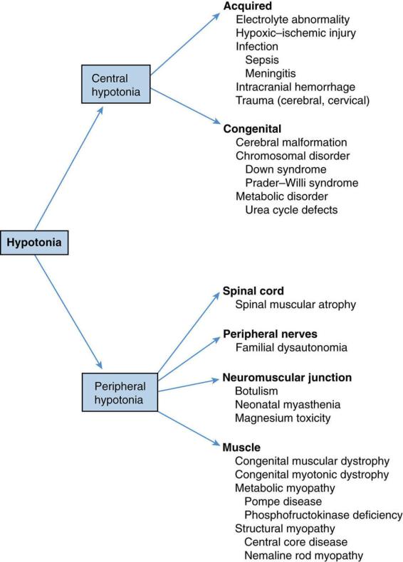
FIGURE 12.1 Differential diagnosis of hypotonia.
457
458
II.Hydrocephalus
A.Definition. Hydrocephalus is an elevation in intracranial pressure (ICP) due to a disturbance in the flow of cerebral spinal fluid (CSF). This can be caused by increased production, blockage of flow, or decreased absorption of CSF. The normal production of CSF is between 400 and 500 mL daily.
B.Types of hydrocephalus
1.Noncommunicating (obstructive) hydrocephalus is due to an obstruction of CSF flow. As a result of this obstruction, the pathways of the ventricular system do not communicate correctly, and there is a buildup of CSF proximal to the blockage.
2.Communicating (nonobstructive) hydrocephalus is due to either an increase in production or a decrease in absorption of CSF. Therefore, all of the pathways of the ventricular system communicate normally, and the problem occurs at the beginning or end of the pathway.
3.Hydrocephalus ex vacuo is not true hydrocephalus, but rather a term used to describe ventricular enlargement caused by brain atrophy.
C.Etiology
1.Congenital causes of hydrocephalus
a.Chiari type II malformation is characterized by downward displacement of the cerebellum and medulla through the foramen magnum, blocking CSF flow. This malformation is often associated with a lumbosacral myelomeningocele.
b.Dandy–Walker malformation is a combination of an absent or hypoplastic cerebellar vermis and cystic enlargement of the fourth ventricle, which blocks the flow of CSF.
c.Congenital aqueductal stenosis is the most common cause of noncommunicating hydrocephalus. Some cases of aqueductal stenosis are inherited as an X-linked trait, and these patients may have thumb abnormalities and other CNS anomalies such as spina bifida (SB).
2.Acquired causes of hydrocephalus include intraventricular hemorrhage (most common in preterm infants), bacterial meningitis, and CSF-producing brain tumors.
D.Clinical features
1.Increasing head circumference that crosses percentile lines, or head circumference >97% for age
2.Infants with open cranial sutures have the following clinical signs:
a.Large anterior and posterior fontanelles and split sutures
b.Setting-sun eyes, a tonic downward deviation of both eyes caused by pressure from the enlarged third ventricles on the upward gaze center in the midbrain
3.Older children with closed cranial sutures have the following symptoms and signs of increased ICP:
a.Headache
b.Nausea and vomiting
c.Unilateral sixth nerve palsy
d.Papilledema
e.Brisk deep tendon refluxes (DTRs), but usually with a downward plantar response
E.Evaluation. Increasing head circumference and signs or symptoms of increased ICP mandate an urgent head ultrasound in infants or head CT scan in older children.
F.Management. Hydrocephalus requires the surgical placement of a ventriculoperitoneal shunt to divert the flow of CSF. Complications of ventriculoperitoneal shunts include shunt infection and shunt obstruction. In older children, acetazolamide may be used to decrease
459
ICP as well.
G.Prognosis. Outcome varies depending on the cause of the hydrocephalus and the success of the intervention.
1.Patients with aqueductal stenosis have the best cognitive outcome.
2.Patients with Chiari type II malformation may have low normal intelligence and language disorders.
3.Patients with X-linked hydrocephalus frequently have cognitive delays.
460
III.Spina Bifida
A.Definitions
1.Spina bifida (SB) is a general term that refers to any failure of bone fusion in the posterior midline of the vertebral column.
2.Neural tube defect is a broad term that includes all forms of failure of neural tube closure, from anencephaly to sacral meningocele.
B.Types of SB
1.SB occulta is an incomplete closure of vertebrae without herniation of tissue through the cleft.
2.Meningocele is the herniation of the meninges only through a bony cleft, most commonly in the lumbosacral region.
3.Myelomeningocele is the herniation of the meninges and spinal cord tissue through the bony cleft, most commonly in the lumbosacral region. Myelomeningocele is more common than meningocele.
C.Epidemiology. The incidence of neural tube defects varies with geographic location. In the United States, the prevalence is approximately 0.5 in 1000; however, in China and India, it is as high as 10 per 1000.
D.Etiology. The multifactorial etiology includes environmental, genetic, nutritional, and teratogenic factors.
1.Taking a prenatal multivitamin preparation that includes folic acid has decreased the incidence of SB in the newborn. The mechanism of action of folic acid is uncertain.
2.Risk factors include a history of previous affected pregnancy, inadequate folic acid intake, and maternal diabetes.
3.Teratogens causing SB include valproic acid, carbamazepine, phenytoin, colchicine, vincristine, azathioprine, and methotrexate.
E.Clinical features
1.SB occulta. The skin on the back (usually lumbosacral region) is epithelialized, and a hairy patch or dimple often covers the area. No neurologic deficits are present.
2.Meningocele. A fluctuant midline mass is present overlying the spine. The mass is filled with CSF but does not contain spinal cord tissue and can be transilluminated. Neurologic deficits are usually not present or are only very mild.
3.Myelomeningocele
a.A fluctuant midline mass is present anywhere along the spine, but most commonly in the lumbosacral region.
b.Neurologic defects are present and depend on the level of the lesion, varying from complete paraplegia (above L3) to preserved ambulation and variable bladder or bowel incontinence (S3 and below).
c.Associated anomalies and complications
1.Hydrocephalus. Ninety percent of lumbosacral myelomeningoceles are associated with Chiari type II malformation and hydrocephalus. Cervical and thoracic myelomeningoceles are not associated with hydrocephalus.
2.Cervical hydrosyringomyelia (accumulation of fluid within the central spinal cord canal and the cord itself)
3.Defects in neuronal migration (e.g., gyral anomalies, agenesis of the corpus callosum)
4.Orthopedic problems (e.g., rib anomalies, deformities of the lower extremities, and lower extremity fractures from loss of sensation)
5.Genitourinary neurologic deficits
461
F.Diagnosis
1.Prenatal diagnosis is common.
a.α-Fetoprotein (AFP), the main serum protein in fetal life, is elevated in amniotic fluid and maternal serum in open neural tube defects, and when measured in maternal serum at 16–18 weeks’ gestation, detects 80% of spinal defects.
b.Fetal sonography is highly sensitive in detecting spinal defects.
2.Diagnosis after birth
a.SB occulta is suggested by finding any skin abnormality overlying the spine and may be confirmed by spinal radiographs.
b.Meningocele is suggested by physical examination findings and is confirmed by MRI of the spinal cord and spine.
c.Myelomeningocele is a clinical diagnosis based on physical examination at birth.
G.Management
1.SB occulta does not require treatment.
2.Meningocele requires surgical repair.
3.Myelomeningocele requires urgent surgical repair within 24 hours of birth, or in some cases in utero, to reduce the morbidity and mortality from infection and to prevent further trauma to the exposed neural tissue.
H.Prognosis
1.SB occulta and meningocele have excellent prognoses as a result of the absence of neurologic deficits.
2.Myelomeningocele. Without treatment, around 85% die within the first year of life. With treatment, 90% of patients survive to adolescence, but many have functional disabilities. Associated problems include wheelchair dependency, bladder or bowel incontinence, seizures, precocious puberty, pressure sores, and fractures.
462
IV. Approach to the Comatose Patient
A.Definition. Coma is a state of unawareness of self and environment in which the patient lies with the eyes closed, without purposeful movement or sleep–wake cycles, and is unarousable by external stimuli.
B.Etiology (Table 12-1). In addition to traumatic brain injury, the most common non-traumatic cause of coma in children is infection.
C.Assessment. The goal of assessment of the comatose patient is to determine the depth of coma, to identify the neurologic signs that indicate the site and cause of the coma, and to monitor the patient’s recovery.
1.Glasgow Coma Scale provides a standard measure to monitor the level of consciousness (see Chapter 20, Table 20-1).
2.Head and neck exam. The patient should be assessed for scalp injuries, breath odors
(for alcohol intoxication or ketosis caused by diabetic ketoacidosis), and nuchal rigidity (caused by meningitis). CSF or blood draining from the nose or external auditory canal may indicate a basilar skull fracture.
3.Abnormal motor responses to stimuli can indicate the location of brain damage.
a.Flaccidity or no movement may suggest severe spinal or brainstem injury.
b.Decerebrate posturing (extension of arms and legs) is seen in diffuse toxic/metabolic disorders or midbrain compression.
c.Decorticate posturing (flexion of arms and extension of legs) is seen in cortical and/or subcortical abnormalities with preservation of brainstem function.
d.Asymmetric responses suggest hemispheric injury.
4.Abnormal respiratory responses may indicate the location of brain injury or its cause.
a.Hypoventilation suggests opiate or sedative overdose.
b.Hyperventilation suggests metabolic acidosis (Kussmaul respirations or rapid, deep breathing may occur), neurogenic pulmonary edema, or midbrain injury.
c.Cheyne–Stokes breathing (alternating apnea and hyperpnea) suggests bilateral cortical injury and is associated with herniation, specifically displacement of the diencephalon (thalamus and hypothalamus).
d.Apneustic breathing (pausing at full inspiration) indicates pons or upper medulla damage.
e.Ataxic or agonal breathing (irregular respirations with no particular pattern) indicates medullary injury and impending brain death.
5.Pupillary size and reactivity may provide clues.
a.Unilateral dilated nonreactive pupil suggests uncal herniation.
b.Bilateral dilated nonreactive pupils suggest topical application of a dilating agent, a postictal state, or irreversible brainstem injury.
c.Bilateral constricted reactive pupils suggest opiate ingestion or pontine injury.
6.Other brainstem reflexes should be assessed to determine the extent of injury to the brainstem.
a.Oculocephalic maneuver (doll’s eyes). When turning the head of an unconscious patient, the eyes normally look straight ahead and then slowly drift back to midline position, because the intact vestibular apparatus senses a change in position. In an injured brainstem, movement of the head does not evoke any eye movement. This is termed a negative oculocephalic maneuver or negative doll’s eyes.
b.Caloric irrigation. When the oculocephalic response is negative or cannot be performed because of possible cervical cord injury, caloric testing should be performed. This involves angling the head at 30° and irrigating each auditory canal
463

with 10–30 mL of ice water. An intact (normal) cold caloric response is reflected by eye deviation to the ipsilateral side, with nystagmus to the contralateral ear. An abnormal response suggests pontine injury.
c.Abnormal corneal and gag reflexes indicate significant brainstem injury.
D.Evaluation. Once the airway, breathing, and circulation are stable, further diagnostic workup may begin.
1.Glucose should be checked immediately in any comatose patient.
2.Urine toxicology screen, serum electrolytes, and metabolic panel should also be evaluated.
3.Head CT scan should be performed to identify mass lesions or trauma.
4.Lumbar puncture (LP) to rule out meningoencephalitis should be considered if the CT scan is negative.
5.Urgent electroencephalography (EEG) should be considered, even in patients without a history of clinical seizures.
Table 12-1
Causes of Impaired Consciousness and Coma in Childhood and Adolescence
Infectious
Meningitis
Encephalitis
Focal infection (abscess, cerebritis)
Inflammatory
Vasculitis
Demyelinating disorders (acute disseminated encephalomyelitis)
Traumatic
Concussion
Abusive head trauma
Diffuse axonal injury
Cerebral hematoma or contusion
Vascular disease
Cerebral infarction
Cerebral hemorrhage
Neoplasm
Hypoxia
Shock
Cardiac failure
Nonfatal drowning
Carbon monoxide poisoning
Metabolic disorders
Fluid or electrolyte imbalance
Hypoglycemia/hyperglycemia
Hyponatremia
Diabetic ketoacidosis
Organic acidemias
Amino acidemias
Hepatic encephalopathy
Urea cycle disorders
Disorders of fatty acid metabolism
Reye syndrome
464

Hyperammonemia
Hypothyroidism or hyperthyroidism
Nutritional
Thiamine deficiency
Pyridoxine deficiency
Folate and vitamin B12 deficiency
Toxins/poisons
Alcohol
Prescription medications
Atropine, scopolamine, benzodiazepines, barbiturates, lithium, opiates, tricyclic antidepressants Over-the-counter medications
Heavy metal poisoning Lead, mercury, arsenic
Illicit drugs*
*Amphetamine, cocaine, and hallucinogens (lysergic acid diethylamide [LSD], mescaline, phencyclidine hydrochloride [PCP]) cause agitation, confusion, delirium, and hallucinations but not coma.
465
V.Seizure Disorders of Childhood
A.Definitions
1.A seizure is a transient occurrence of signs and/or symptoms due to abnormal excessive or synchronous neuronal activity in the brain.
2.Epilepsy is diagnosed if any of the following three scenarios occur:
a.The patient has at least two unprovoked seizures >24 hours apart
b.The patient has one unprovoked seizure and the probability of further seizures occurring over the next 10 years (i.e., the patient has another diagnosis that predisposes to an increased risk of seizures, such as a brain tumor)
c.The patient is diagnosed with an epilepsy syndrome (e.g., childhood absence epilepsy)
3.Status epilepticus is defined as 5 or more minutes of continuous seizure activity, or repetitive seizures without recovery of consciousness.
B.Epidemiology
1.One percent of children have a single unprovoked seizure before 16 years of age.
2.After a single unprovoked seizure, approximately 40% of children will have a second seizure.
3.Prevalence of epilepsy in children is 0.5–0.8%.
C.Etiology
1.The seizure discharge is caused by an imbalance between excitatory and inhibitory input within the brain, or abnormalities in the membrane properties of individual neurons.
2.In some children, the cause of seizures is known (Table 12-2); however, in many, the cause remains unknown.
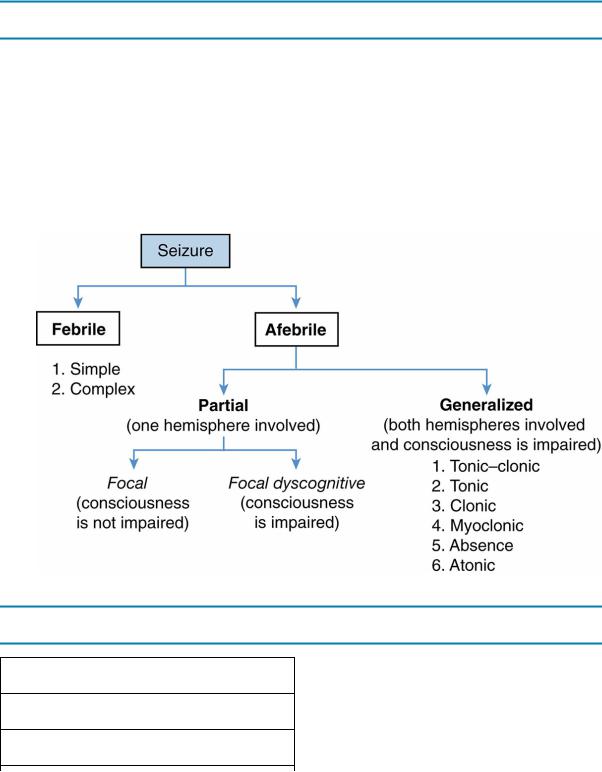
D.Classification of seizures. Criteria for the classification of seizures include the presence or absence of fever, the extent of brain involvement, whether consciousness is impaired, and the nature of the movements (Figure 12-2).
1.Febrile seizures are a common but benign type of seizure associated with fever [see section V.K].
2.Afebrile seizures are either generalized (starting throughout the entire brain) or partial (starting from a single part or “focus” in the brain).
a.Generalized seizures are caused by the discharge from a group of neurons in both cerebral hemispheres and are associated with an alteration of consciousness. Two common types are tonic–clonic and absence seizures.
1.Tonic–clonic seizures are the most common type of generalized seizure.
These seizures are characterized by increased thoracic and abdominal muscle tone, followed by clonic movements of the arms and legs, eyes rolling upward, incontinence, decreased consciousness, and a postictal state of variable duration.
2.Absence seizures are brief staring spells that occur without loss of posture and with only minor motor manifestations (e.g., eye blinking or mouthing movements). The seizure lasts <15 seconds, and there is no postictal state.
3.Other generalized seizures include tonic, clonic, myoclonic, and atonic.
b.Focal (partial) seizures are caused by the discharge from a group of neurons in one hemisphere. Seizure symptoms may have predominately motor, sensory, or psychomotor features. There are two types:
1.In focal seizures (prior term: simple partial seizure), consciousness is not impaired.
2.In focal dyscognitive seizures (prior term: complex partial seizure),
466
consciousness is altered.
E.Classification of epilepsy. Classification may be based on either the predominant seizure type, the site of origin of the epileptic discharge or the genetic mutation.
F.Differential diagnosis of seizure-like events (Table 12-3)
G.Diagnosis. Epilepsy is diagnosed on the basis of history and physical examination, as well as testing for genetic mutations. Other studies (EEG, MRI) may be useful.
1.EEG is useful to evaluate the background rhythms as well as seizures, particularly in determining the location of onset of focal seizures. However, an abnormal EEG is not required for the diagnosis of epilepsy (in particular, focal seizures may have a normal EEG during the interictal period between seizures).
2.Video-EEG monitoring is a useful tool when clinical information is inadequate or incomplete (e.g., when patients are <3 years of age, when events concerning for seizures occur during sleep, or when the history is unclear). Video-EEG monitoring is also useful to capture and characterize focal seizures for possible surgical intervention.
3.Neuroimaging studies (primarily MRI) may be useful in children with epilepsy. Children diagnosed with absence seizures or benign rolandic epilepsy [see sections V.L.4 and V.L.5] do not require imaging.
H.Evaluation of an acute seizure
1.Initial treatment starts with assessment of the patient’s airway, breathing, and circulation.
2.For a first nonfebrile seizure, routine EEG is recommended; however, laboratory studies, LP, and neuroimaging are based on clinical circumstances and typically are unnecessary in a child with a normal neurologic exam.
3.For a simple febrile seizure evaluation should be focused on determining the cause of the child’s fever [see section V.K]. Meningitis should be considered, and LP should be performed if there are concerns for a CNS infection. Further evaluation, including lab studies, EEG, or neuroimaging, is usually not required.
I.Management
1.Treatment of status epilepticus requires intravenous anticonvulsants, such as a shortacting benzodiazepine (e.g., lorazepam or diazepam) followed by a loading dose of either phenobarbital or phenytoin.
2.Treatment of epilepsy
a.Pharmacotherapy. Once the type of seizure has been determined, single-drug therapy is started with the antiepileptic drug that has the best combination of high efficacy and low toxicity. Examples of commonly recommended drugs for the following seizure types:
1.Generalized epilepsy: valproic acid, lamotrigine, levetiracetam
2.Absence epilepsy: ethosuximide, valproic acid, lamotrigine
3.Focal epilepsy: carbamazepine, lamotrigine, levetiracetam
4.Lennox–Gastaut: felbamate, rufinamide, lamotrigine, topiramate
b.Surgery
1.For medically intractable epilepsy, surgery to remove epileptic tissue may be an option.
2.The best prognosis is for patients with temporal lobe lesions, >75% of whom have complete seizure control or remission after surgery.
c.Vagal nerve stimulator is a pacemaker-sized device that sends an electrical impulse to the vagus nerve. A common side effect is hoarseness.
d.Ketogenic diet (a high-fat, low-carbohydrate diet) is thought to suppress seizure activity by producing a state of ketosis.
J.Prognosis. Epilepsy is not necessarily a lifelong disorder. About 70% of epileptic children can
467
be weaned off their medications after a 2-year seizure-free period and normalization of the EEG.
K.Febrile seizures
1.Definition. Seizures that occur in children between 6 months and 6 years of age accompanied by a fever (temperature > 38°C), in whom CNS infection, metabolic imbalance, and history of afebrile seizure are excluded.
2.Epidemiology. Febrile seizures are the most common childhood seizure type, occurring in 2–4% of all children.
3.Etiology
a.The pathophysiologic mechanism is unknown.
b.Febrile seizures can be inherited, and several gene mutations have been found.
4.Classification
a.A simple febrile seizure lasts less than 15 minutes and is generalized.
b.A complex febrile seizure lasts more than 15 minutes, has focal features, or recurs within 24 hours.
5.Risk factors for first febrile seizure include having a firstor second-degree relative with a history of febrile seizures, neonatal hospital stays >30 days, developmental delay, attendance at daycare, high frequency of febrile episodes, peak body temperature and recent vaccination.
6.Diagnosis
a.The diagnosis of a febrile seizure is based on history, a normal neurologic examination, and the exclusion of CNS infection.
b.An LP is necessary if meningitis is suspected at any age. For a child 6–12 months of age, an LP should be seriously considered if the child is deficient in vaccination for
Haemophilus influenzae type b (HIB) or Streptococcus pneumoniae or when immunization status cannot be determined.
c.Neither neuroimaging nor EEG is needed unless the neurologic examination is abnormal.
7.Management
a.First-time or occasional febrile seizures are not treated with anticonvulsants.
b.Antipyretic treatment during subsequent febrile illnesses is unlikely to prevent febrile seizures.
c.Frequent, recurrent, or prolonged febrile seizures do pose a risk and may require additional treatment with daily anticonvulsant prophylaxis or the use of rescue medications (e.g., rectal diazepam).
8.Prognosis. Approximately 30% of patients will have a recurrence. Recurrence risk decreases with increasing patient age. The risk of epilepsy is low (2–10%).
L.Epileptic syndromes
1.Definition. Epileptic syndromes are epileptic conditions characterized by a specific age of onset, seizure characteristic, and EEG abnormality.
2.Classification. There are many types of epilepsy syndromes recognized. Each has its own seizure type, severity, and prognosis. Three common types are infantile spasms, absence epilepsy of childhood, and benign rolandic epilepsy.
3.Infantile spasms
a.Epidemiology. Age of onset is typically 4–7 months. Infantile spasms are rare in children older than 2 years of age.
b.Etiology. Although infantile spasms are frequently cryptogenic (unknown etiology), there are a number of associations, including hypoxic–ischemic injury, tuberous sclerosis, malformations of cortical development, Down syndrome, neonatal hypoglycemia, meningitis, trauma, phenylketonuria, intraventricular
468
hemorrhage, pyridoxine deficiency, and infection. Tuberous sclerosis is the most commonly identified cause of this disorder.
c.Clinical features
1.Brief tonic extension of the extremities and flexion of the trunk (jackknife or salaam seizures) lasting 1–2 seconds each, occurring in clusters of 5–10 seizures spread over 3–5 minutes
2.Hypsarrhythmia is a characteristic EEG pattern consisting of a high amplitude, disorganized background with multifocal spike-wave discharges.
3.West syndrome is a triad of infantile spasms, hypsarrhythmia, and developmental delay or regression.
d.Management
1.Adrenocorticotropic hormone (ACTH) intramuscular injections for a 4- to 6- week period are effective in 50-70% of affected patients.
2.Vigabatrin is frequently effective for patients with infantile spasms associated with tuberous sclerosis and is often used in combination with ACTH.
3.Other treatments include valproic acid, topiramate zonisamide, ketogenic diet, pyridoxine, and surgery.
e.Prognosis. Outlook is poor. Despite the success of these different medications in suppressing seizures, children often develop moderate to severe intellectual disability.
4.Absence epilepsy of childhood
a.Epidemiology. Age of onset is between 4 and 8 years of age and is more common in girls.
b.Etiology. There is a presumed genetic cause, and some cases may have an autosomal dominant inheritance pattern.
c.Clinical features of absence seizures
1.Brief, lasting 5–10 seconds
2.Frequent, tens to hundreds per day
3.May be accompanied by automatisms, such as eye blinking, chewing, and incomprehensible utterances.
4.Loss of posture, urinary incontinence, and a postictal state do not occur.
d.Diagnosis. The EEG shows the characteristic generalized 3-Hz spike-and-wave discharges. Hyperventilation for 2–3 minutes frequently provokes absence seizures.
e.Management. Treatment includes ethosuximide, valproic acid, or lamotrigine.
f.Prognosis. Outlook is very good; the seizures usually resolve by adolescence without cognitive impairment.
5.Benign epilepsy with centrotemporal spikes (BECTS), which is also known as benign rolandic epilepsy
a.Definition. BECTS involves nocturnal focal seizures occasionally with secondary generalization. Focal spike discharges in the centrotemporal region is seen on EEG.
b.Epidemiology
1.Benign rolandic epilepsy is the most common partial epilepsy during childhood, accounting for 15% of epilepsy.
2.It commonly presents at 3–13 years of age. Peak incidence is at 6–7 years of age. Boys are more likely to be affected.
c.Etiology. Inheritance is autosomal dominant with age-dependent penetrance.
d.Clinical features
1.Seizures typically occur in the early morning hours when patients are asleep with oral–buccal manifestations (i.e., moaning, grunting, pooling of saliva).
469
2.Seizures may spread to the face and arm and then secondarily generalize into tonic–clonic seizures.
e.Diagnosis. The EEG shows biphasic spike and sharp-wave discharges in the midtemporal and central regions.
f.Management. Treatment includes valproic acid or carbamazepine.
g.Prognosis. Outcome is excellent. Seizures typically remit spontaneously during adolescence. There are reports of associated learning disabilities associated with BECTS.
Table 12-2
Causes of Acute Seizures During Childhood
Head |
Cerebral contusion, subdural hematoma |
trauma |
|
Brain tumor |
Astrocytoma |
Toxins |
Amphetamines, cocaine |
Infections |
Meningitis, encephalitis, brain abscess, neurocysticercosis |
Vascular |
Cerebral infarction, intracranial hemorrhage |
Metabolic |
Hypocalcemia, hypoglycemia, hypomagnesemia, hypoor hypernatremia, pyridoxine deficiency |
disturbances |
|
Systemic |
Hypertension, hypoxic–ischemic injury, inherited metabolic disorder, liver disease, renal failure, neurocutaneous |
diseases |
disorders (e.g., tuberous sclerosis, neurofibromatosis and Sturge-Weber syndrome) |
FIGURE 12.2 Classification of seizures.
Table 12-3
Differential Diagnosis of Seizure-like Events
Breath-holding spells (in infants)
Gastroesophageal reflux disease (Sandifer syndrome)
Syncope
470
Migraine
Vertigo
Movement disorder (e.g., tics, chorea)
Sleep disturbances (e.g., night terrors, somnambulism)
Transient ischemic attack
Rage attacks
Psychogenic nonepileptic events (pseudoseizures)
471
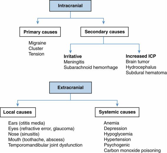
VI. Headaches in Childhood
A.Etiology. Headaches may have intracranial or extracranial causes (Figure 12-3).
1.Intracranial causes
a.Primary headaches are more common and are idiopathic, not occurring for a specific reason, or the result of an underlying disease. These are likely due to a combination of genetic, developmental, and environmental risk factors. Migraine, tension-type, and cluster are common primary headaches.
b.Secondary headaches are caused by an underlying disease process such as increased intracranial pressure (e.g., hydrocephalus), meningeal irritation (e.g., meningitis, subarachnoid hemorrhage), or inflammatory processes.
2.Extracranial causes
a.Local causes include sinusitis, perioral abscess, toothache, chronic otitis media, or refractive errors.
b.Systemic causes include anemia, hypoglycemia, depression, and hypertension.
B.Important clinical information about a patient’s headache helps to determine the cause.
1.Quality of pain. Throbbing or pounding pain suggests migraine headaches, whereas a sensation of squeezing or pressure is more common in tension-type headaches.
2.Location and radiation. Migraine headaches are frequently unilateral (although commonly bilateral in children) and may begin in the periorbital area and spread to the forehead and occiput, whereas tension-type headaches are often generalized or bitemporal.
3.Time of onset. Tension headaches occur toward the end of the day, whereas headaches from increased ICP occur in the morning.
4.Duration. The shorter the headache duration, the less likely a serious disorder is responsible.
5.Red flags indicating more serious pathology may include an abnormal neurologic examination, a new and unusual headache (especially when described as “a sudden worst headache of one’s life”), a patient with an immunocompromised state, fever, or stiff neck, and blurring of optic disc margins on examination.
C.Migraine headaches
1.Definition. Migraine headaches are prolonged (often 30–60 minutes, but may last up to 72 hours), unilateral (may be bilateral in children) headaches that are associated with nausea, vomiting, or visual changes and are caused by changes in cerebral blood flow.
2.Epidemiology
a.Migraines are the most common cause of headaches in children and adolescents.
The prevalence increases from 3% in 7-year-olds, to 11% in 11-year-olds, and up to 20% in adolescents.
b.Before puberty, incidence is higher in males; after puberty, incidence is higher in females.
3.Etiology
a.Seventy to eighty percent of children with migraines have at least one affected parent.
b.The pathophysiology of migraines is complicated and evolving. Serotonin (5-HT), calcitonin gene related peptide (CGRP), inflammatory markers (substance P, vasoactive intestinal peptide), the trigeminal nerve and changes in cerebral blood flow all appear to play a role.
4.Classification
a.Migraine without aura is the most common form of migraine in children.
472
Headaches occur in the absence of any warning symptoms.
b.Migraine with aura. The onset of the headache is preceded by transient visual changes or unilateral paresthesias or weakness. The visual changes may include blurred vision, small areas of decreased vision (scotomata), streaks of light, or hemianopsia.
c.Migraine equivalent. In young children, the headache itself may be absent, but there is a prolonged, albeit transient, alteration of behavior that manifests as cyclic vomiting, cyclic abdominal pain, or paroxysmal vertigo.
d.Migraines associated with focal neurologic signs
1.Ophthalmoplegic migraine. Unilateral ptosis or cranial nerve III palsy accompanies this headache.
2.Basilar artery migraine. Vertigo, tinnitus, ataxia, or dysarthria may precede the onset of this headache.
5.Precipitating factors. There is no obvious precipitating cause, although many migraine sufferers have triggers such as red wine, cheese, preserved meats, and chocolate. Some patients note that stress, fatigue, menstruation, dehydration, skipping meals, or exercise induces the headache.
6.Clinical features
a.A prolonged, throbbing, unilateral headache starts in the supraorbital area and radiates to the occiput. In young children, the headache is often bifrontal.
b.Nausea and vomiting may occur. A history of motion sickness is common.
c.Visual disturbances include blurred vision, scotomata, and jagged streaks of light that take on the outline of old forts (fortifications).
d.Photophobia or phonophobia occurs. Many patients treat themselves by lying in a dark, quiet room.
e.Symptoms are improved by sleep.
f.Neurologic examination is normal.
7.Diagnosis. Diagnosis is made by history and the presence of a normal neurologic examination.
8.Management. Treatment includes rest and elimination of known triggers. Medications may be very helpful.
a.Abortive treatment includes nonsteroidal anti-inflammatory drugs (NSAIDs) and triptans, selective 5-HT agonists, available in injectable, intranasal, and oral forms.
b.Preventive treatment options include propranolol, amitriptyline, topiramate, valproic acid and cyproheptadine.
9.Prognosis. Migraines can be a lifelong disorder with a waxing and waning course.
D.Tension headaches
1.Definition. Tension headaches are bifrontal or diffuse, dull, aching headaches that are often associated with muscle contraction.
2.Epidemiology. Tension headaches are more commonly seen in children older than 7 years with a prevalence as high as 25%.
3.Clinical features
a.Pain is described as dull, aching, and rarely throbbing, and it increases in intensity during the day.
b.The pain is usually bifrontal but may be diffuse.
c.Isometric contraction of the temporalis, masseter, or trapezius muscle often accompanies the headache.
d.No vomiting, visual changes, or paresthesias occur.
4.Diagnosis. Clinical presentation provides a clue to the diagnosis, and no laboratory or imaging study is diagnostic. Tension headaches are rare in young children, and
473
therefore, other diagnoses (e.g., migraines) should be preferentially considered.
5.Management. Treatment includes reassurance and pain control (e.g., acetaminophen, ibuprofen). Stress and anxiety reduction may provide long-term relief.
E.Cluster headaches. These headaches are extremely rare during childhood.
1.Diagnostic criteria includes the following:
a.Attacks of severe unilateral facial/orbital pain
b.At least five attacks that last between 15 minutes and 3 hours
c.Sense of restless agitation
d.Ipsilateral conjunctival injection, eyelid edema, lacrimation, nasal congestion, rhinorrhea, and forehead sweating.
2.Treatment includes abortive therapy with oxygen or triptans. Prophylactic treatments include calcium-channel blockers and valproic acid.
FIGURE 12.3 Causes of headache. ICP = intracranial pressure.
474
VII. Approach to Unsteady Gait
A.Definition. Ataxia is a disturbance in smooth coordination of movements and often manifests as unsteady gait. Ataxia can be the result of cerebellar or proprioceptive dysfunction (sensory loss ataxia).
B.Differential diagnosis. A variety of neurologic problems can give the appearance of an unsteady gait.
1.Cerebellar dysfunction. Children with a cerebellar gait have an unsteady, wide-based stance with irregular steps, also known as a “drunken gait.” See Table 12-4 for the causes of cerebellar ataxia.
2.Weakness. Any cause of muscle weakness or sensory loss, such as spinal cord lesions or acute disorders of the motor unit [e.g., Guillain–Barré syndrome, see section VII.D], can lead to an unsteady gait.
3.Encephalopathy as a result of infection, drug overdose, or recent head trauma may cause decreased levels of consciousness, which may affect gait.
4.Seizures. During a seizure, or while in the postictal period, the patient’s gait may be irregular and unsteady.
5.Vision problems can mimic the appearance of an unsteady gait.
6.Vertigo from migraines, acute labyrinthitis, and brainstem tumors may lead to unsteady walking.
C.Acute cerebellar ataxia of childhood
1.Definition. Acute cerebellar ataxia is an unsteady gait secondary to a presumed autoimmune or postinfectious cause.
2.Epidemiology
a.Acute cerebellar ataxia is the most common cause of ataxia in children.
b.Age of onset is between 18 months and 7 years. Acute cerebellar ataxia rarely occurs in children older than 10 years.
3.Etiology
a.Common preceding infections include varicella, coxsackievirus, Epstein–Barr virus (EBV), and mycoplasma. The ataxia usually follows a viral illness by 2– 3 weeks.
b.The postulated cause is thought to be molecular mimicry, where molecular similarity between antigens from the infectious entity and the patient (e.g., cerebellar structures) triggers an autoimmune response.
4.Clinical features
a.Truncal ataxia with deterioration of gait is characteristic. Young children may refuse to walk for fear of falling.
b.Slurred speech and nystagmus are often present, and hypotonia and tremors are less common.
c.Fever is absent.
5.Diagnosis. Diagnosis is by history and physical examination and by exclusion of other causes of ataxia. For patients with clear acute cerebellar ataxia, neuroimaging is generally not necessary. If there are atypical features or concern for other causes of ataxia, however, neuroimaging is necessary (e.g., to rule out acute life-threatening causes such as tumors or hemorrhage in the posterior fossa). If head CT is obtained to rule out acute life-threatening causes, it is normal in acute cerebellar ataxia. MRI provides better detail to visualize the posterior fossa for evaluation of other causes of ataxia.
6.Management. Treatment is supportive. Complete resolution of symptoms generally occurs in 2–3 weeks. Physical therapy is often needed.
475

D.Guillain–Barré Syndrome (acute inflammatory demyelinating polyneuropathy)
1.Definition. Guillain–Barré syndrome is a demyelinating polyneuritis characterized by ascending weakness, areflexia, and paresthesias.
2.Etiology. The most commonly associated infectious agent is Campylobacter jejuni, which causes a prodromal gastroenteritis. Many other infectious agents have been associated with Guillain–Barré syndrome, such as Mycoplasma pneumoniae, cytomegalovirus, EBV, herpes zoster virus, influenza, varicella, and coxsackievirus.
3.Pathophysiology
a.The principal sites of demyelination are the ventral spinal roots and peripheral myelinated nerves.
b.Injury is triggered by a cell-mediated immune response to an infectious agent that cross-reacts to antigens on the Schwann cell membrane.
4.Clinical features
a.Ascending, symmetric paralysis may progress to respiratory arrest.
b.No sensory loss occurs, although low-back or leg pain may be present in 50% of patients.
c.Cranial nerve involvement. Facial weakness occurs in 40–50% of patients and is often bilateral.
d.Dysautonomia, arrhythmia, orthostatic hypotension, and urinary retention may occur.
e.Miller Fisher syndrome, a variant of Guillain–Barré syndrome, is characterized by ophthalmoplegia, ataxia, and areflexia.
5.Diagnosis
a.LP shows albuminocytologic dissociation (i.e., increased CSF protein in the absence of an elevated cell count), which is usually evident 1 week after symptom onset.
b.EMG demonstrates decreased nerve conduction velocity or conduction block.
c.Spinal MRI may be necessary in children younger than 3 years to rule out compressive lesions of the spinal cord, because the sensory examination in children of this age is often difficult to evaluate.
6.Management. Treatment should be initiated as soon as the diagnosis is established because of the risk of respiratory muscle paralysis.
a.Intravenous immune globulin (IVIG), given for 2–4 days, is the preferred treatment for children because of its relative safety and ease of use.
b.Plasmapheresis removes the patient’s plasma along with the presumed antimyelin antibodies and is performed over a 4- to 5-day period.
7.Prognosis. Complete recovery is the rule in children but depends on the severity and extent of the weakness. Physical therapy may be necessary for several weeks or longer to aid recovery.
Table 12-4
Differential Diagnosis of Cerebellar Ataxia
Brain tumors |
Pilocytic astrocytoma (occurring in the cerebellum) |
|
Medulloblastoma (occurring in posterior fossa) |
|
Neuroblastoma (opsoclonus myoclonus ataxia syndrome) |
Trauma |
Cerebellar contusion |
|
Subdural hematoma |
Toxins |
Ethanol |
|
Antiepileptic medications |
Vascular |
Cerebellar infarction or hemorrhage |
Infections |
Meningitis |
|
|
476
|
Encephalitis |
Inflammatory |
Acute cerebellar ataxia of childhood |
Demyelination |
Acute disseminated encephalomyelitis (ADEM) |
|
Multiple sclerosis |
Migraine syndromes |
Basilar migraines and familial hemiplegic migraines |
|
Benign paroxysmal vertigo (may have history of migraine) |
477
VIII. Movement Disorders
A.Sydenham chorea (St. Vitus dance)
1.Definition. Sydenham chorea is a self-limited autoimmune disorder and is one of the major Jones criteria for diagnosis of rheumatic fever (see Chapter 16, section VI). It presents with chorea (involuntary, brief, purposeless movements of the limbs and upper body) and emotional lability.
2.Epidemiology
a.Sydenham chorea occurs in approximately 25% of patients with rheumatic fever.
b.Onset is most common between 5 and 13 years of age.
3.Pathophysiology. Sydenham chorea occurs secondary to antibodies that cross-react with membrane antigens on both group A β-hemolytic streptococcus and basal ganglia cells.
4.Clinical features
a.Immunologic response usually follows streptococcal pharyngitis by 2–7 months.
b.Children appear restless. The face, hands, and arms are mainly affected, and the movements appear continuous, quick, and random. The chorea may begin as clumsiness of the hands.
c.Speech is also affected and can be jerky or indistinct.
d.Patients are unable to sustain protrusion of the tongue (chameleon tongue).
e.The wrist is held flexed and hyperextended at the metacarpal joints (choreic hand). On gripping the examiner’s fingers, patients are unable to maintain the grip (milkmaid’s grip).
f.Emotional lability (impulsivity, obsessive compulsive symptoms, aggression) is common and may precede signs of chorea.
g.Gait and cognition are not affected.
5.Differential diagnosis. Other conditions that may cause chorea include many acquired and congenital conditions, including N-methyl-d-aspartate (NMDA) receptor encephalitis, kernicterus, systemic lupus erythematosus, Huntington disease, and Wilson disease.
6.Diagnosis. There is no single confirmatory test for Sydenham chorea. The diagnosis is made clinically based on presumptive evidence of rheumatic fever and the exclusion of other likely causes of chorea.
a.Elevated antistreptolysin O (ASO) or anti-DNase B (ADB) titer may indicate a recent streptococcal infection.
b.Neuroimaging
1.Head MRI may show increased signal intensity in the caudate and putamen on T2-weighted sequences.
2.Single-photon emission computed tomography (SPECT) may demonstrate increased perfusion to the thalamus and striatum.
7.Management. Treatment should be initiated when chorea is leading to significant impairment of motor function. Treatment options include corticosteroids, haloperidol, valproic acid, or phenobarbital.
8.Prognosis. Symptoms may last from several months to 2 years. Generally, all patients recover.
B.Tourette syndrome
1.Definitions
a.Tics are brief, stereotypical behaviors that are initiated by an unconscious (premonitory) urge that can be temporarily suppressed.
478
b.Tourette syndrome is defined as having two or more motor tics and at least one vocal tic (do not have to occur at the same time), for at least 1-year duration and beginning before 18 years of age. In addition, symptoms cannot be secondary to other illnesses or medications.
2.Epidemiology. The prevalence of Tourette syndrome is 1 in 1000 live births. However, tics occur in 3% of children.
3.Etiology. The cause of Tourette syndrome is unknown. In some patients, there is a genetic predisposition.
4.Clinical features
a.Motor tics can be simple (e.g., eye blinking, head or shoulder shaking) or complex (e.g., bouncing, jumping, kicking).
b.Vocal tics can be simple (e.g., coughing, grunting, humming, clearing the throat) or complex (e.g., echolalia, which is the repetition of heard words or phrases).
c.Tics must be present ≥1 year, although their severity and frequency waxes and wanes.
d.Absence of any signs of a neurodegenerative disorder
e.Coprolalia, the utterance of obscene words, is a dramatic symptom that occurs in <10% of patients.
f.Associated findings include mood disorders, anxiety, attention deficit/hyperactivity disorder, and obsessive–compulsive traits.
5.Differential diagnosis. Disorders that may cause tics include Wilson disease, Sydenham chorea, partial or myoclonic seizures, pediatric autoimmune neuropsychiatric disorders associated with streptococcal infection (PANDAS or PANS disorder is an autoimmune neuropsychiatric disorder which follows infection with wide variety of agents, including streptococcal or mycoplasma infection, and is characterized by dramatic and acute onset of obsessive compulsive behaviors and/or severe restricted food intake, with associated neuropsychiatric symptoms which may include tics, anxiety, emotional lability, deterioration in school performance or behavioral regression), or simple habits. (Habits differ from tics in that habits are situation-dependent and are under voluntary control.)
6.Diagnosis
a.Tourette syndrome is a clinical diagnosis based on history and neurologic findings.
b.No laboratory or imaging tests confirm the diagnosis.
7.Management
a.For tics that are mild and nondisabling, no treatment is recommended.
b.Nonpharmacologic treatment includes habit reversal therapy and comprehensive behavioral intervention for tics.
c.Pharmacologic treatment includes alpha-adrenergics (clonidine, guanfacine) and both typical and atypical neuroleptics (pimozide, haloperidol, risperidone). Botulinum toxin injection may be effective for some focal motor tics.
8.Prognosis
a.Tics wax and wane over a lifetime and tend to decrease in adulthood.
b.Pharmacotherapy is generally successful, but side effects from the medications may be limiting.
479
IX. Duchenne and Becker Muscular Dystrophies (DMD, BMD)
A.Definition. DMD and BMD are progressive, X-linked myopathies characterized by myofiber degeneration. DMD is more severe than BMD.
B.Epidemiology
1.These worldwide disorders occur in all ethnic groups.
2.Prevalence is 1 in 10,000 live births.
3.Onset of symptoms is between 2 and 5 years of age.
C.Etiology. These X-linked disorders are caused by a deletion in the dystrophin gene.
D.Pathophysiology
1.Dystrophin is located on the plasma membrane of muscle fibers and provides mechanical reinforcement and stabilization. It is a high–molecular weight cytoskeletal protein that associates with actin and other structural membrane elements.
2.The absence of dystrophin causes weakness and eventually rupture of the plasma membrane, leading to injury and degeneration of muscle fibers.
E.Pathology. Both DMD and BMD have the same appearance on light microscopy.
1.Degeneration and regeneration of muscle fibers
2.Infiltration of lymphocytes into the injured area and replacement of damaged muscle fibers with fibroblasts and lipid deposits
F.Clinical features
1.Slow, progressive weakness affecting the legs first
2.In DMD, children lose the ability to walk by around 12 years of age. In BMD, patients lose the ability to walk by 20 or more years of age.
3.Pseudohypertrophy of calves is present because of the excess accumulation of lipids, which replace the degenerating muscle fibers. This is more common in DMD than in BMD.
4.Gowers sign is present. Because of the weakness of pelvic muscles, patients arise from the floor in a characteristic manner by extending each leg and then “climbing up” each thigh until they reach an upright position.
5.Cardiac involvement (e.g., cardiomegaly, tachycardia, or cardiac failure) occurs in 50% of patients.
6.Mild cognitive impairment may occur in DMD, but normal intelligence is present in BMD.
G.Diagnosis
1.The presence of enlarged calf muscles in a young boy with muscle weakness suggests the diagnosis.
2.CK levels are VERY high, even before muscle weakness.
3.EMG shows small, polyphasic muscle potentials with normal nerve conductions.
4.Muscle biopsy shows the typical dystrophic pattern.
5.Absent or decreased dystrophin levels are present on immunocytochemistry or Western blot assay of muscle.
6.DNA testing may reveal the gene deletion in >90% of patients.
H.Management. There is no cure, but oral steroids can improve strength transiently and prolong duration of ambulation when the disease is in the early stages. Current research is evaluating gene replacement therapy to convert DMD to the more favorable BMD.
I.Prognosis
1.In DMD, patients are wheelchair dependent by 12 years of age and often die in their late teens from respiratory failure or cardiomyopathy. Assisted ventilation may help individuals to live longer.
480
2.In BMD, patients become wheelchair dependent in their twenties. Life expectancy is beyond the age of 30 years.
481
X.Myasthenia Gravis
A.Definition. Myasthenia gravis is an autoimmune disorder that presents with generalized weakness, fatigability of muscles, ptosis, and diplopia.
B.Etiology. Myasthenia gravis is caused by antibodies against the acetylcholine receptor
(AChR) in the postsynaptic membrane of the neuromuscular junctions.
C.Classification
1.Neonatal myasthenia is a transient weakness in the newborn period secondary to transplacental transfer of maternal AChR antibodies from a mother affected with myasthenia gravis.
2.Congenital myasthenia is familial and is not transferred by the mother. There are no maternal antibodies, and this disorder consists of multiple subtypes.
3.Juvenile myasthenia gravis presents in childhood secondary to AChR antibody formation.
D.Epidemiology. Juvenile myasthenia gravis affects girls two to six times more frequently than boys.
E.Clinical features
1.In neonatal myasthenia, hypotonia, weakness, feeding problems, and weak cry are the most common findings.
2.In juvenile myasthenia gravis, several findings are characteristic.
a.Bilateral ptosis is the most common presenting sign.
b.Characteristic increasing weakness occurs later in the day and with repetitive or sustained muscle activity.
c.Diplopia secondary to decreased extraocular movements may be the only manifestation.
d.DTRs are preserved.
e.Other autoimmune disorders, including juvenile rheumatoid arthritis, diabetes mellitus, and thyroid disease, may coexist.
F.Diagnosis. Diagnosis is made by the following:
1.Tensilon test. Intravenous injection of edrophonium chloride, a rapidly acting cholinesterase inhibitor, produces transient improvement of ptosis.
2.Decremental response to low-frequency (3–10 Hz) repetitive nerve stimulation
3.Presence of AChR antibody titers
G.Management
1.In neonatal myasthenia, treatment is supportive because the disorder is self-limited. Small feedings by nasogastric tubes and assisted ventilation are provided if needed. Cholinesterase inhibitors can be used to aid with feeding and respiratory support.
2.In juvenile myasthenia gravis, treatment involves the following:
a.Cholinesterase inhibitors are the mainstay of treatment. Pyridostigmine bromide is the drug of choice.
b.Immunotherapy
1.Corticosteroids are used when cholinesterase inhibitors fail.
2.Plasmapheresis lowers the level of AChR antibodies. It is useful when symptoms worsen, when respiratory effort is compromised, or when the patient is unresponsive to other therapies.
3.IVIG may also be effective.
c.Thymectomy is performed if there is evidence of thymoma (there is also some evidence for thymectomy even in the absence of thymoma)
H.Prognosis
482
1.In neonatal myasthenia, symptoms are mild and generally resolve within 1–3 weeks.
2.In juvenile myasthenia gravis, remission of symptoms can be as high as 60% after thymectomy.
483