LATER POSTEMBRYONIC PHASE |
289 |
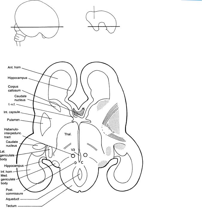
Figure 24–30. 75 mm (approximately 12 weeks). This is a horizontal section through the interventricular foramina. The longitudinal fissure is evident rostrally, and the tectum caudally. Many features are already arranged more or less as in the adult, e.g., the sequence of corpus striatum, internal capsule, thalamus, and third ventricle, as well as the sequence of lateral and medial geniculate bodies, and tectum. The aqueduct is still relatively large, and the corpus callosum is limited to the rostral region. Several structures show the characteristic C-shaped disposition; e.g., the lateral ventricle, hippocampus, and caudate nucleus are each sectioned twice. The lateral ventricle (shown also in the upper right-hand drawing) is sectioned in its anterior and inferior horns. The atrium of the lateral ventricle is the trigonal junction of the central part with the inferior and posterior horns. The key drawings are at 88 mm (left) and 78 mm (right). The section is based on a photomicrograph in Feess-Higgins and Larroche (1987), and the outline of the lateral ventricle is after Westergaard (1971).
290 |
C h a p t e r 2 4 : LATER POSTEMBRYONIC PHASE |
A
B
C
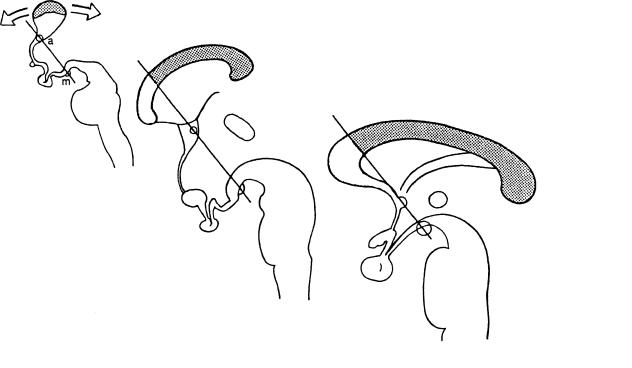
Figure 24–31. Because the trunk of the corpus callosum is sometimes found in the absence of a genu, it has been proposed by Kier and Truwit (1996) that the first part of the corpus callosum to become visible is the front portion of the trunk, which then develops bidirectionally, thereby forming the genu and the splenium. The genu was found always to project in front of a reference line from the mamillary body through the anterior commissure and corpus callosum. Their interpretation is shown. (A) 13 weeks (105 mm), (B) 15 weeks (125 mm), and (C) adult. A and B are modified from Hochstetter. Stippling, trunk. Yellow, genu a, anterior commissure. m, mamillary body.
Callosal development involves (1) a “subcallosal sling,” (2) a median “glial wedge” and glia of the indusium griseum, and (3) pioneering axons from the cingulate cortex (Ren et al., 2006).
LATER POSTEMBRYONIC PHASE |
293 |
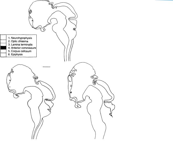
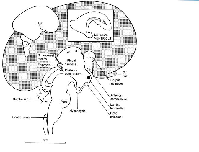
Figure 24–33. 100 mm (approximately 13 weeks). This is a median section at the end of trimester 1. The medial surface of the left cerebral hemisphere is stippled. The corpus callosum is still small, so that the roof of the third ventricle is exposed at the bottom of the longitudinal fissure. The pineal region and its associated recesses and commissures are well-developed. The aqueduct is still relatively wide. (A graph of its cross-sectional area correlated with age is given by Lemire et al., 1975, p. 98.) The vermis is small. (The number of folia seen in the vermis increases greatly during trimesters 2 and 3, as shown in a graph by Lemire et al., 1975, p. 150.) The tegmental portion of the brain stem is voluminous. Although the forebrain is at an angle (of approximately 116◦ ) with the brain stem, the flexures characteristic of the embryonic period proper have now become difficult to detect. The parts marked a and b are described with Figure 26–3. The section of the brain stem is based on Hochstetter (1919), and the inset showing the lateral ventricle is from a cast by Day (1959).
Details of the medial surface of the prosencephalon at 95 mm are given in Figure 26-3B.
294 |
C h a p t e r 2 4 : LATER POSTEMBRYONIC PHASE |
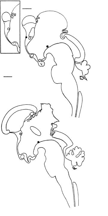
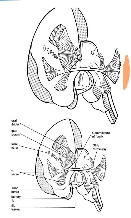
Figure 24–34. 95 mm (approximately
13 weeks). Anteromedial view of some major fiber tracts. The right half of the brain stem has been preserved intact. In the rostral part of the telencephalon everything has been removed except the fiber tracts, thereby exposing the corpus callosum, the column and commissure of the fornix, two divisions of the anterior comissure, and the internal capsule, which last subdivides the corpus striatum into the caudate and lentiform nuclei. On the left side of the brain, the connection from the stria terminalis to the anterior commissure is shown. Various features are identified in the key drawing. Based on Streeter in Keibel and Mall (1912).
It is to be noted that fibers in the corpus callosum have already crossed the median plane.
The development of the fornix can be summarized as follows. The earliest components of the fornix, present already at stage 20, are the precommissural fibers from the septal nuclei to the (as yet poorly developed) hippocampus (Fig. 20–13). Early in the fetal period (at about 9-11 weeks)
the columns arise as connections between the hippocampal system and the mamillary nuclei (Figs. 25–1 and 26–9B). The commissure of the fornix develops as the corpus callosum becomes identifiable (55 mm, 10 weeks, Rakic and Yakovlev, 1968; 105 mm, 13 weeks, Hochstetter, 1929).
LATER POSTEMBRYONIC PHASE |
295 |
Adult
Fetal
C
B
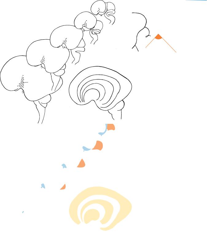
Figure 24–35. Lengthening and “rotation” of the olfactory bulb during the fetal period. (A) Showing the olfactory bulb at stage 23. (B) Near the end of trimester 1. (C) At the beginning of trimester 2.
The above-left diagram shows the forebrain angle (of Hirako), which seems to increase during trimester 1 but decrease later (Koya, 1963).
25 mm
M
38 mm
A
53 mm
68 mm
96 mm
|
|
B |
|
|
96 |
|
Insula |
68 mm |
|
53 |
|
|
|
|
38 |
|
|
25 |
|
|
mm |
Figure 24–36. Outlines of the brain during trimester 1, drawn to the same scale, showing the considerable enlargement.
(A) Left lateral views. The diencephalon and mesencephalon become increasingly covered by the cerebral hemispheres. Correspondingly the insula becomes more and more apparent. (B) Superimposed views of the cerebral hemisphere (based on Hochstetter, 1919).
296 |
C h a p t e r 2 4 : LATER POSTEMBRYONIC PHASE |
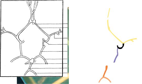
Ant. communicating
Ant. cerebral
Middle cerebral
Int. carotid
Post. communicating
Post. cerebral
Sup. cerebellar
Basilar
Vertebral
Figure 24–37. The circulus arteriosus (of Willis) at 65 mm (11 weeks). Courtesy of Dr. Alexander Barry, University of California, Davis.
C H A P T E R 25
CH |
TRIMESTER 2 |
GL |
Approximately 100–250 mm in Greatest Length; |
|
|
Approximately 13–26 Postfertilizational Weeks |
26W
Sulci begin to appear on the surface of the cerebral hemisphere at about the middle of prenatal life. The data of Larroche have been schematized by
Lemire et al. (1975, pp. 235 and 236) and further details are given by Gilles et al. (1983, pp. 96–99). The progression is well displayed in a series of photographs in Feess-Higgins and Larroche (1987, pp. 14 and 15). Cerebral asymmetry is present at the latest during trimester 2. The maturation of the fetal brain can be correlated with the microscopical development of the renal glomeruli (Dorovini-Zis and Dolman, 1977). Synapses increase significantly in number at the beginning of trimester 2 (Kostovic´ and Rakic, 1990). By the middle of trimester 2 at least two subcortical afferent systems “wait” (Rakic, 1977) in the subplate, namely thalamocortical fibers and those of the basal forebrain. Electrical activity can be detected in the hippocampal region and in the diencephalon.
The supracallosal portion of the hippocampus begins to regress early in trimester 2 and later becomes the indusium griseum. At about the same period, the characteristic interlocking C formation of the hippocampus and the dentate gyrus becomes noticeable, at approximately 150 mm.
The crura cerebri, although identifiable during trimester 1, have become prominent bundles on the ventral surface of the midbrain by the middle of prenatal life.
Myelination, as detected by light microscopy, begins in the CNS at about 20 weeks; by electron microscopy, evidence in the spinal cord can be found late in trimester 1. The data of Yakovlev and LeCours have been schematized by Lemire et al. (1975, pp. 44–46), and numerous tables and graphs are provided by Gilles et al. (1983, Chapter 12).
The pyramidal tracts do not begin to be myelinated until shortly before birth, although myelinated fibers are present at the level of the pyramidal decussation by the middle of prenatal life (Wozniak´ and O’Rahilly, 1982).
A solid reconstruction of a brain at 145 mm has been illustrated by Velasco-Villamar (1967).
A basal view of the brain at 150 mm (about 17 weeks) is shown in Figure 25-5.
Functional cortical innervation of spinal gray matter is probably in place by 22–26 postfertilizational weeks (Dr. G.J. Clowry, Newcastle-upon-Tyne, personal communication, 1998).
At 22–26 postovulatory weeks, afferent fibers accumulate in the subplate; subsequently they penetrate the cortical plate. Cortical responses occur and depend on deep synapses and deep dendritic maturation (Kostovic´ and Judas, 2002).
For a long period, the cerebral wall is irrigated by blood vessels that consist of simple endothelial channels. A vascular network within the cortex develops between 22 and 24 weeks. A muscularis is acquired only near term and postnatally (Nelson et al., 1991).
Projecting callosal neurons are present both in the cortical plate and in the subplate at 25–32 postovulatory weeks. Those in the cortical plate develop earlier and are the first to project axons through the corpus callosum (de Azevedo et al., 1997). It is believed that median glial structures guide callosal axons to cross the median plane and, together with pioneering axons in the cingulate cortex, are important in the development of the corpus callosum (Ren et al., 2006).
The Embryonic Human Brain: An Atlas of Developmental Stages, Third Edition. By O’Rahilly and Muller¨ Copyright C 2006 John Wiley & Sons, Inc.