

Книги по МРТ КТ на английском языке / The Embryonic Human Brain An Atlas of Developmental Stages. Third Edition. 2006. By Ronan O'Rahilly-1
.pdf158 C h a p t e r 1 9 : CHOROID PLEXUS OF THE FOURTH VENTRICLE AND THE MEDIAL ACCESSORY OLIVARY NUCLEUS
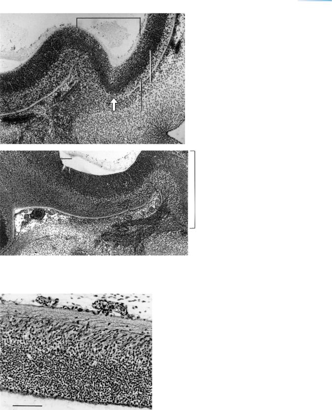
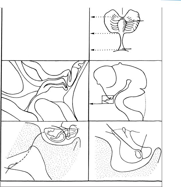
Figure 19–10. Still another section, to show the medial ventricular eminence and the ventral thalamus. Preoptico-hypothalamic (lower
Thal. v arrowhead) and mamillotegmental (upper arrowhead) tracts are identifiable.
Med. em.
Figure 19–11. Transverse section through part of one of the nasal sacs. The (blue) basement membrane is interrupted where emigrating crest cells leave the nasal epithelium.
|
CHOROID PLEXUS OF THE FOURTH VENTRICLE AND THE MEDIAL ACCESSORY OLIVARY NUCLEUS |
159 |
|||||
A |
|
|
|
|
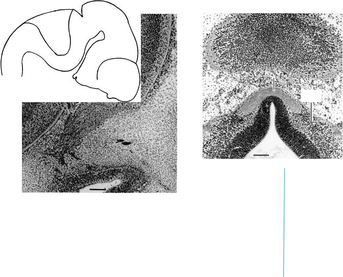
Figure 19–12. Olfactory and vomeronasal nerves |
||
|
|
|
|
in lateral to medial silver-impregnated sagittal |
|
||
|
Olfactory bulb |
|
|
|
|||
|
|
|
sections. (A) The olfactory fibers accompanied by |
||||
|
|
|
|
|
|||
|
|
|
|
|
olfactory ensheathing cells enter the olfactory bulb |
||
|
|
|
|
|
and form the external fibrous layer of the bulb. At |
||
|
|
|
|
|
the “pole” the intermediate layer of the bulb |
|
|
|
|
|
|
|
consists of only one lamina of tangentially arranged |
||
|
|
|
|
|
cells. (B) The vomeronasal nerve enters the |
|
|
|
|
|
|
|
olfactory bulb above the olfactory nerve (which |
||
|
|
|
|
|
differs in rodents). The area between the olfactory |
||
|
|
|
|
|
bulb and the medial ventricular eminence is the |
||
|
|
|
|
Ventricular |
forebrain septum. |
|
|
|
|
|
|
|
From Muller¨ |
and O’Rahilly (2004c, Cells Tissues |
|
|
|
|
|
|
Organs) by permission of S. Karger AG, Basel. |
|
|
|
"Pole" |
|
|
Intermediate |
|
|
|
|
Olf. n. |
|
|
|
|
|
|
|
|
|
|
|
|
|
|
B |
Med. eminence |
|
|
|
O |
|
|
|
|
|
|
|
|
||
|
|
|
|
l |
|
|
|
|
|
|
|
|
|
|
|
|
|
|
|
|
f |
|
|
|
|
|
|
|
a |
|
|
|
|
|
|
|
c |
|
|
|
|
|
|
|
t. |
|
|
|
|
|
|
|
b |
|
|
|
|
|
|
|
u |
|
|
|
|
|
|
|
l |
|
|
|
|
|
|
|
b |
|
|
|
|
. |
|
|
|
|
|
|
.n |
|
|
|
|
|
|
|
V.n |
|
|
|
|
|
|
|
|
|
|
. |
|
|
|
|
|
|
n |
|
|
|
|
|
|
|
. |
|
|
|
|
|
|
Olf |
|
|
|
|
|
|
|
|
|
|
|
|
|
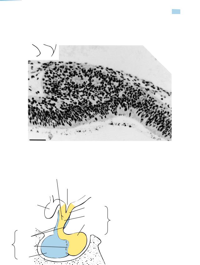
Stria medullaris thalami
Habenulo-interpeduncular tract
Lateral habenular nucleus
Figure 19–13. The lateral habenular nucleus in a sagittal section, an example of a far-advanced epithalamic area. The arriving fibers are of the stria medullaris thalami, the leaving fibers form the habenulo-interpeduncular tract (Fig. 19–23).
160 C h a p t e r 1 9 : CHOROID PLEXUS OF THE FOURTH VENTRICLE AND THE MEDIAL ACCESSORY OLIVARY NUCLEUS
Olf. ventricle
N. |
|
|
|
|
M |
|
|
terminalis |
|
|
|
|
|
|
|
|
|
|
|
|
×4 |
||
Isthmic nucleus
Figure 19–14. Relationships of olfactory tubercle and bulb, showing the entry of the nervus terminalis into the olfactory tubercle, and of the olfactory nerve fibers into the olfactory bulb. The olfactory ventricle is evident. Both tubercle and bulb possess an intermediate layer, and multiple nerve fibers are present in the tubercle. The sulcus circularis is marked by an arrowhead. The neural crest of earlier stages had developed into vomeronasal and terminal ganglia, but only after a certain degree of development of the central structures (tubercle and bulb). Both ganglia are medial, but the terminalis is more rostral, although its nerve fibers enter the brain caudal to the sulcus circularis. The ganglion of the nervus terminalis, although not yet found in the previous stage, is almost constantly present in stage 19. The fibers of the nervus terminalis appear later than the ganglion, as described in a fetus of 38 mm by Pearson (1941).
Figure 19–15. The isthmic nucleus and the commissure of the trochlear nerves. The level of this coronal section is shown in the key drawing. The bilateral isthmic nuclei are ventrolateral and rostral to the trochlear decussation (X4). Details have been provided by O’Rahilly et al. (1988).
CHOROID PLEXUS OF THE FOURTH VENTRICLE AND THE MEDIAL ACCESSORY OLIVARY NUCLEUS |
161 |
I |
0.1 mm |
II
A
III
IV
Habenular commissure
V
Intermediate
zone
Commissures
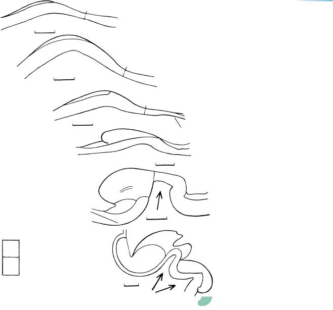
Figure 19–16. The development of the epiphysis cerebri. Based on Turkewitsch (1933), whose stadia are given Roman numerals. See Table 17–1.
A, Vorderlappen
P, Hinterlappen
Posterior commissure
A
A
A
P
VI
Pineal recesses
162 C h a p t e r 1 9 : CHOROID PLEXUS OF THE FOURTH VENTRICLE AND THE MEDIAL ACCESSORY OLIVARY NUCLEUS
Figure 19–17. A sagittal section to show the epiphysis, which is now a rostrally directed outgrowth of the roof of the synencephalon (Fig. 19–16D). This is the Vorderlappen of Stadium IV of Turkewitsch (1933). It projects towards the left side of the photomicrograph and forms a characteristic “step” (Stufe). Two so-called follicles are visible, as observed also by Hochstetter (1923). The site of the epiphysial evagination is marked by the pineal recess. Bar: 0.05 mm.
|
Infundibulum |
|
|
Infundibular |
|
|
recess |
|
|
NEUROHYPOPHYSIS |
|
Optic |
|
|
chiasma |
|
|
|
Median |
|
|
eminence |
|
|
Infundibular |
|
|
stem |
|
ADENOHYPOPHYSIS |
Infundibular |
|
process |
||
|
||
Pars |
(neural lobe) |
|
tuberalis |
|
|
Pars |
|
|
intermedia |
|
|
Pars |
|
|
distalis |
|
|
ANTERIOR |
POSTERIOR |
Figure 19–18. The terminology of the hypophysis cerebri based on
Rioch et al. (1940). Cf. Table 17–2.
|
CHOROID PLEXUS OF THE FOURTH VENTRICLE AND THE MEDIAL ACCESSORY OLIVARY NUCLEUS |
163 |
||
|
|
|
|
|
A |
B |
Infundibular notch |
|
|
|
|
|
||
|
|
|
Pars |
|
|
|
|
distalis |
|
|
|
Solid |
|
|
|
|
stem |
|
|
|
|
|
Pharyngeal |
|
|
|
Pharynx |
hypophysis |
|
|
|
|
|
|
C |
D |
|
|
|
|
3rd ventricle |
|
|
|
|
Chiasma |
|
|
|
|
Basi |
|
|
|
|
- |
|
|
|
T |
Pharynx |
ongue |
|
|
E |
Stem |
cranium |
|
F
Pars |
Pars |
|
tuberalis |
||
intermedia |
||
|
Basi-
cranium
Dorsum |
Pars |
|
distalis |
||
sellae |
||
|
Hypophysial
fossa
Pharyngeal hypophysis
Figure 19–19. Development of the hypophysis as seen in median sections. (A), (C), and (D) The adenohypophysial stalk is closed and is embedded in the developing sphenoid cartilage. The attachment of the stalk to the roof of the pharynx constitutes the pharyngeal hypophysis. The wall of the neurohypophysis is characteristically folded. (B) The adenohypophysial pouch seen from in front. (E) At stage 23. The remains of the stalk are shown by an interrupted line. (F) At 60 mm. The greater part of the organ lies in the hypophysial fossa of the (still cartilaginous) sphenoid bone. The (white) cleft represents the cavity of the original adenohypophysial pouch.
164 C h a p t e r 1 9 : CHOROID PLEXUS OF THE FOURTH VENTRICLE AND THE MEDIAL ACCESSORY OLIVARY NUCLEUS
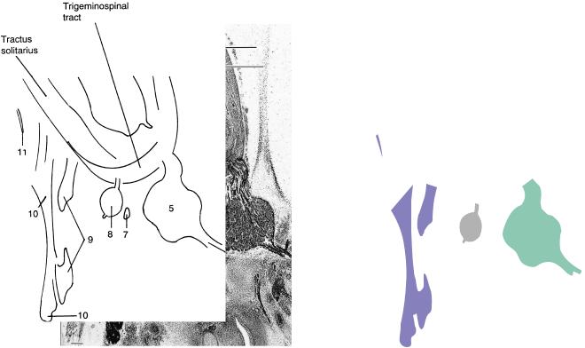
Mes. tract of 5
Inf. cerebellar peduncle
Figure 19–20. The area of the pontine flexure with cranial nerves 5, 7, 8, 9, and 10, silver-impregnated. Many tracts are identifiable, e.g., the mesencephalic tract of the trigeminal nerve, the trigeminospinal tract, and the tractus solitarius. Among the cerebellar connections are found the inferior cerebellar peduncle (vestibulocerebellar and trigeminocerebellar fibers) and even components of the superior peduncle (dentatorubral fibers, which pass lateral to the nucleus isthmi). The porus duralis of the trigeminal nerve is visible. The internal carotid artery, sectioned below the trigeminal ganglion, is filled with blood and possesses a thick arterial wall. Bar: 0.2 mm. From Muller¨ and O’Rahilly (1990a).
In monkeys at a comparable stage, cerebellar neuronal production is at a peak. Two migratory waves participate in the formation of the cerebellum: from the rhombic lip, the migrating cells being at the surface, to which their nuclei are parallel; and from the ventricular layer, the cells lying at a right angle to the surface of the cerebellum. The cells of the two migrations become partly mixed, as is true in the dentate nucleus.
Parasympathetic ganglia appear in the head between stages 15 and 19 (Wozniak´ and O’Rahilly, 1980).
CHOROID PLEXUS OF THE FOURTH VENTRICLE AND THE MEDIAL ACCESSORY OLIVARY NUCLEUS |
165 |
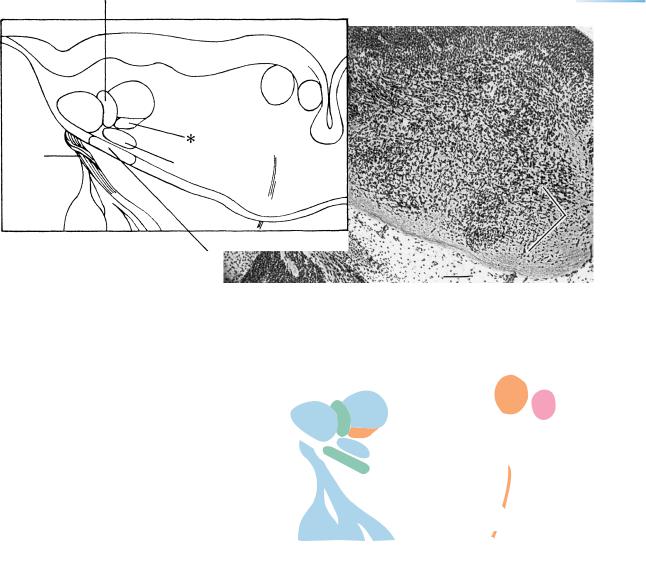
Inferior cerebellar peduncle
|
|
6 |
|
8v |
7 |
|
|
|
|
8c |
|
Cochlear |
|
|
nerve |
8v |
6 |
|
Trigeminospinal tract
Figure 19–21. The cochlear nerve and nuclei. The cochlear nerve has a spiral pattern like a rope from stage 19 onwards. It pierces a dense mass of cells (the ventral cochlear nucleus), where it enters the rhombencephalon. Both cochlear nuclei are lateral to the inferior cerebellar peduncle, whereas the vestibular nuclei are medial. An asterisk indicates the tractus solitarius in the key. Bar: 0.1 mm.
166 C h a p t e r 1 9 : CHOROID PLEXUS OF THE FOURTH VENTRICLE AND THE MEDIAL ACCESSORY OLIVARY NUCLEUS
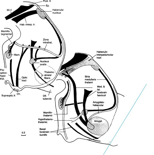
Fig. 19–24
Figures 19–22 and 19–23. Tracts of the forebrain in a median and a right lateral view, respectively. Some of the nuclei are shown as stippled areas, and superficially situated tracts are also stippled. New tracts in stage 19 include the thalamostriatal tract (Stammbundel¨ of His or lateral prosencephalic fasciculus), around which the internal capsule continues to develop (but does not yet reach the pallium) (Fig. 19–24), the stria medullaris thalami, the fibers of the zona intrathalamica, and the amygdalo-habenular tract. The tract of the zona limitans intrathalamica later becomes the lamina medullaris externa. The sequence of appearance of the tracts is given in Appendix 4.
The fibers of the catecholamine neurons seem to penetrate no further than the ventricular eminences (Zecevic and Verney, 1995). At about this stage it has been found in the rat that septohippocampal projections reach the hippocampus, and that they are preceded by hippocamposeptal projections.
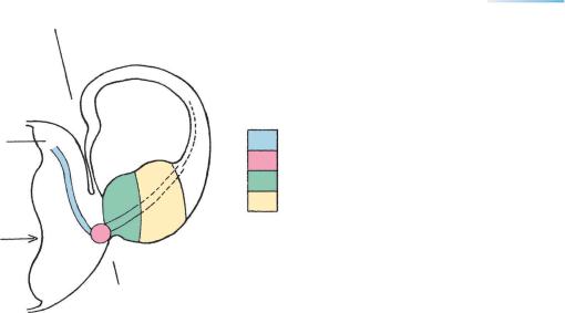
CHOROID PLEXUS OF THE FOURTH VENTRICLE AND THE MEDIAL ACCESSORY OLIVARY NUCLEUS |
167 |
|
Di. |
Tel. |
|
Dorsal |
|
|
thalamus |
Striothalamic fibers |
|
Lat. forebrain bundle
Med. ventricular eminence
Lat. ventricular eminence
Hypothalamic
sulcus
Figure 19–24. Beginning development of the internal capsule. Thalamostriatal fibers end abruptly in the medial ventricular eminence (cf. Fig. 5 in Richter, 1965), after having changed their direction at the ditelencephalic boundary (hemispheric stalk), where the fibers contribute to the lateral forebrain bundle (Stammbundel¨ of His, 1904). Corticothalamic fibers appear in stage 21 (Meyer et al., 2000). Their outgrowth (in the mouse) is influenced by Pax 6-positive cells at the corticostriatal boundary. Gene mutations (Pax 6 Sey/sey, Gbxc-2, f.ws.) cause defects of the internal capsule (Table 2 in Molnar´ and Butler, 2002). The level of this section is indicated in Figure 19–23.