208 |
C h a p t e r 2 2 : THE INTERNAL CAPSULE AND THE OLFACTORY BULBS |
Figure 22–4. Median view to show elements that are important in understanding the internal capsule. The lateral ventricular eminence, which gives rise to the future corpus striatum, is projected onto the medial surface to show its extent. The fibers of the lateral prosencephalic fasciculus (Stammbundel¨ of His) arise in the neopallial wall and descend lateral to the lateral ventricular eminence. The fibers, having traversed the hemispheric stalk, ascend in the diencephalic wall and spread into, among other structures, the dorsal thalamus. All the features indicated are shown as if the prosencephalon were transparent. The stria terminalis “arising mainly in the amygdaloid complex” follows the curvature of the sulcus terminalis towards the interventricular foramen and here, near the anterior commissure, various components of the stria terminalis become separated (Kuhlenbeck, 1977).
It has been found (in the hamster) that thalamocortical fibers (stage 21) are present before corticothalamic fibers become visible in the internal capsule.
Lateral forebrain bundle & internal capsule
Stria terminalis
THE INTERNAL CAPSULE AND THE OLFACTORY BULBS
A C
Nerves
C1
2
3
4
5
6
7
8
T1
Foramen Notochord transversarium
209
Vertebrae
C1
2
3
4
5
6
7
T1
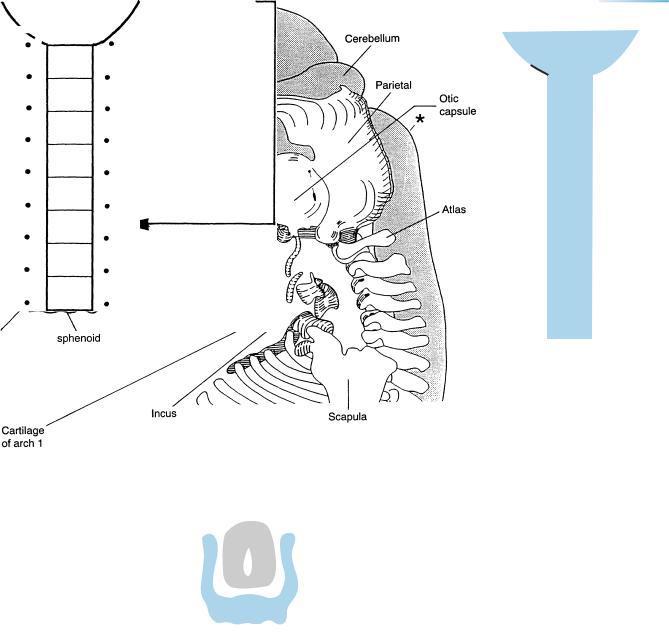
Figure 22–5. Development of the skull and vertebrae. (A) Left lateral view of a reconstruction of the brain and chondrocranium. The asterisk indicates the junction with the spinal cord. The basal portions of the skull are present in cartilage. The basolateral and lateral walls are still far from complete, but the following are discernible: the greater and lesser wings of the sphenoid; the otic capsule; and the parietal plate, which is not yet fused with the plate of the other side. Hence the foramen magnum has not yet developed. The chondrocranium of stage 22 is compared with that of stage 23 in Figure 23–8. (B) The temporary, normal spina bifida. Cf. Fig. 20–22. (C) Relationship of spinal nerves to vertebrae.
Cervical nerve 1 emerges below the skull (otherwise it would be a cranial nerve!) and above the atlas. Cervical nerves 2 to 7, therefore, leave the vertebral canal above the correspondingly numbered vertebrae. Cranial nerve 8 emerges below cervical vertebra 7 and above thoracic vertebra 1.
All the remaining spinal nerves leave below the correspondingly numbered vertebrae.
210 C h a p t e r 2 2 : THE INTERNAL CAPSULE AND THE OLFACTORY BULBS
|
|
Stria |
|
B |
medullaris |
|
thalami |
|
|
|
|
|
A
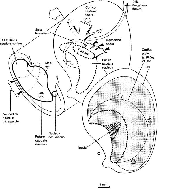
Figure 22–6. A schematic representation of the development of the internal capsule based on reconstructions. (A) Dorsal view of the opened right hemisphere showing the internal capsule in relation to the lateral ventricular eminence. Neopallial fibers (between the black arrowheads), which are an essential component of the definitive internal capsule, are shown on their way from the neopallium, lateral to the ventricular eminence and running to the lateral prosencephalic fasciculus (cf. Fig. 22–11). The first fibers can be traced to an area comparable to the later postcentral gyrus. The lateral ventricular eminence has grown more rapidly than the medial, which is adjacent to the hemispheric stalk. A faint groove called the intereminential sulcus by the present writers is distinguishable between the eminences.
The neurons for the neostriatum arise during stages 21–23, as has been shown in rhesus. It has been found that the histochemical development of the nucleus accumbens resembles that of the lateral ventricular eminence. (B) A right lateral view showing the internal capsule in more detail. The neopallial fibers (black arrowheads) between the primordia of the future caudate nucleus medially, and the putamen laterally, accumulate in the thick lateral prosencephalic fasciculus (Stammbundel¨ of His) before entering the diencephalon. Finally they continue in three main groups: epithalamic, thalamic, and mesencephalic (white arrows). The main bundle is arched over by the stria terminalis, which is composed chiefly of fibers that can be traced to the medial part of the amygdaloid nuclei. (C) Also a right lateral view: the cortical plate at stage 22 (stippled area) extends over half the surface of the neopallium, and its increase from stage 22 to stage 23 is shown.
THE INTERNAL CAPSULE AND THE OLFACTORY BULBS |
211 |
22 – 7, 8
Lat. eminence
Ivf
Hippocampus
V3
Tegmental fibers
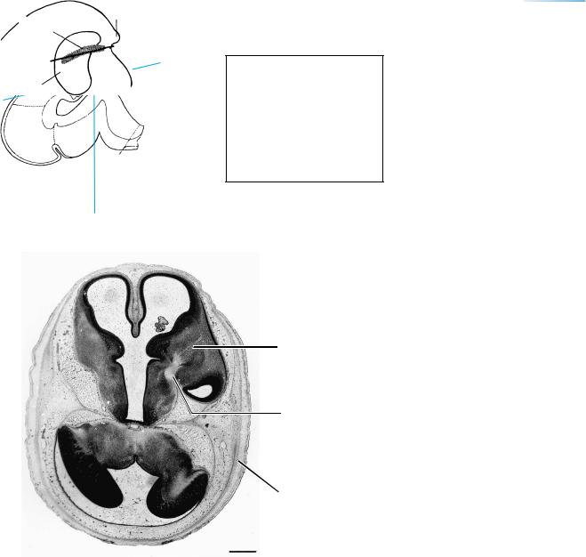
Med. longutidinal fasciculus
Inf. colliculus
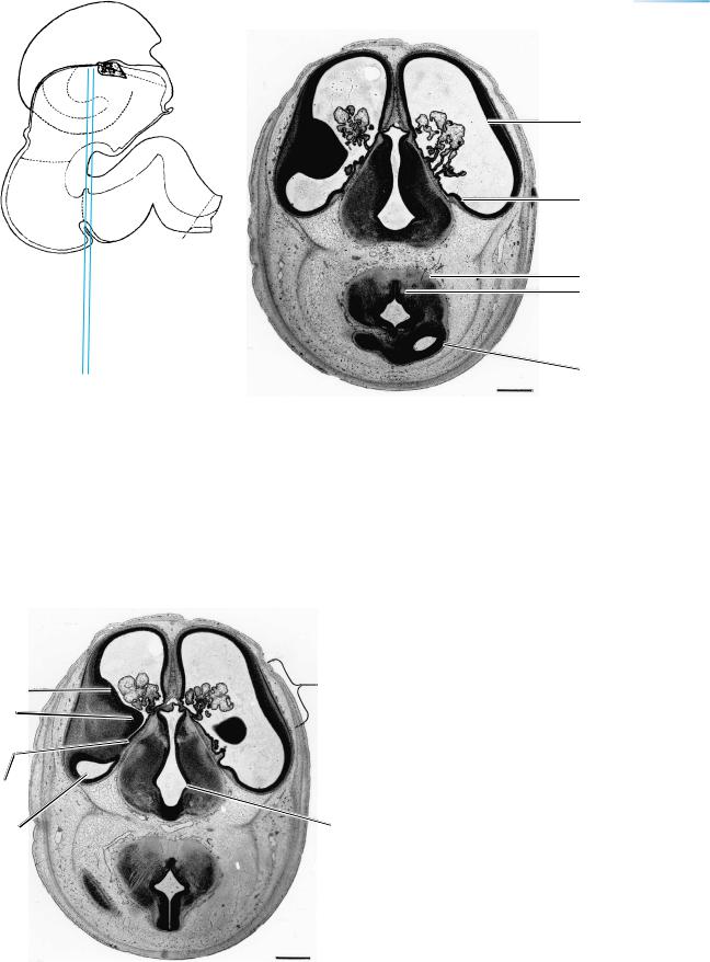
Figure 22–7. The lateral ventricular eminence, hippocampus, and choroid plexus are visible. The folded roof of the diencephalon is adjacent to the stria medullaris thalami on each side. The dorsal thalami appear dark. The mesencephalic ventricle (future aqueduct) and its prolongation into the right inferior colliculus are evident. The fiber masses in the tegmentum are increasing. The medial longitudinal fasciculus is visible on each side. Dorsal to the main mesencephalic ventricle, the white lines are fibers leading to the trochlear decussation.
Figures 22–7 to 22–11. The levels of these sections are indicated also in Figure 22–3. The left side is cut at a deeper level than the right. The sections of Figures 22–7 to
22–9 and of Figure 22–11 were selected by Bartelmez and Dekaban (1962), who completed their study with stage 22. The bars represent 1 mm.
Lat. & med.
em.
Sulcus terminalis
Inf. horn
 Insula
Hypo-
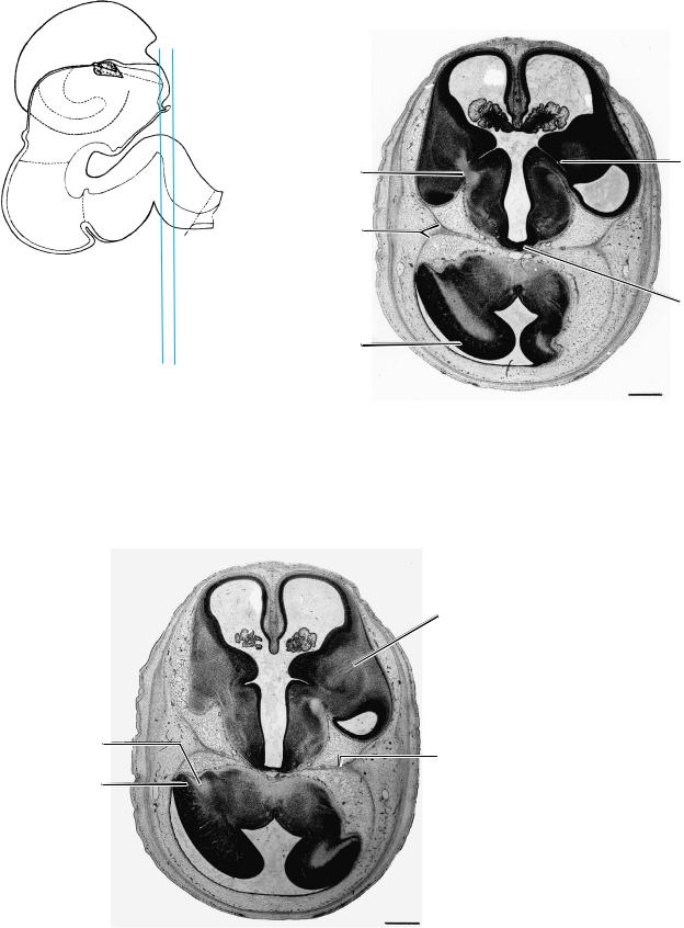
thalamic Figure 22–8. The cortical plate is evident on each side sulcus and is mostly opposite the ventricular eminences at the
site of the future insula. A faint groove can be seen between the medial and lateral eminences. The sulcus terminalis is medial, towards the diencephalon. The inferior horn of the left lateral ventricle is evident. The diencephalic area dorsal to the hypothalamic sulcus is mainly ventral thalamus. In the isthmus, the tegmentum is filled with fibers, including the medial longitudinal fasciculus. A cerebellar plate is visible to the left of the isthmus.
212 |
C h a p t e r 2 2 : THE INTERNAL CAPSULE AND THE OLFACTORY BULBS |
|
TEL. |
Lat. |
Sulcus |
prosenc. |
terminalis |
fasciculus |
|
|
DI. |
Tentorium |
|
|
Mamillary |
|
area |
Internal |
|
cerebellar |
|
swelling |
|
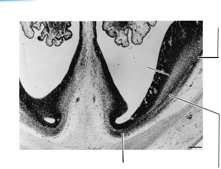
Figure 22–9. This section, at the level of the interventricular foramina, shows the lateral prosencephalic fasciculus (Stammbundel¨ of His) on the left side. The sulcus terminalis is the internal boundary between telencephalon and diencephalon. The mamillary region and the mamillothalamic tract are visible. The internal cerebellar swellings almost fill the fourth ventricle. The fibers penetrating it (on the left side) belong mainly to the inferior cerebellar peduncle. The lateral portions of the tentorium cerebelli are evident here as well as in the other sections.
Inf. cerebellar peduncle
Flocculus
Corticothalamic fibers
Tentorium
Figure 22–10. At the right, fibers can be seen arising in the neopallium and running towards the Stammbundel¨ , which is visible at the di-telence-phalic borderline (hemispheric stalk). The stalk lies between the sulcus terminalis on the ventricular and the di-telencephalic sulcus on the external surface. The mamillary region still shows. At the rostral surface of the left cerebellar plate, the lighter triangular area is the inferior cerebellar peduncle. To its left is the flocculus, and to its right is the area of the dentate nucleus.
216 C h a p t e r 2 2 : THE INTERNAL CAPSULE AND THE OLFACTORY BULBS
|
A |
Figure 22–13 (Continued ). The level of A and |
|
the areas A and B in the photomicrographs are |
|
|
|
|
indicated. |
B
A
|
|
THE INTERNAL CAPSULE AND THE OLFACTORY BULBS |
217 |
|
|
|
|
|
|
A |
B |
|
C
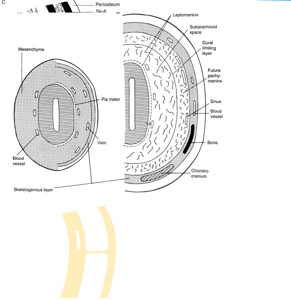
Figure 22–14. Schematic representation of the development of the cranial meninges. (A) At approximately 3 12 postfertilizational weeks, mesenchyme from various sources surrounds the brain, and blood vessels are present. The sources probably include prechordal plate, unsegmented paraxial mesenchyme, segmented paraxial (somitic) mesenchyme, ectomesenchyme (neural crest), and neurilemmal cells (neural crest). The pia mater develops between the blood vessels and the cerebral wall. A dense skeletogenous layer forms at the periphery and veins are situated on its internal aspect. As the meningeal vessels are transformed into arteries and veins, they become progressively isolated from the CSF compartment (Mar´ın-Padilla, 1988b). (B) Approximately 7 postfertilizational weeks. The dural limiting layer appears between the pia and the skeletogenous layer. Large meshes form in the future subarachnoid space, smaller meshes between the dural limiting and skeletogenous layers (in the pachymeninx). Cartilage and intramembranous bone are forming within the skeletogenous layer. The veins (dural sinuses) on the internal aspect of the skeletogenous layer are now intradural in position. The leptomeningeal cells establish perivascular spaces around the arachnoidal and pial vessels, and these drain into the perivascular lymphatic system of the extracranial vasculature (Mar´ın-Padilla, 1988b).
(C) Scheme of the meninges in the adult. So-called subdural hemorrhage is within the dural border layer and hence is intradural. Based largely on Schachenmayr and Friede (1978) and Alcolado et al. (1988).
The development of the cranial meninges was investigated by Hochstetter (1939) among others, and the complicated origin of these membranes from several sources (including the prechordal plate, parachordal mesoderm, and neural crest) was studied by O’Rahilly and Muller¨ (1986). The loose mesenchyme around most of the brain at stage 13 (Fig. 13–5) is termed the primary meninx. At stage 17 the dural limiting layer begins to form basally (Fig. 17–5) and the skeletogenous layer of the head soon becomes visible (Fig. 22–14). At stage 23 the pachymeninx and the leptomeninx are distinguishable in the head (Fig. 23–9).
The spinal meninges have been investigated particularly by Hochstetter (1934) and Sensenig (1951). The future pia mater, derived from neural crest cells, can be detected at stage 11 and, at stage 15, the primary meninx is represented by a loose zone between the vertebral primordia and the neural tube. At stage 18 the mesenchyme adjacent to the vertebrae becomes condensed as the dural lamella. At stage 23 the dura lines the vertebral canal completely and the future subarachnoid space is becoming cell-free. The arachnoid, however, does not appear until either trimester 3 or postnatally, although a much earlier artifact has frequently been mistaken for it.