

Книги по МРТ КТ на английском языке / The Embryonic Human Brain An Atlas of Developmental Stages. Third Edition. 2006. By Ronan O'Rahilly-1
.pdf258 |
C h a p t e r 2 3 : THE BRAIN AT THE END OF THE EMBRYONIC PERIOD |
Figure 23–36. A comparison of the brain at 6 weeks with that at 8 weeks. A median section at stage 17 (in mauve) has been superimposed on one at stage 23, drawn to the same scale and centered on the floor of the mesencephalon. In the intervening fortnight (1) the brain has increased in size (approximately 30%),
(2) the cervical and pontine flexures have become more Stage 17 pronounced, and (3) the roof of the diencephalon has become
almost completely covered by the cerebral hemispheres.
17
Stage 23
The Cerebellum
Develops from the alar plate of both the isthmic and the first rhombencephalic neuromeres. The rhombic lip, which is a part of the alar plate, is not the sole origin of the cerebellum.
At Stage 23:
The two intraventricular cerebellar components grow towards the median plane and begin to fuse rostrally.
Two areas of fusion are visible: (1) that adjacent to the superior velum would appear to be the nodule and (2) that beginning at the level of the cerebellar hemispheres.
The roof of the isthmus is the superior medullary velum.
Medulloblastoma. The external granular layer of the cerebellum, which appears at this stage and disappears within 1–2 years after birth, is generally believed to be the origin of so-called medulloblastoma, most instances of which occur in the vermis.


THE SPINAL CORD NEUROTERATOLOGY |
261 |
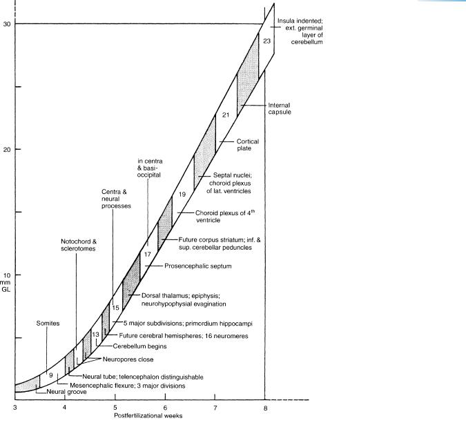
Figure 23–37. Summary of some of the major morphological events in terms of stages in a graph of the greatest embryonic length plotted against postfertilizational age.
Chondrification

262 |
C h a p t e r 2 3 : THE BRAIN AT THE END OF THE EMBRYONIC PERIOD |
|
|
||
TABLE 23–2. Principal Research Publications on the Nervous System in Staged Human Embryos |
||
Stages |
Major Topic |
References |
|
|
|
8 |
Neural folds |
O’Rahilly and Muller¨ (1981) |
|
Prechordal plate |
Muller¨ and O’Rahilly (2003a) |
9 |
Divisions of brain |
Muller¨ and O’Rahilly (1983) |
|
Primitive streak |
Muller¨ and O’Rahilly (2004a) |
|
Otic primordium |
O’Rahilly (1963) |
10 |
Fusion of neural folds |
Muller¨ and O’Rahilly (1985) |
|
|
O’Rahilly and Muller¨ (2002) |
|
Optic primordium |
O’Rahilly (1966) |
|
Caudal eminence |
Muller¨ and O’Rahilly (2004a) |
11 |
Rostral neuropore |
Muller¨ and O’Rahilly (1986) |
|
|
O’Rahilly and Muller¨ (1989; 2002) |
12 |
Caudal neuropore |
Muller¨ and O’Rahilly (1987) |
|
|
O’Rahilly and Muller¨ (2002) |
|
Hypoglossal nerve |
O’Rahilly and Muller¨ (1984) |
|
Somites |
O’Rahilly and Muller¨ (2003) |
|
Segmentation |
Muller¨ and O’Rahilly (2003b) |
13 |
Closed neural tube |
Muller¨ and O’Rahilly (1988a) |
14 |
Cerebral hemispheres |
Muller¨ and O’Rahilly (1988b) |
|
Neuromeres complete |
Muller¨ and O’Rahilly (1997) |
15 |
Zoning in diencephalon |
Muller¨ and O’Rahilly (1988c) |
16 |
Neurohypophysial evagination |
Muller¨ and O’Rahilly (1989a) |
17–23 |
Olfactory system |
Muller¨ and O’Rahilly (1989b; 2004b) |
|
Amygdaloid complex |
Muller¨ and O’Rahilly (2006) |
18–20 |
Choroid plexuses |
O’Rahilly and Muller¨ (1990) |
21–23 |
Cortical plate; cerebellum |
Muller¨ and O’Rahilly (1990b) |
|
Ventricular eminences |
Muller¨ and O’Rahilly (1990b) |
|
Ventricles and choroid plexuses |
O’Rahilly and Muller¨ (1990) |
|
Meninges |
O’Rahilly and Muller¨ (1986) |
23 |
Rhombencephalon |
Muller¨ and O’Rahilly (1990a) |
|
Occipitocervical segmentation |
Muller¨ and O’Rahilly (1994, 2003a) |
|
|
|


EARLY POSTEMBRYONIC PHASE |
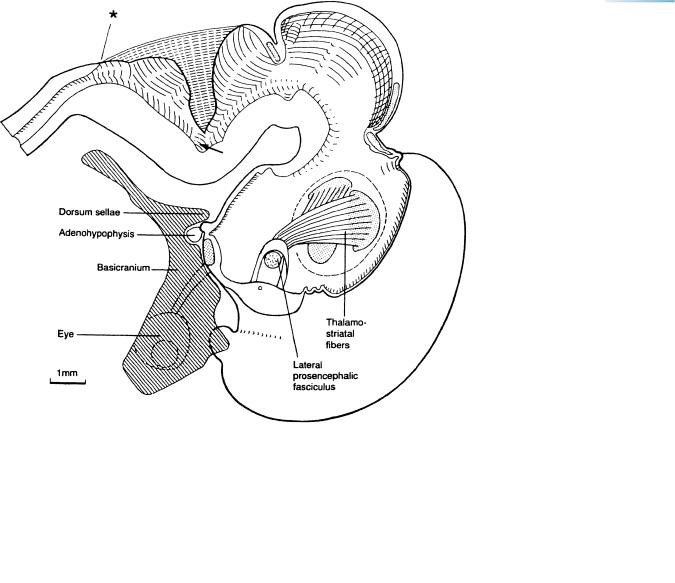
267 |
Figure 24–2. Graphic reconstruction from sagittal sections to show a median view of the brain at 33 mm. The asterisk marks the junction with the spinal cord. The commissural plate is now impressive. The rostral part of the roof of the third ventricle is folded. The entrance of the lateral prosencephalic fasciculus (Stammbundel¨ ) into the diencephalon is shown, as is the stria terminalis arching over it at the di-telencephalic border. Corticipetal fibers that participate in the formation of the primordial plexiform layer arrive early (ca. stage 16, Marin-Padilla, 1988a, b). In experimental mammals corticifugal fibers have also been observed before the cortical plate forms. Three nuclear areas (stippled) are beginning to be outlined in the thalamus. (See also Fig. 24–3.) The thalamostriatal and striatothalamic fibers are connected mostly with what may later become the dorsomedial nucleus. The facial colliculus (arrow) is noticeable on the floor of the rhombencephalon. The central stem of the chondrocranium (hatched) has been included, as well as the left optic nerve and the eye.