Книги по МРТ КТ на английском языке / The Embryonic Human Brain An Atlas of Developmental Stages. Third Edition. 2006. By Ronan O'Rahilly-1
.pdf178 C h a p t e r 2 0 : THE CHOROID PLEXUS THE LATERAL VENTRICLES, THE OPTIC AND HABENULAR COMMISSURES
Intermediate layer Septal nucleus
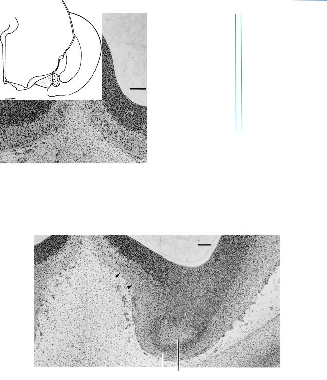
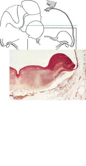
Figure 20–14. A graphic reconstruction of the medial septal nucleus, which consists of one separate mass in this embryo. Two additional fiber bundles are shown: a, between the olfactory bulb and the medial septal nucleus, and b, between the rostral and caudal septal areas.
Figures 20–13 to 20–17 show the nuclei of the prosencephalic septum. The septal nuclei are cellular agglomerations at the periphery of the intermediate layer. The cholinergic neurons of the nuclei at the base of the forebrain are believed to be important in the degenerative processes leading to Alzheimer disease. Septo-hippocampal cholinergic fibers reach their target (in the rat) at what would correspond to stage 19.
The basal part of the medial telencephalic wall (the septum verum) forms as the cerebral hemispheres expand beyond the lamina terminalis, and it extends between the commissural plate and the olfactory bulb.
Figure 20–15. Detail of the septal area of the right hemisphere in a coronal section. The cellular agglomeration at the periphery of the intermediate layer of the medial wall of the telencephalon represents the primordium of a septal nucleus.
Figures 20–15 to 20–17. Sections at the levels indicated in the key on the next page. The bars represent 0.1 mm.
THE CHOROID PLEXUS THE LATERAL VENTRICLES, THE OPTIC AND HABENULAR COMMISSURES |
179 |
Commissural |
20 – 15, |
17 |
|
16 |
|||
plate |
|||
|
|||
|
|
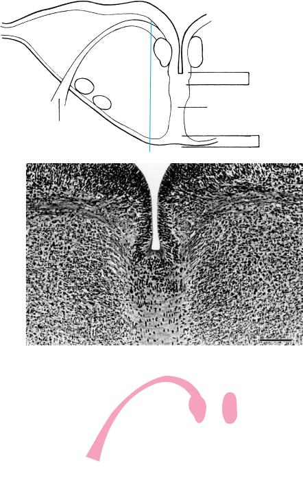
Figure 20–16. A more rostral section showing the right and left sides of the septal area. The connection is the commissural plate, which at this time consists mainly of ventricular layer. The cellular agglomeration in the septal area is evident.
Olfactory bulb
Olfactory nerve fiber layer
Figure 20–17. A coronal section at the level of the olfactory bulbs. Fibers from the developing external fibrous layer of the olfactory bulb ascend (arrowheads) to the medial septal nucleus.
180 C h a p t e r 2 0 : THE CHOROID PLEXUS THE LATERAL VENTRICLES, THE OPTIC AND HABENULAR COMMISSURES
Figure 20–18. A coronal section through the rhombencephalon in the region of the facial nucleus. The septum medullae, which is derived from the floor plate, is present in the epinotochordal region and extends as far rostrally as the diencephalon (to the boundary between caudal and rostral parencephalon). The septum is a specialized grid formed by intersecting sagittal and transverse fibers, between which are migrating cells from the floor plate. These cells form the raphe nuclei, which, because of their neurotransmitters, are essential for the development of the brain. Moreover, they are related to the locus caeruleus, the substantia nigra, the red nucleus, and (after stage 20) to the cortical plate. Bar: 0.1 mm.
At their first appearance the visceromotor nuclei are ventromedial in position (Fig. 14–9). Subsequently, the neurons of cranial nerves 5 and 9–11 migrate laterally. The facial nucleus, however, remains medially and is now also dorsal in position. Migration of the facial neurons laterally and rostrally occurs relatively late (Windle, 1970): in the late embryonic or early fetal period. The facial nucleus becomes better defined at the beginning of the fetal period (Pearson, 1946; Jacobs, 1970).
The migration of the facial nucleus and the unexpected course of its fibers around the abducent nucleus are an example of neurobiotaxis, a term introduced in 1907 by C. U. Ariens¨ Kappers (1932) for the retention of a neuron as close as practicable to its main source of stimulation, and the migration that may occur in the direction from which the greatest density of stimuli arises, achieved by elongation of the axon. The mechanism of this phenomenon remains obscure. Another example is provided by the nucleus ambiguus.
7
Floor plate
Septum
medullae
Motor fibers
from 7
Floor plate
THE CHOROID PLEXUS THE LATERAL VENTRICLES, THE OPTIC AND HABENULAR COMMISSURES |
181 |
Hypoglossal nucleus
Dorsal efferent vagal nucleus
Nucleus intercalatus
12
|
Med. |
Septum |
accessory |
medullae |
olivary |
|
nucleus |
|
12 |
20 – 18
20 – 19
Med. accessory olivary nucleus
Nucleus
solitarius
Inferior cerebellar peduncle
Spinothalamic tract
Somatic & visceral afferent
Visceral efferent
Somatic efferent
Figure 20–19. A coronal section of the rhombencephalon. The medial accessory olivary nucleus arises between the hypoglossal roots of the two sides. This nucleus is the first of five (Muller¨ and O’Rahilly, 1990c) that will be present by the beginning of the fetal period (Fig. 24–4). The hypoglossal nucleus is visible dorsally. Migratory material has accumulated at the ventral surface of the section. It arises in the rhombic lip and was first described in the human embryo by Essick (1912). His term “olivo-arcuate migration” is unsatisfactory, however, because it has since been shown (in the monkey) that the olivary nuclei arise mainly from the ventricular layer. Tall cells present in the raphe are contacted by fibers from the nuclei of nearby cranial nerves, and are important in the production of neurotransmitters. Bar: 0.2 mm.
182 C h a p t e r 2 0 : THE CHOROID PLEXUS THE LATERAL VENTRICLES, THE OPTIC AND HABENULAR COMMISSURES
Cerebellum
Roof plate |
4th ventricle |
Alar lamina
Basal lamina
Floor plate
Basilar a.
Figure 20–20. A transverse section through the hindbrain showing the internal cerebellar swellings. The alar and basal laminae, which are lateral and medial in this region, are separated from each other on their ventricular surface by the sulcus limitans.
THE CHOROID PLEXUS THE LATERAL VENTRICLES, THE OPTIC AND HABENULAR COMMISSURES |
183 |
A |
Middle |
|
plexus |
Rostral
plexus
B |
Tentorial |
|
plexus |
Sagittal
plexus
Inf. cerebral v.
Sup. petrosal sinus
Ophthalmic v.
Primary capital v.
Otic vesicle
Caudal
plexus
Future jugular foramen
Precardinal v.
Transverse
sinus
Caudal
plexus
Sigmoid
part
Int. jugular
v.
Cavernous sinus
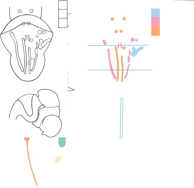
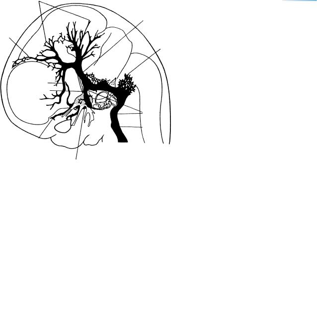
Figure 20–21. The venous system of the head. (A) At stage 17 the tributaries of the primary capital vein are arranged as three dural plexuses. The internal jugular vein has begun its development. (B) At stage 20. The sigmoid sinus has begun to form. From now on the venous system of the head is asymmetrical. From Streeter (1918). The venous system from stage 14 to 80 mm GL has been studied in detail by Padget (1957).
184 C h a p t e r 2 0 : THE CHOROID PLEXUS THE LATERAL VENTRICLES, THE OPTIC AND HABENULAR COMMISSURES
Ventricular
Intermediate Zones
Marginal
Spinal ganglion
Neural
process
Leptomeninx
Centrum Notochord
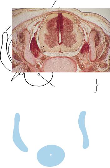
Figure 20–22. Section through the spinal cord and a centrum. The neural processes of the two sides do not yet meet each other dorsally, thereby constituting the normal spina bidfida occulta that is characteristic of the embryonic and early fetal periods.

NEUROTERATOLOGY |
185 |
NEUROTERATOLOGY SPINA BIFIDA OCCULTA
Spina bifida occulta is possible from the first sign of the appearance of vertebrae at stage 15 until completion of the neural arches early in fetal life (Fig. 20–22). It is important to keep in mind that before ossification of the spinous processes has taken place, a normal spina bifida occulta may
be said to be present throughout the length of the vertebral column. This is the condition at birth. Postnatally a radiologically detectable failure in the completion of one or more neural arches may be merely a lack of ossification dorsomedially, especially in the lumbosacral region. This is a normal finding in children of 2 years of age and in part of the sacrum it persists in about one-fifth to one-quarter of adults.

186 C h a p t e r 2 0 : THE CHOROID PLEXUS THE LATERAL VENTRICLES, THE OPTIC AND HABENULAR COMMISSURES
TABLE 20–1. Features of the Longitudinal Zones of the Diencephalon: Details for the General Scheme Given in Table 18–1
Zone |
Feature |
Stage of Primordium |
Fig. |
|
|
|
|
1. Epithalamus |
Habenular nucleus |
15 |
19–13 |
|
Epiphysis |
16 |
16–7F |
|
Posterior commissure |
17 |
19–4 |
2. Dorsal thalamus |
Nucleus of posterior commissure |
15 |
|
|
Lateral geniculate body (dorsal part) |
Fetal period |
|
|
Sulcus medius (marginal ridge) |
15–16 |
16–7F |
|
|
|
17–16 |
|
|
|
18–6 |
|
|
|
21–14 |
3. Ventral thalamus |
Lateral geniculate body (ventral part) |
21 |
21–14 |
|
Hypothalamic sulcus |
15 |
15–4 |
|
|
|
16–7E |
|
|
|
17–16 |
4. Subthalamus |
Subthalamic nucleus |
16 |
21–12 |
|
Globus pallidus externus |
19–20 |
21–15 |
|
Globus pallidus internusa |
21 |
21–14 |
|
Faint, unnamed sulcus |
15 |
15–4 |
5. Hypothalamus sensu lato |
Chiasmatic plate |
10 |
10–7 |
|
|
|
12–5 |
|
|
|
15–9 |
|
Hypothalamic cell cord |
14 |
16–7C |
|
Preoptico-hypothalamotegmental tract |
14 |
19–10 |
|
Interstitial nucleus |
14 |
14–5 |
|
Medial ventricular eminence |
14 |
16–7B |
|
Neurohypophysial diverticulum |
15–16 |
17–17 |
|
|
|
21–15 |
|
Mamillary nuclei |
15–16 |
21–11 |
|
|
|
22–9 |
|
|
|
|
This table provides details for the general scheme given in Table 18–1. The figure references are to photomicrographs.
a Entopeduncular nucleus.