

Griffith's experiment: transformation of pneumococci.
P
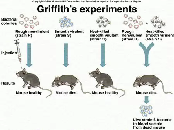
Griffith found that mice injected with live strain S soon died, but those injected with live strain R survived. Mice injected with dead strain S bacteria (killed by heat) аll survived. The results of this series of experiments were as expected. However, the results of Griffith's next series of experiments were thoroughly baffling: mice injected with a mixture of heat-killed strain S and live strain R died. Moreover, Griffith recovered live strain-S bacteria from the dead mice.
After many careful experiments, Griffith concluded that hereditary material had passed from the dead bacteria to the live bacteria. This changed harmless strain R bacteria into virulent strain S pathogens. This process is called transformation.
Avery's experiment: DNA was the transforming agent.
I n
the 1940s, Oswald T. Avery, Colin MacLeod, and Maclyn McCarty showed
that DNA was responsible for transformation.
n
the 1940s, Oswald T. Avery, Colin MacLeod, and Maclyn McCarty showed
that DNA was responsible for transformation.
They used enzymes that hydrolysed polysaccharide, DNA, RNA, and protein on samples of the disease-causing strain-S pneumococci.
Different samples had different parts of their cells destroyed by these enzymes.
The researchers then exposed strain-R pneumococci to the treated samples of strain S.
The transformation of strain R to strain S was blocked only when the DNA in the sample was destroyed.
These results provided strong evidence that DNA carried genetic information for transformation. However, many scientists remained unconvinced.
Hershey and Chase: the role of DNA on the T2 phage life cycle
I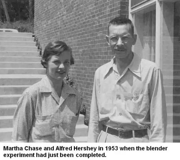 n
1952, Alfred D. Hershey and Martha Chase performed several
experiments with T2 bacteriophage, a virus that infects bacteria.
Their results convinced even the sceptics that DNA, and not protein,
was the genetic material.
n
1952, Alfred D. Hershey and Martha Chase performed several
experiments with T2 bacteriophage, a virus that infects bacteria.
Their results convinced even the sceptics that DNA, and not protein,
was the genetic material.
Electron micrographs indicate that T2 bacteriophage infects Escherichia coli by injecting its DNA into the bacterium while leaving its protein coat on the outside. The phage takes over the genetic machinery of the host cell to make new phages. Eventually, the bacterial cell bursts (a process called lysis), releasing new phages to infect other bacteria (figure 1).
Hershey and Chase wanted to test the hypothesis that only the viral DNA entered the bacterium. They made use of the fact that DNA contains phosphorus but not sulphur, whereas protein contains sulphur but not phosphorus.
With some T2 phages, they labelled the viral DNA with a radioactive isotope of phosphorus (32P). With other T2 phages, they labelled the viral protein coat with a radioactive isotope of sulphur (35S).
They added the viruses to a culture of E. coli and gave them enough time to infect their host cells (but not enough time to reproduce).
The viral coats were then separated from the infected bacteria by shaking the mixture vigorously in a blender.
When E. coli was infected with a T2 phage containing 35S (labelled Protein), little radioactivity occurred within the bacterial cells.
With a T2 phage containing 32P (labelled DNA), the bacterial cells were radioactive. Moreover, when the bacterial cells burst open, the new viruses that emerged were radioactively labelled with 32P. When the protein was labelled, new viruses were only slightly radioactive.