

новая папка / Integrative Human Biochemistry_ A Textbook for Medical Biochemistry (PDF).pdf
.pdf2.1 The Basics of Chemistry in Cells and Tissues |
31 |
hydroxide anion. These chemical species (OH−, H3O+, and H2O) are all related and equilibrium among them may be reached:
2H2O !H3O+ + OH-
The equilibrium constant for this reaction is:
|
|
|
éH |
O+ ù éOH- ù |
|
K |
|
= |
ë |
3 |
û ë û |
eq |
|
|
[H2O]2 |
||
|
|
|
|
||
Because [H2O] (molar concentration of water) is constant at any given temperature and pressure, the so-called ionic product of water, Kw, is used instead for its simplicity:
K |
w |
= éH |
O+ ù éOH- ù |
|
ë 3 |
û ë û |
Nearly at 25 °C, Kw = 1 × 10⁻¹⁴ mol2 dm−6 (i.e., 1 × 10⁻¹⁴ M2). This may seem rather pointless at a first glance, but it is from here that one can conclude that in pure water, pH value is 7. In pure water one ion of H3O+ is formed for each OH−, therefore:
éH |
O+ ù éOH- ù = 1´10-14 M2 Û |
|
ë |
3 |
ûeq ë ûeq |
éH |
O+ ù2 = 1´10 -14 M2 |
|
ë |
3 |
ûeq |
éH |
O+ ù = 1´10-7 M |
|
ë |
3 |
ûeq |
Hence: |
|
|
éH |
O+ ù = 1´10-7 M Û |
|
ë 3 |
ûeq |
|
- log éH |
O+ ù = 7 Û |
|
|
ë 3 |
ûeq |
pH = 7
So, in pure water, at temperature nearly 25 °C, the pH is 7.
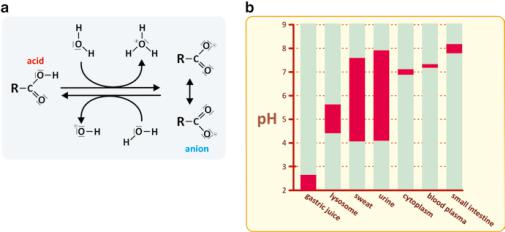
The cytoplasm of cells is approximately at pH 7, although the pH could vary in certain cellular organelles. In the human body as a whole, the pH values of different environments are very diverse, from the extremely acidic gastric juice to the basic intestinal lumen medium (Fig. 2.8).
Other kinds of reactions such as addition elimination or nucleophilic substitutions are also very frequent, but acid–basic will continue to be the focus of our attention for the importance the control of pH has in homeostasis. Variations in pH cause variations in the protonation/deprotonation of proteins and other biological molecules, which in turn affect their function. Take the example of enzymes: protonation or deprotonation of chemical groups on the structure of the protein
32 |
2 The Chemistry and Physics of Life |
Fig. 2.8 (a) Protonation (right to left) and deprotonation (left to right) of a carboxylic acid. The pH of the medium depends on this kind of reactions. (b) A high variety of environments, having different pH can be found in the human body
causes variation in charge, leading to new sets of attractions and repulsions between different parts of the molecule that may cause that some segments of the protein contract and others become looser, impairing or facilitating the optimal function of enzymatic catalysis (this will become more clear in Sect. 3.3, where we address the structure of proteins in detail). The same impact on molecular structure and function applies for other biological molecules, such as polysaccharides. Therefore, stabilizing pH in order to guarantee proper structure and function of biological molecules is very important. This is not saying that the pH should be the same in all tissues or in all cells of the same tissue or in all organelles of the same cell. pH is actively controlled in different anatomical, histological, and cellular environments, but it is not the same in all cases. Blood plasma pH, for instance, is very strictly controlled and only allowed to vary in a very restricted range around 7.4. This is not surprising because the efficiency with which hemoglobin transports O2 is very much dependent on pH. Nevertheless, CO2, which is a molecule that has the potential to largely impact on pH (see next reaction scheme), diffuses freely in the blood.
CO2 + H2O ®! H2CO3 " HCO3- + H+ , H+ + H2O ® H3O+
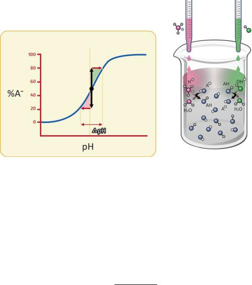
Then, how is it possible for our body to cope with CO2 diffusion and still maintain a blood plasma pH tightly controlled, centered at pH 7.4? The answer resides on a deceivingly simple mechanism of pH control named “pH buffering” (Fig. 2.9).
pH buffers are no more than mixtures of weak acids or weak bases with their conjugated bases or acids, respectively. In practice, an aqueous solution of a weak acid or weak base in equilibrium is a pH buffer because these chemical species dissociate to a moderate extent forming mixtures that are pH buffers. The reason why these mixtures work in a way to maintain pH constant is related to the basic
2.1 The Basics of Chemistry in Cells and Tissues |
33 |
Fig. 2.9 How a pH buffer works. A pH buffer solution is a mixture of a weak acid (HA) with its conjugated base (A−) or vice versa. When an acid is added to the solution, the deprotonated species (A−) reacts with the added acid to form the protonated form (HA). Part of the H3O+ is thus consumed and the pH drop is thus attenuated. When the added solution is a base, part of the OH- is consumed by reaction with HA, the acidic form of the buffer, and the rise in pH is thus attenuated. Additions of significant amounts of acids or bases to buffered solutions result in modest variation in pH as long as protonated (HA) and deprotonated (A−) buffer species coexist
principles of chemical equilibrium. Consider a generic weak acid, represented by HA for the sake of simplicity, in aqueous solution:
HA + H2O !A- + H3O+
éA- ù éH O+ ù Keq = ë [û ë 3] û
HA
Upon the addition of a strong acid, HA! (by definition strong acids have nearly complete dissociation), the concentration of H3O+ raises:
HA¢ + H2O ® A¢- + H3O+
The newly formed H3O+ have an impact on the equilibrium of the weak acid, which will progress in the reverse order (formation of HA) in order to consume part of the H3O+. The extent of this consumption of H3O+ can be calculated based on the equilibrium constant, which remains unaltered: the concentrations of H3O+ and HA increase and the concentration of A− decreases down to the point where Keq is kept (see Box 2.1).

34 |
2 The Chemistry and Physics of Life |
Box 2.1: pH Buffers and the Origin of the Henderson–Hasselbalch Equation
The initial situation, before strong acid addition is:
HA + H2O !A- + H3O+
éA- ù éH O+ ù Keq = ë [û ë 3] û
HA
After strong acid (HA!) addition [H3O+] raises:
HA¢ + H2O ® A¢- + H3O+
But HA dissociation equilibrium is the same in both cases, so:
|
|
|
|
|
|
éA- ù éH |
O+ ù |
éA- ù éH |
O+ ù |
|||||||||
Keq,i = Keq, f |
Û |
ë |
|
ûi ë |
3 |
|
ûi |
= |
ë ûf ë 3 |
ûf |
Û |
|||||||
|
|
[HA]i |
|
|
|
[HA]f |
|
|||||||||||
|
|
|
|
|
|
|
|
|
|
|
|
|
|
|
||||
éH |
|
O+ ù |
|
éA- ù |
éA- ù |
|
|
|
|
|
|
|
||||||
ë |
3 |
ûf |
= |
ë |
ûi |
/ |
ë |
ûf |
|
|
|
|
|
|
|
|||
ëH3O ûi |
[ |
|
|
[ |
|
]f |
|
|
|
|
|
|
|
|||||
|
HA |
]i |
HA |
|
|
|
|
|
|
|
||||||||
é |
|
+ ù |
|
|
|
|
|
|
|
|
|
|
|
|
|
|||
|
|
|
|
|
|
|
|
|
|
|
|
|
|
|
|
|
|
|
2.1 The Basics of Chemistry in Cells and Tissues |
35 |
Box 2.1 (continued)
is no more than a simple rewriting of the equilibrium constant (here named Ka to stress it refers to an acid):
|
|
éA- ù éH |
3 |
O+ ù |
|
|
+ |
|
|
[HA] |
|
|
|
|
|
|
|
|||
|
ë |
û ë |
û |
|
|
|
|
|
|
|
|
|
|
|
||||||
K = |
|
|
|
|
|
|
Û |
éH O |
ù = K |
|
|
Û |
|
|
|
|
|
|
||
|
|
[ |
|
] |
ëA |
û |
|
|
|
|
|
|
||||||||
a |
|
|
HA |
|
ë 3 |
|
û |
|
|
|
|
|
|
|
|
|||||
|
|
|
|
|
|
|
|
a é |
- ù |
|
|
|
|
|
|
|
||||
- log éH O+ ù = - log K - log [HA] |
|
|
|
|
|
éA- ù |
||||||||||||||
Û pH = pK |
|
+ log |
ë |
û |
||||||||||||||||
|
[ |
|
|
] |
||||||||||||||||
|
ë |
3 û |
|
|
|
a |
|
ëA |
û |
|
|
|
a |
|
HA |
|||||
|
|
|
|
|
é |
- ù |
|
|
|
|
|
|
||||||||
A graphical schematic representation of this equation (see figure) shows that pH varies very little when strong acids or bases are added to buffers, mainly when pH is within the pKa ± 1 range. This range is centered around the point where [A−] = [HA], as implied by the Henderson–Hasselbalch equation when pH = pKa. Naturally, the efficiency of the buffer increases with [HA] because higher [HA] imply that more H3O+ of OH− can be added to the solution before [A−] reaches zero, a point where the buffer mechanism becomes exhausted.
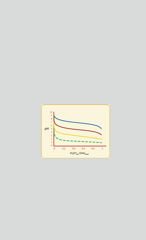
Changes in pH in a buffered solution are much less severe than they would be in the unbuffered solution. Examples of pH variation in a solution initially at pH 9 upon addition of a strong acid, with a total concentration of HA of 0.1 M, when the pKa of HA is 7 (blue), 5 (red), or 3 (yellow). The dashed green curve indicates the pH drop if HA was absent (unbuffered solution, 0 < [H3O+]ad < 0.1 M). The concentration of the H3O+ added to the solution is [H3O+]ad and is represented relative to the total concentration of HA used in the buffer
([HA]total). The buffering zone (nearly horizontal pH variation lines) is mainly in the pH range that lies within pKa ± 1, and the buffering capacity (i.e., the limits of [H3O+]ad in the
buffer zone) depends on [HA]total. This example illustrates how big amounts in added H3O+ cause modest changes in pH in the range pKa ± 1. The case in which a base is added to an
acidic solution is similar. OH− concentration raises, which is concomitant with a decrease in H3O+, and the situation is symmetrical to the one obtained here (raise in pH instead of drop)
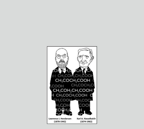
Although the Henderson–Hasselbalch equation is central in understanding fundamental pH buffer chemistry, its historical root is in medicine. Lawrence
(continued)
36 |
2 The Chemistry and Physics of Life |
Box 2.1 (continued)
Henderson was a doctor interested in pH plasma alterations in pathology. He created the concept of acid–base equilibrium. Karl Hasselbalch studied the effect of CO2 in hemoglobin with Christian Bohr (whom the Bohr effect was named after—see Sect. 3.3.3). The works of both doctors opened the way for the scientific study of respiratory and metabolic perturbation of blood plasma pH equilibrium, a significant breakthrough in the early twentieth century physiology.
2.1.1Principal Biological Buffers
Generalization of the previously mentioned concepts and equations should be made cautiously for several reasons:
(a)From the point of view of scientific accuracy, activity coefficients should be used in addition to concentrations in all the abovementioned equations. Activity coefficients are used in thermodynamics to account for deviations from ideal behavior in a mixture of chemical substances. For practical reasons, it is assumed that activity coefficients do not alter equilibrium constants in the experimental conditions addressed.
(b)Water dissociates to form H3O+; therefore, H3O+ ions are always present in aqueous solutions. In the abovementioned equations and reasoning, the ionic product of water was never considered, which is valid in circumstances where
the acidic species are present in concentration much higher than those involved in the ionic product of water (éH O+ ù = 10-7 M).
ë3 û

2.1 The Basics of Chemistry in Cells and Tissues |
37 |
(c) We have explored the concept of buffers in situations in which chemical equilibrium exists. This is not always the case in biological situations.
In spite of all these limitations, the general concept of buffer still applies to cells and organs. Equations should be applied judiciously to chemical problems in living systems, but the concept of pH buffer in vivo is still valid. Wherever weak acids or bases are present in cells or body fluids, they contribute to form a buffer. Ions such as H2 PO-4 / HPO24- (pKa = 6.8) are very important to buffer the cytoplasmic pH (notice that the pH of cytoplasm is about 7.0, well within the buffering range of H2 PO-4 / HPO24- = pKa ±1 = 6.8 ±1) but not plasma. H2CO3 / HCO3- is much more important to buffer plasma pH. The –COO− and –NH3+ groups present in plasma proteins also contribute to pH buffering but not as much as the hydrogenocarbonates (H2CO3 / HCO3- ): while proteins contribute to 24 % of the buffering capacity in plasma, H2CO3 / HCO3- contribute with 75 % (the remaining 1% is a modest contribution of HPO24- / H2 PO-4 ) . The prevalence of the H2CO3 / HCO3- is not surprising because CO2 diffuses freely in plasma, regardless of a certain ability hemoglobin has to bind CO2. Carbon dioxide is extensively converted to di-hydrogen carbonate by enzyme carbonate–dehydratase in erythrocytes:
CO2 + H2O ®! H2CO3 .
H2CO3 is then present in plasma, where it is in equilibrium with hydrogen carbonate (also known under the popular name of “bicarbonate”), HCO3- :
H2O + H2CO3- !HCO3- + H3O+ pKa = 6.1 ( plasma)
HCO3- exists in plasma with typical concentrations in the 22–26 mM range.
It should be stressed that the H2CO3 / HCO3- equilibrium in vitro has pKa = 3.8, quite different from the pKa in plasma (6.1), which is said to be an apparent pKa because it is under the influence of the enzymatic production of H2CO3. To account for the change in pH due to fluctuation in the plasma levels of CO2, one can apply the Henderson–Hasselbalch equation to the multiple equilibria.
CO2 + H2O !H2CO3
H2CO3 + H2O !HCO3- + H3O+
CO2 + 2H2O !HCO3- + H3O+
|
|
éHCO- ù éH |
O+ ù |
||
Ka,apparent |
= |
ë |
3 û ë |
3 |
û |
|
[CO2 |
] |
|
||
|
|
|
|
||
The water concentration is constant and is thus incorporated in Ka,apparent; the variables are on the right-hand side of the equation, and the left-hand side of the
equation is constant. It would be an unnecessary complication to have [H2O], which is constant, in the right-hand side of the equation.
38 |
2 The Chemistry and Physics of Life |
Now the question arises as to what is the most appropriate way to express [CO2] because, at the pressure of 1 atmosphere and usual temperatures, CO2 is a gas and molar units are not well suited for gases. [CO2] should then be converted to partial pressure, pCO2:
[CO2 ] = 0.03´ pCO2 |
|
|
|
||||||
for [CO2] in mM and pCO2 in mmHg. Thus: |
|
|
|
|
|
|
|||
|
éHCO- |
ù éH |
O+ ù |
|
|
|
|||
Keq,app = |
ë |
3 |
û ë 3 |
û |
Û |
|
|
|
|
|
|
|
|
|
|
|
|
||
|
0.03pCO2 |
|
|
|
|||||
|
|
éHCO- ù |
|
|
|
||||
pH = 6.1+ log |
ë |
|
3 û |
|
|
|
|
|
|
0.03pCO2 |
|
|
|
||||||
|
|
|
|
|
|||||
This equation helps by anticipating the drop in pH when pCO2 |
increases, but it |
||||||||
|
|
|
|
|
|
|
é |
- ù |
, |
should be emphasized that an increase in pCO2 also causes an increase in ëHCO3 û |
|||||||||
mitigating the impact of the fluctuation of plasma levels of CO2 in pH. |
|
|
|||||||
2.2More than Only Chemistry: There Is Physics Too
Given the importance of chemical reactions in cells and in the organisms as a whole, one tends to forget the importance of physical processes in life. They are also important to understand life at the molecular level. Apart from all the issues related to the solubility of molecules in aqueous media, which were discussed in the beginning of this chapter, the interaction of light and biomolecules, for instance, is of utmost importance. This is clear when studying the vision of animals, the photosynthesis in plants or the bioluminescence of microorganisms, just to name a few examples. Regarding human biochemistry in the health sciences field, the topics of election are the early events of vision and the effects of ultraviolet (UV) light in tissues such as skin and hair. Other topics, such as the effects of radioactivity and other nonoptical radiation on humans, are left out of this book, although they are interesting and very relevant in particular situations.
When radiation interacts with matter, different kinds of phenomena may occur, such as absorption, scattering, or diffraction. Absorption implies that the energy of radiation matches the energy needed to change molecular states. This means that the energy of radiation is used in the change of the molecular configuration of nuclei and/or electrons so that the radiation extinguishes in the process (it is “absorbed”). This happens when a photon enters our eyes and triggers a radical change in the conformation of retinol in the retina, thereby initiating the physiological process that ends with visual perception. Scattering relates to the radiation that interacts with molecules and is not absorbed, but causes the molecules to emit secondary
2.2 More than Only Chemistry: There Is Physics Too |
39 |
radiation, having the same energy or not. This is what happens when light impinges on matter and is not absorbed: the radiation causes oscillations on the electronic cloud of molecules that in turn causes simultaneous emission of radiation, the scattered light. Scattering is responsible for the white color of milk, blue color of the sky, or reddish color of the sunset. Excessive light scattering in human eyes causes blurring in vision. Diffraction is a particular kind of scattering that occurs when radiation impinges on matter that has voids of size comparable to wavelength of the radiation. X-rays, for instance, are diffracted by molecular crystals because the distances between the nuclei, the chemical bonds length, are similar to its wavelength, i.e., in the order of tenth of nanometer. The spatial pattern of diffracted X-rays can be analyzed to reveal the 3D structure of crystallized molecules, even if they are quite big and complex, such as proteins (Fig. 2.10).
The complete range of frequencies (i.e., energies) of radiation known so far constitutes the so-called electromagnetic spectrum (Fig. 2.11a). It includes low-energy (low frequency, long wavelength) radiation, such as radio and television radiation, as well as high-energy (high frequency, short wavelength) radiation, such as X- and gamma rays, capable of ionizing molecules and disrupting covalent bonds. Very low-energy radiation hardly causes changes in molecules and therefore is not prone to produce effects in living organisms. High-energy radiation (starting from the far, high-frequency, UV radiation) is able to disrupt molecules by destroying chemical bonds and is therefore a potential hazard to the chemistry of life (an amazing exception is presented in Box 2.2). It is not surprising, therefore, that natural phenomena in living beings involving radiation occur in a limited range of energies, from microwaves to near (lower frequencies) UV. Even so, one has to wonder why many animal eyes, including human, uses a much smaller fraction of radiation, from about 400 nm (blue light) to about 800 nm (red light). The answer is simple and can be found in molecular evolution toward the optimized use of environmental resources: the 400–800 nm range accounts for the most abundant sun radiation reaching the surface of planet Earth (Fig. 2.11b). In addition, this radiation penetrates water down to tenths of meters. Naturally, this dictated the course of evolution of vision. The same happened with photosynthetic organisms.
In the human eye, rhodopsin, a membrane protein (opsin) that exists in retina rod cells, has a covalently bound retinal molecule, which adopts two stable isomeric forms: cis and trans (Fig. 2.12). When the protein-attached cis-retinal residue absorbs light, it converts to trans-retinal, this being the triggering event of vision. Not only retinal changes in shape, from a bent structure to a nearly linear arrangement, but it also detaches from opsin. All-trans-retinal (i.e., the “linear” retinal, with all double bonds in the trans-configuration) does not fit the pocket formed by the opsin transmembrane helices as the cis isomer does, causing tensions in the protein structure, which has to change conformation (shape) to adapt. A reduction in cyclic GMP results from this process, which in turn triggers a series of reactions that cause an electric current along the cell. The intensity of this current is proportional to the intensity of light that reached the retina. So, an electric impulse is created that is detected by ganglion cell and then the optic nerve. Nerve fibers reach the
40 |
2 The Chemistry and Physics of Life |
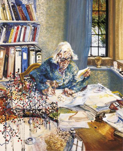
Fig. 2.10 Dorothy Mary Crowfoot Hodgkin in her study at home portrayed by Maggi Hambling (oil on canvas, 1985). The four-handed scientist representation symbolizes her unusual working and entrepreneurial capacity. Dorothy Hodgkin made extremely valuable contributions to the advancement of biochemistry. She revealed the structure of cholesterol, penicillin, insulin, and vitamin B12, among other molecules. She also contributed to the refinement of X-ray diffraction spectroscopy to unravel the structure of proteins. A structural model of insulin is on the left. Reprinted with the permission of the National Portrait Gallery, London, UK
back of the brain (occipital lobe), where images are created. This region is called the primary visual cortex. Some of the visual fibers extend to other parts of the brain to help to control eye movements, the responses of the pupils and iris, and behavior.
The effects of UV light on the skin and hair constitute other examples on how the radiation–matter interaction is important in natural processes. In practical terms, UV is divided in three categories, A to C, according to their biological effects. UV-A is the lowest energy class (320–400 nm). UV-C is the highest energy range (200–290 nm) and is, to a great extent, filtered by the ozone layer in the atmosphere.