

новая папка / Integrative Human Biochemistry_ A Textbook for Medical Biochemistry (PDF).pdf
.pdf82 |
3 The Families of Biological Molecules |
Fig. 3.22 The extracellular matrix has hyaluronic acid in its composition. (a) Histological preparations of cock crest highlighting the hyaluronic acid matrix (left, conventional electron microscopy; center, platinum–carbon replica; right, preserved blue-dyed hyaluronic and extracellular heavily glycosylated proteins, the “proteoglycan matrix”). Figure reprinted with the permission of Instituto de Histologia e Biologia do Desenvolvimento, Faculdade de Medicina, Universidade de Lisboa, Portugal. (b) Schematic representation of the molecular organization of extracellular matrix, which is composed of gel-forming saccharides attached to a backbone of hyaluronic acid that is intertwined among collagen fibrils. Polysaccharides form gels due to the high density of H bonds. These gels confer a moderately rigid structure and mechanical protection to cells and retain water, which prevents desiccation of the tissues
3.2 Saccharides and Their Polymers and Derivatives |
83 |
3.2.2Molecular Conjugates of Monosaccharides
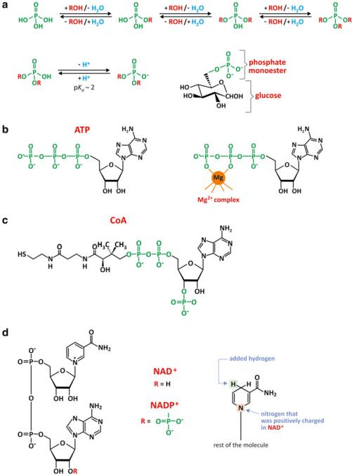
We have seen in previous sections that saccharide monomers offer diverse possibilities of reaction and so they are molecules that form many conjugates in nature. The most important derivatives are phosphate esters. Phosphoric acid is able to form up to three ester bonds (Fig. 3.23), although the tri-esters are not commonly found in nature. Yet, diesters are important and enable saccharide phosphates to form polymers (e.g., nucleic acids) or bridge saccharides with other organic molecules.
Phosphate groups forming esters are anionic in aqueous environment in the most common biological pH ranges. This means that neutral molecules, such as glucose, become charged when esterified with a phosphate. The consequence is an increase in solubility in water and a decrease in the ability to cross lipid bilayers, for instance. This is deemed important as glucose metabolism starts by forming glucose phosphate (Fig. 3.23a).
Phosphate ester hydrolysis is a spontaneous but very slow process, which makes it under enzymatic control in cells. In addition, many chemical processes occurring in cells, such as condensation of polymers with formation of water, are unfavorable processes (there is “excess” water in most cell environments); enzymes speed the reaction but do not shift the equilibrium toward condensation. The use of phosphate derivatives of the monomers in the process of condensation facilitates the reaction as phosphates are so-called good leaving groups: they alter the reactivity of transient chemical species in the course of the mechanism of reaction.
ATP (adenosine triphosphate, Fig. 3.23b) is among the biological molecules that are saccharide derivatives and involves a phosphate ester. A phosphate diester bond bridging two other phosphates is another interesting characteristic of this molecule. The energy involved in the phosphate–phosphate bonds makes this molecule pivotal in energetic metabolism. Divalent cations such as Mg2+ are usually associated to ATP and other molecules having diphosphate groups. This reduces electrostatic repulsion between the oxygen atom of water and the negative charge of phosphate groups, facilitating the hydrolysis of phosphate derivatives.
Probably not so famous as ATP, but equally important in biochemistry, is coenzyme A (Fig. 3.23c). This is a relatively small but complex molecule. Amazingly, coenzyme A has phosphate and saccharide groups but owes its reactivity to a terminal thiol (–SH) group. This thiol group may bind an acetyl residue through a thioester bond, but may also bind a fatty acid, forming acyl-CoA, which is involved in lipid metabolism.
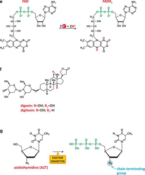
Nicotinamide adenine dinucleotide (NAD+) is another interesting case of saccharide derivative that also contains phosphates. NAD+ intervenes in redox reactions as it may accept and donate electrons, changing from NAD+ to NADH + H+ or vice versa. One extra phosphate group turns NAD+ into NADP+, which has similar redox properties but can only bind to specific enzymes that usually do not bind NAD+. This implies that there are specific metabolic roles for NADP+, distinct from NAD+. The phosphates are involved in enzyme recognition but not in the redox activity itself (Fig. 3.23d). The same happens with flavin adenine dinucleotide (FAD and FADH2; Fig. 3.23e).
Nucleotides themselves deserve closer attention because they polymerize to form the so-called nucleic acids. They will be left for further discussion in the next section. To finalize, it should be stressed that many therapeutic drugs are also sac-
84 |
3 The Families of Biological Molecules |
Fig. 3.23 Phosphates are able to form esters or diesters bridging two organic molecules (a). Phosphate confers an anionic charge to the newly formed chemical entity because the ionization of the phosphate group occurs at pH > 2, increasing its solubility in aqueous medium and decreasing its ability to translocate across lipid membranes. This is the case for glucose-6-phosphate, which is “trapped” in the cytosol of cells, where it will be processed in different metabolic pathways. Adenosine triphosphate, ATP (b), and coenzyme A, CoA (c), are important biological molecules with a saccharide residue bound to a phosphate group. ATP also contains a phosphodiester bond, very important for its reactivity in cells. CoA has a couple of phosphate groups bound to
3.2 Saccharides and Their Polymers and Derivatives |
85 |
Fig. 3.23 (continued) each other, but its reactivity in cells is dictated by the sulfhydryl group (-CSH). Nicotinamide adenine dinucleotide (NAD+) is another saccharide derivative with saccharide residues bound to phosphates. NAD+ may be reduced to NADH (d). Redox reactions of NAD+/ NADH take place in a specific cyclic residue of the molecule, involving a nitrogen atom (right). NAD+/NADH phosphate, NADP+/NADPH (d), also participates in redox reactions in human metabolism. NADH and NADPH cannot be distinguished by their reducing properties because the phosphate group that distinguishes them does not interfere with the nitrogen atom that affords the redox properties. Yet enzymes use specifically NADH or NADPH and so there is no redundancy between these molecules. FAD and FADH2 (e) are molecules similar to NAD+ and NADH in that both constitute adenine nucleotides and their role in metabolic redox reactions. Digoxin and digitoxin (f) are examples of drugs with monosaccharides in their structure; more specifically, three residues are specifically combined as part of a unique structure. Another example of drug that is a saccharide derivative is azidothymidine (AZT), which is converted to a triphosphate in cells (g) and is able to insert in the active center of the reverse transcriptase of HIV because it is structurally similar to the natural substrate. However, the natural substrate does not have the N3 group. The presence of this group blocks the conversion of the viral RNA into DNA
86 |
3 The Families of Biological Molecules |
charide derivatives, such as digoxin (Fig. 3.23f), used in the treatment of heart conditions. Azidothymidine (AZT) is another example. It is an analogue of thymidine that may inhibit the action of reverse transcriptase of HIV. It was the first drug used in the treatment of AIDS. Cellular enzymes convert AZT into the effective 5-triphosphate form (Fig. 3.23g). Once bound to reverse transcriptase, the azide group, N3, is responsible for chemical inhibition. Inspired by the success of AZT (Fig. 3.24), many nucleosides are now under development to create new inhibitors of HIV reverse transcriptase to fight AIDS.
3.2.3Molecular Conjugates of Oligosaccharides
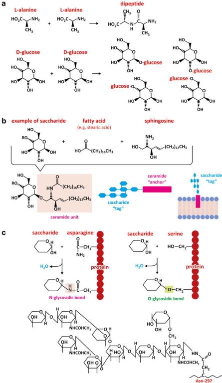
It is worth stressing that the combination of saccharide monomers may generate a big diversity of products when compared to amino acids (Fig. 3.25). Two glucoses, for instance, can bind via 6 carbons in each monomer, thus being able to form 36
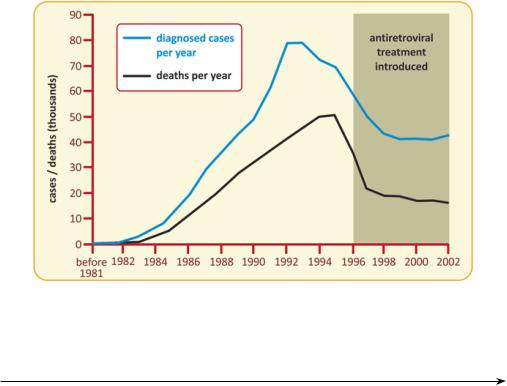
Fig. 3.24 Preventive campaigns highlighting the need to change risk behaviors had a strong impact in the spreading of AIDS in the USA, with a marked decrease in the number of diagnosed cases and deaths per year after 1993-95. The use of AZT and other drugs had a very positive additional effect in the reduction of AIDS-caused mortality
Fig. 3.25 (continued) Saccharide tags are covalently bound to lipids, usually rigid lipids such as ceramide for a better anchoring to the membrane (b). Glycolipids (i.e., associations of saccharides and lipids) determine blood groups, for instance (see Box 3.5). The same principle applies to oligosaccharides attached to proteins, i.e., glycoproteins (c). The side chain of the amino acid asparagine may react with a saccharide by dehydration forming an N-glycosidic bond (analogous to an O-glycosidic bond, but involving N instead of O). Likewise, the side chain of the amino acid serine may react with a saccharide forming an O-glycosidic bond. The oligomeric sequences of saccharides attached to protein IgG is shown in panel (c) bottom as an example
3.2 Saccharides and Their Polymers and Derivatives |
87 |
Fig. 3.25 A combination of two amino acids generates one single dimer, but there are several ways that two monosaccharides can combine to form a disaccharide (a). Saccharides are better suited to form highly specific structures at the surface of cell membranes (b) or proteins (c).
88 |
3 The Families of Biological Molecules |
different molecules. Considering the anomers, the diversity increases. It is not surprising that oligosaccharides are present in the surface of cells as receptors of unique structure (see an example in Box 3.5) while amino acids form polymers (proteins) having domains with few restricted well-defined structures. Moreover, monosaccharides or oligosaccharides are frequently found in nature attached to proteins.
Box 3.5: The ABO Blood Groups
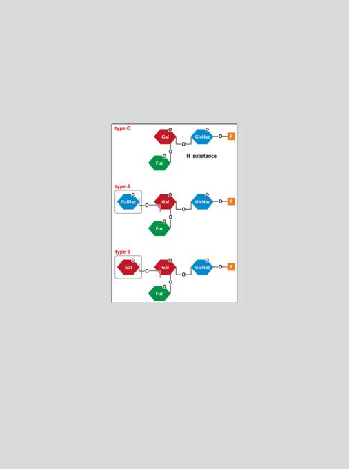
There are different blood groups according to different immunogenic molecules present in erythrocytes. The most important classification of blood groups is based on three antigens, A, B, and O that form 4 groups: A, B, O, and AB—the ABO blood groups. The ABO blood group antigens are oligosaccharide chains attached to proteins and lipids located in the outer surface of erythrocytes. One single residue of a small oligosaccharide determines whether the antigen is A, B, or O (see figure).
Fuc represents the monosaccharide fucose, Gal, galactose, GalNAc, N-acetylgalactosamine, and GlcNAc represents N-acetylglucosamine
The immune system of an individual produces antibodies against the ABO antigens not present in his own erythrocytes. Individuals in A group will have antibodies against B and vice versa. Type O, the most common, does not contain the last residue, which is the antigen, in its structure (in fact the original nomenclature was 0—zero—but became the letter O). So, individuals in blood group O will have both anti-A and anti-B antibodies. Individuals in blood group AB are rare and, naturally, have no anti-A and no anti-B antibodies. This has tremendous implications in blood transfusions as a patient cannot receive erythrocytes against which he/she has antibodies. AB individuals
(continued)

3.2 Saccharides and Their Polymers and Derivatives |
89 |
Box 3.5 (continued)
can, in principle, receive blood from any donor; O individuals can donate blood to any individual; A and B individuals can only donate and receive blood to/from individuals belonging to the same blood group.
It is believed that ABO antibody production is stimulated when the immune system contacts in foods or in microorganisms with the saccharide antigens that are absent in the erythrocytes. The functions of the ABO blood group antigens are not known. Individuals who lack the A and B antigens are healthy, suggesting that any function the antigens have is not important, at least not in modern times.
Hemolytic disease of the newborn (HDN) is a serious medical problem that occurs almost exclusively in infants of blood group A or B who are born to group O mothers. This is because the anti-A and anti-B formed in group O individuals tend to be of the IgG type, which can cross the placenta. HDN tends to be relatively mild mainly because fetal erythrocytes do not express adult levels of A and B antigens. However, the precise severity of HDN cannot be predicted.
3.2.4Polymers of Saccharide Conjugates: Nucleic Acids
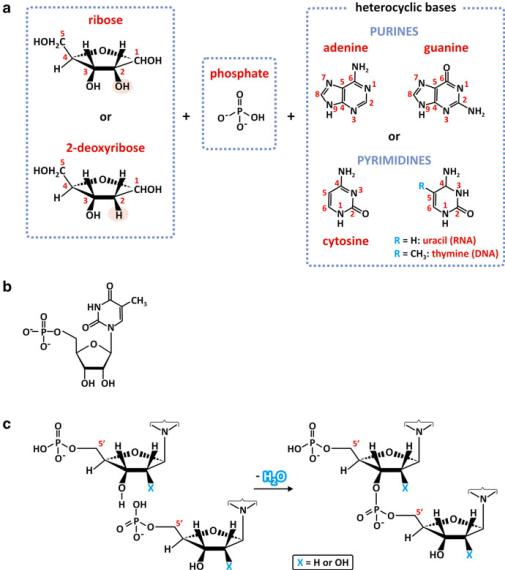
Nucleotides that compose deoxyribonucleic acid (DNA) and ribonucleic acid (RNA) are formed by 2-deoxyribose or ribose, respectively, linked to a heterocyclic base, a purine (adenine, guanine) or a pyrimidine (cytosine and uracil or thymine), and a phosphate group attached to carbon 5 of the ribose residue. To avoid ambiguity with numbering of carbons of the heterocyclic base, the carbon numbers of the ribose and deoxyribose are identified with a prime: phosphate ester linkage occurs at C5& (Fig. 3.26). The physical and chemical characteristics of the heterocyclic bases are extremely important as they are determinant for the way nucleotide polymers (nucleic acids) organize. The bases are planar, cyclic, aromatic molecules with N and O atoms able to participate in hydrogen bonding in the plane of the ring. The bases are low polarity groups poorly solvated, so both faces of the plane of the base rings will be fairly hydrophobic and thus subject to significant entropic effects.
There are four different possible nucleotides in RNA and DNA. RNA is formed by adenosine-5&-monophosphate (AMP), guanosine-5&-monophosphate (GMP), cytidine-5&-monophosphate (CMP), and uridine-5&-monophosphate (UMP). DNA is formed by deoxyribose, which is denoted by the prefix d in dAMP, in dGMP, in dCMP, and in dTMP. dTMP stands for thymine-5&-monophosphate using deoxyribose; DNA does not contain dUMP.
Because nucleotides are phosphate monoesters, they can form additional ester links to other alcohols, such as the OH groups in other nucleotides. In other words, they can polymerize by dehydration reactions. Nucleic acids are formed by phosphodiester bonds between C5&of one nucleotide and C3& of another nucleotide (Fig. 3.26c). The result is a linear polymer having the heterocyclic bases and the
90 |
3 The Families of Biological Molecules |
Fig. 3.26 Nucleotides are formed with ribose or 2-deoxyribose, phosphate and a heterocyclic base, purine (adenine or guanine) or pyrimidine (cytosine, uracil, or thymine) (a). The phosphate group forms a phosphodiester bond in C5 and the heterocyclic base binds to C1. The nucleotide deoxythymidine phosphate is shown as an example (b). A dimer of nucleotides may be formed by dehydration, which creates a phosphodiester linkage between the monomers via C5& and C3& (c). In RNA X=OH, in DNA X=H
phosphate groups in opposing sides, the phosphate groups being anionic (Fig. 3.27). Some simplified representations of nucleic acids pinpoint this characteristic (e.g., Fig. 3.27b), which remains elusive when the nucleic acid is simply represented by a sequence of letters identifying the nucleotides (T, thymine; C, cytosine; G, guanine; A, adenine; U, uracil). By convention, nucleic acid sequence is written from the C5& to the C3& endings, 5& ' 3& (Fig. 3.27).
Unlike polysaccharides, nucleic acids are amphiphilic molecules (Fig. 3.27) so the entropic effect will be a significant driving force for folding in aqueous environ-

3.2 Saccharides and Their Polymers and Derivatives |
91 |
Fig. 3.27 Natural polymers of nucleotides are named nucleic acids. Desoxyribonucleic acid (DNA) has 2-deoxyribose residues and uses guanine, cytosine, adenine and thymine but not uracil (a). Ribonucleic acid (RNA) has ribose residues and uses guanine, cytosine, adenine and uracil but not thymine. Both polymers are formed by C3&–C5& phosphodiester bonds. For the sake of simplicity, the chemical structure of the monomers is usually omitted, and other forms of presenting the nucleotide residues sequence are preferred. The simplest and most common form represents the nucleotides by a one-letter code (the first letter of the base name: G, A, C, T or U). Which end is the free, C5& or C3&, is not explicitly mentioned but it is established by convention that the sequences are presented in the directions 5& to 3& (b)
ment. The heterocyclic bases will tend to nucleate to minimize their contact with water molecules. The crystal structure of transfer RNA (tRNA) shows that the bases stack parallel to each other, which is favored by their strictly planar structure. In addition, the nucleic acid tends to twist along its major axis forming a helix that exposes the phosphates to the aqueous medium and has the bases stacking in its core. In addition, most of the helical regions in tRNA consist of two sequences of the same RNA chain running in opposite directions with bases in opposite sequences contacting each other close enough and with the adequate stereochemical arrangement to establish hydrogen bonding between them. This adequate arrangement only occurs if purines pair with pyrimidines, as in pairs A–U and G–C (Fig. 3.28). This