

новая папка / Integrative Human Biochemistry_ A Textbook for Medical Biochemistry (PDF).pdf
.pdf102 |
3 The Families of Biological Molecules |
Fig. 3.36 Secondary-level structures such as helices interact intramolecularly or intermolecularly through electrostatic forces (3), hydrogen bonding (2) or entropic effect factors that result in exposure of polar groups such as –COO− and –NH3+ to aqueous solvent, and association of hydrophobic groups with minimal exposure to the aqueous environment (1). Two Cys residues in contact may react through the thiol groups (–SH) in the side chains forming disulfide bonds (S–S) that strongly contribute to the stabilization of the structure of proteins (see as an example the structure of insulin in Fig. 3.37)
global geometry of the protein that form fairly independent and separable parts, frequently having specific dynamics and specific functions. These are known as domains. An upper level exists for proteins that associate with other proteins, equal or not, to form organized protein assemblies: the quaternary-level structure. The different levels for protein structure are illustrated in Fig. 3.37, using as example insulin, whose structure was discovered by Dorothy Hodgkin, whom also discovered the structure of cholesterol (see Fig. 2.10).
Hydrogen bonding is frequently the strongest non-covalent factor in keeping the tertiary and quaternary levels of the structure of proteins. Enolase is a good example. Although there are no covalent bonds between both proteins in the dimer,
3.3 Amino Acids and Their Polymers: Peptides and Proteins |
103 |
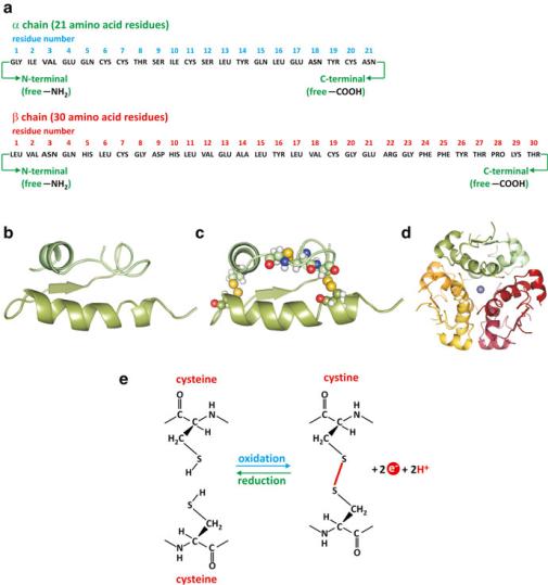
Fig. 3.37 The hormone insulin, from primary to quaternary-level structure. (a) Amino acid (threeletter code) sequence, the primary-level structure. (b) Segments engaging !-helical secondarylevel structure are represented as helical ribbons. (c) The protein folds into a tertiary-level structure that is stabilized by disulfide bonds (yellow in the protein structure). Disulfide bonds are the result of oxidation of two thiol (–SH) groups to form an S–S bond (e). It is common that Cys residues react this way in extracellular proteins. (d) Six insulin monomers associate forming a homohexamer, the quaternary-level structure. The quaternary-level structure is stabilized by the presence of two zinc ions (central sphere) and due to contacts between hydrophobic surfaces of monomers (entropic effect). Insulin is stored in the pancreatic beta cells and secreted into the bloodstream in the form of aggregates of these compact hexamers. Upon dilution in the blood, insulin dissociates, and the active form is believed to be the monomer
hydrogen bonds are frequent (Fig. 3.38). Altogether, the sum of all hydrogen bonds creates a strong network of adhesion forces in the contact surface of the proteins. Hydrogen bonds are directional; they occur in a well-defined direction between chemical groups at a definite distance; this further contributes to maintain the structure of proteins. The extreme contribution of hydrogen bonding to polymer structure
104 |
3 The Families of Biological Molecules |
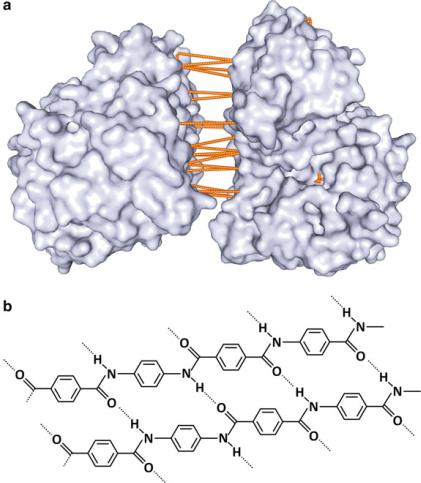
Fig. 3.38 Examples of hydrogen bonding contribution to polymer structure. (a) Enzyme enolase (PDB 1IYX) is a dimer in which both subunits are attached by a dense array of hydrogen bonds in the contact surface between them. (b) The structure of an amide polymer (such as proteins) commercially known as Kevlar. It involves a dense network of hydrogen bonds, which confers high resistance, and Kevlar is used in protective materials such as helmets and bullet proof vests. Compare the molecular-level details of Kevlar and "-sheets in proteins; there is a parallelism between the resistance of Kevlar and the extreme stability of aggregates formed by the juxtaposition of "-sheets in amyloid plaques (see Box 3.6)
may not be intuitive, but one should bear in mind that Kevlar, an extremely resistant material used in protective items such as bulletproof vests, owes its properties in part to hydrogen bonding (Fig. 3.38).
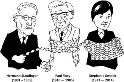
Kevlar was named after its inventor, the chemist Stephanie Louise Kwolek, whom had planned to attend a medical school but started a temporary job in chemistry and finally quit a medical career. The historic parallelism between artificial polymeric materials and biological molecules dates back to 1920, when Hermann Staudinger proposed that rubber and other polymeric molecules such as starch, cellulose, and proteins are long chains of short repeating molecular units linked by covalent bonds, a disruptive concept at that time. Staudinger used the term macromolecule
3.3 Amino Acids and Their Polymers: Peptides and Proteins |
105 |
(“makromoleküle”) for the first time, a term now very popular among biochemists. Paul Flory, a chemist pioneer of the studies of three-dimensional organization of polymers and its relation to dynamics, also worked for the rubber industry during certain periods of his career. His work opened the field to the establishment of struc- ture–function relationships in macromolecular biochemistry. Flory was awarded the Nobel Prize in Chemistry in 1974 “for his fundamental achievements, both theoretical and experimental, in the physical chemistry of macromolecules.”
Thinking of natural protein fabrics, such as silk and spider webs, and artificial fabrics made of nylon and other polymers helps us realize that in molecular world the boundaries between nature and human artifacts are extremely tenuous.
As mentioned before, there are also covalent contributions to the tertiary level of structure of proteins, namely, disulfide bonds (or “bridges”) and attachment of metal or other non-proteic groups to more than one amino acid residue. Disulfide bonds are formed by oxidation of two contacting Cys thiol (-SH) groups originating an S–S bond between the Cys residues (cystine). Cell cytosol is a relatively strong reducing environment, and the contribution of disulfide bonds to the stabilization of cytosolic proteins is limited. However, in other circumstances, disulfide bridges form and are strong stabilizers of protein structure at the tertiary level. Insulin, a peptide hormone, is an example (see Fig. 3.37).
Metals can bind multiple ligands and covalently link different amino acid residues in a protein, therefore also contributing to stabilize a tertiary-level structure. Frequently, metals bind to the thiol group of Cys. In the electron transfer chain proteins, several metallic complexes are present, which in addition to chemical functions also contribute to the stability of the proteins (Fig. 3.39; see also Sect. 6.2.2).
When one refers to quaternary-level structure, one usually refers to proteins that associate with high specificity and well-defined function, such as hemoglobin. This does not include pathological cases in which aggregation of proteins leads to loss of function and increase in toxicity. Extensive tertiary-level alterations are observed when amyloid fibers form upon aggregation of proteins or when prions trigger

106 |
3 The Families of Biological Molecules |
Fig. 3.39 Metal complexes, such as iron–sulfur centers (a), are common among the proteins of the electron transfer chain. Iron complexes with sulfur atoms but also with the thiol group of the side chain of Cys residues, resulting in stabilization of the structure of the proteins where they insert. The nitrogen atoms in the side chains of His are also prone to complex formation with metals. Panel (b) shows a detail of complex formation with metals in complex IV (PDB 1OCC) of the electron transfer system (see also Sect. 6.2.3). An iron ion (red sphere) complexes simultaneously the N atoms of two His side chains (blue), stabilizing the tertiary-level structure of the protein. It also binds to a nonprotein molecule, the heme a (red organic structure), which is also associated to complex IV
conformational changes of native proteins (see Box 3.6), for instance. These are referred to as protein-folding diseases. Folding is the expression used to comprise secondaryand tertiary-level structure altogether.
3.3.2Structure and Function in Proteins
Proteins can adopt many different structures at the tertiary level, from extended rods to compact globules. Extended proteins may associate in fibers and globular proteins may have flexible domains able to bind other molecules. This gives the impression that proteins with extended conformation, like keratin, collagen, or silk fibroin, are adequate to maintain the structure of tissues or biomaterials, whereas globular proteins intervene in dynamical processes, which explains why enzymes

3.3 Amino Acids and Their Polymers: Peptides and Proteins |
107 |
Box 3.6: Amyloids and Prions: When Misfolding Turns into Disease
The relationship between the structure and function of proteins has been one of the main issues of modern biochemistry for decades. Mutated proteins may have important changes in their structure and may thus display a defective function. However, the knowledge that proteins without mutations can fold in diverse forms, some of them pathogenic, is recent. Protein folding is the key to important diseases such as Alzheimer’s, in which massive stacks of "-sheet- folded proteins accumulate in the brain. These stacks form plaques of insoluble protein in the extracellular tissue, which cannot be broken down by enzymes. When these plaques were found for the first time, they were described as related to saccharides and named amyloids. Although the chemical nature of the plaques is now known not to be related to saccharides, the name “amyloid” is still used and the group of diseases is known as amyloidoses.
Amyloid plaques grow with an ordered structure forming long filaments (fibrils). There are about 20 different proteins that can act as the building blocks of these fibrils, each of which is associated with a different disease. In so-called systemic amyloidoses, the precursors of these plaques are transported through the bloodstream from their point of origin to their point of deposition. Localized amyloidoses are of great clinical significance, as they mainly affect the central nervous system, the extracellular tissue of which is particularly susceptible to damage.
Transmissible spongiform encephalopathies (TSEs), which include mad cow disease (bovine spongiform encephalopathy; BSE) and Creutzfeldt– Jakob disease (CJD) in humans, are forms of amyloidoses in which the diseased brain degenerates to a porous sponge-like structure. These diseases appear when human proteins called prions misfold. The human prion is a component of the membrane of healthy nerve cells (called PrPc) that may misfold in a particular way. Amazingly, the misfolded prion may induce misfolding in a neighboring prion if contact among both molecules occurs. This has the appearance of an infection-like process in which the misfolded molecule “infects” the “healthy” molecule. “Infectious” prions can be transmitted in the diet, triggering a domino effect on healthy prions.
In Alzheimer’s disease "-amyloid plaques are formed by cleavage of the amyloid-precursor protein (APP) by two different enzymatic activities, which release peptide fragments that are 40 or 42 amino acids long. When these peptides fold into "-sheets and aggregate, fibrils are formed, surrounding neurons and causing damage. This does not happen when the same peptides fold differently. It is only in "-sheets that hydrophobic amino acids are exposed, and they rapidly bind to hydrophobic groups of other peptides due to the entropic effect. The "-sheet structure, being highly ordered, is prone to regular stacking, ultimately leading to fibril formation.
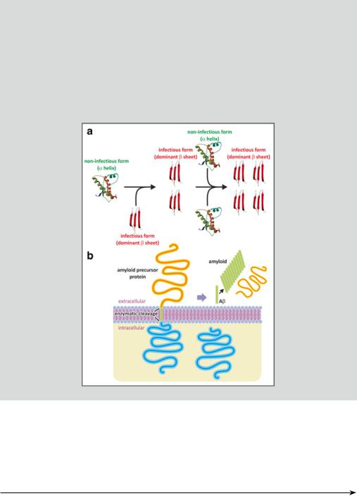
In panel (a) of the following figure, a prion protein domain PrP(121–231) is shown in the noninfectious form (mouse, PDB 1AG2). The infectious form, having a structure not known in detail but dominated by beta strands
(continued)
108 |
3 The Families of Biological Molecules |
Box 3.6 (continued)
(tentatively depicted; red), may induce conformational changes in the noninfectious protein to produce a replica of itself. The process may amplify and aggregates of the infectious form thus accumulate in pathological conditions. In Alzheimer’s disease, "-amyloid plaques are not formed by an infectionlike propagation of the proteins dominated by beta-strand domains. Panel B shows cleavage of the APP with concomitant release of peptides (green) that fold into "-sheets and aggregate.
are globular proteins. While this is generally true, it should also be acknowledged that the frontier between both is not always clear. For instance, actin is a globular protein that binds other actin molecules to form quaternary-level fibers in the cytoskeleton and muscle contractile system. Actin is an example of a globular protein having structural functionality (Fig. 3.40; see also Sect. 10.1.1). Another interesting
Fig. 3.40 (continued) exposed in collagen and form cross-links that strengthen the collagen fibers. The chemical process of this cross-linking reaction between the endings of the Lys side chains depends on vitamin C (ascorbic acid). Lys residues are also involved in cross-links of elastin (d), a connective tissue protein of unusual elasticity. The cross-links involve four Lys residues forming a desmosine arrangement. Actin (e) is a globular protein that self-associates forming fibers important for muscle contraction. Myosin (f), on the other hand, is a combination of an extended domain with a globular domain (“head”) having catalytic activity. The extended domain associates to other proteins forming fiber-like oligomers responsible for muscle contraction (see Sect. 10.1.2) Hyp - Hydroxyproline
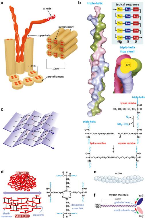
Fig. 3.40 Proteins such as keratin (a) and collagen (b, PDB 1BKV) adopt string-like conformations (very extended helices) that associate to form fibers that stabilize structures such as hair or nails (keratin), or the connective tissue (collagen). Silk fibroin (c) forms very extensible Glyand Ala-rich "-sheets that associate in an antiparallel fashion forming a very resistant biomaterial such as spider silk. It is important to realize that collagen has a Gly residue at every third position of its amino acid sequence. Gly is a very small amino acid because the side chain is H. This creates a line along the helix surface where two other similar helices may dock in close contact. Lys side chains are

110 |
3 The Families of Biological Molecules |
molecule that challenges the classical dichotomy of structural/extended vs. functional/globular proteins is lung elastin. When relaxed, elastin is a globular protein but it stretches allowing lung expansion. Elastin molecules are interconnected covalently by the side chain of four Lys residues: cross-linked desmosine bonds (Fig. 3.40). In this way, the continuous alteration between the globular and extended conformation of elastin confers to the lung the ability to expand and contract without histological lesions. Finally, one should stress the fact that there are also proteins that have both extended and globular domains. This is the case of myosin, another central protein in muscle contraction, in which an extended domain forms oligomeric fibers (Fig. 3.40; see also Sect. 10.1.1).
Besides tight parallel packing in collagen fibers, proteins bind covalently to other proteins in different fibrils of collagen through side chains of Lys residues (Fig. 3.40). When these strong covalent meshing of collagen is disrupted, the properties of the connective tissue are very much affected causing diseases (Box 3.7). Likewise, mutations of the Gly residues impact dramatically on collagen structure and connective tissue function. This has a particular effect on bones from early age: They loose resistance and break easily. This disease is known as osteogenesis imperfecta, Lobstein syndrome, or, more commonly, “brittle bone disease.”
Box 3.7: Scurvy: An Example of a Pathology Directly Associated to Protein Structure
Scurvy is a pathology characterized by fatigue, anemia, gingivitis (gum disease), and skin hemorrhages caused by diets with a prolonged deficiency of ascorbic acid (vitamin C). It was a frequent disease of sailors in long voyages during the pioneering intercontinental discoveries of the fifteenth century. Many men died until it was discovered that scurvy could be cured and prevented by consuming citrus, such as oranges, lemons, and limes. The Portuguese sailor Vasco da Gama was the first European to lead a fleet that reached India by sea, linking Europe and Asia and the Atlantic and the Indic. The drama of scurvy in his first trip to India (1497–99) is eloquently described in an epic poem by Luís de Camões, in The Lusiads (1572):
“And ‘twas that sickness of a sore disgust, the worst I ever witness’d, came and stole the lives of many; and far alien dust buried for aye their bones in saddest dole.
Who but eye-witness e’er my words could trust? of such disform and dreadful manner swole
the mouth and gums, that grew proud flesh in foyson till gangrene seemed all the blood to poyson: “Gangrene that carried foul and fulsome taint, spreading infection through the neighbouring air: No cunning Leach aboard our navy went,
much less a subtle Chirurgeon was there;
but some whose knowledge of the craft was faint strove as they could the poisoned part to pare,
(continued)
3.3 Amino Acids and Their Polymers: Peptides and Proteins |
111 |
Box 3.7 (continued)
as though ‘twere dead; and here they did aright; — all were Death’s victims who had caught the blight.
“The Lusiads” (Canto V, 81 and 82) version reproduced here was translated to English in 1880 by Richard Burton
Nearly two thirds of the sailors of the entire fleet of Vasco da Gama died during the trip, although documents from that time clearly show that the by Portuguese sailors knew that a diet based on fruits and other unprocessed foods was a treatment for scurvy. A manuscript from a pilot in the fleet of Pedro Álvares Cabral, discoverer of Brazil in 1500, says that a diet of fresh foods, including sheep, chicken, ducks, lemons, and oranges, was used to heal scurvy.
It was only in the eighteenth century that the Scottish doctor James Lind related scurvy to diets poor in citrus on a reasonably scientific way. Ascorbic acid was discovered by the biochemist Albert Szent-Gyorgyi (Born Hungarian, later US citizen), who was awarded the Nobel Prize in Medicine or Physiology in 1937 (Szent-Gyorgyi also performed important studies on muscle contraction; see Box 10.1). Ascorbic acid takes part of in biochemical pathways, the synthesis of collagen being one of them. Specifically it is mandatory in protein hydroxylation, which is a posttranslational modification in which a hydroxyl group (–OH) is added to a protein residue. Collagen is naturally hydroxylated in healthy individuals. Ascorbic acid is also mandatory in the biosynthesis of carnitine. Impaired synthesis of carnitine and collagen accounts for the common symptoms of scurvy.
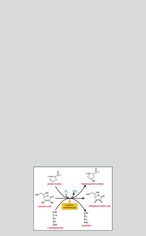
The primary defects behind rotten or loose teeth, rigid tendons, or cartilage fragility observed in scurvy reside in the connective tissue. Without ascorbic acid, collagen is not hydroxylated, and a nonfibrous, defective incomplete collagen is formed instead of fibrous collagen due to the impairment of crosslinking. The enzyme prolyl 4-hydroxylase, for instance, hydroxylates a Pro residue using an iron atom that is oxidized in the process. Ascorbic acid is needed to reduce the iron and make the enzyme active again (see figure).
(continued)