

новая папка / Integrative Human Biochemistry_ A Textbook for Medical Biochemistry (PDF).pdf
.pdf122 |
3 The Families of Biological Molecules |
high amounts in the blood of men who have prostate cancer. A short number of other diseases cause moderate increased levels of PSAP, but only direct lesion of the prostate such as that provoked by tumors in this organ causes high levels of the protein in plasma. PSAP is then classified as a marker for prostate cancer.
Being highly irrigated (see Box 8.1) and particularly exposed to the action of drugs and other exogenous chemicals and viruses, the liver is an organ that suffers frequent insults that lead to the presence of hepatic enzymes in plasma. Two transaminases, alanine transaminase (ALT) and aspartate transaminase (AST), are markers of lesions frequently assayed in blood samples when hepatitis, poisoning, or alcoholic liver disease is suspected. However, one should bear in mind that these enzymes are also present in other organs, albeit in smaller concentrations. A full diagnosis is composed of data that takes into consideration not only biochemical analysis but also the symptoms, the history, lifestyle, and other diagnostic results.
The enzymes lactate dehydrogenase (LDH) and creatine kinase (CK) are of particular interest because they are markers of heart muscle lesion. Although these enzymes also exist in muscles other than the heart, there are differences in the amino acid composition that enable detection of the heart variants. Enzymes with similar activity and extensive structure homology are known as isoenzymes. For instance, three CK isoenzymes have been discovered: CK-MM or CK3, found mostly in skeletal muscle; CK-BM or CK2, found mostly in myocardium; and CK-BB or CK1, which is concentrated in the lungs and brain. Because of this distribution of CK isoenzymes, a pulmonary embolism is associated with elevated levels of CK-BB. On the other hand, an acute myocardial infarction is associated with elevated levels of CK-MB, and injuries of skeletal muscle cause elevated levels of CK-MM. LDH isoenzymes are tetramers, both in the heart and other muscles. These tetramers may disassemble and reassemble in the form of mixed heterogeneous tetramers because the structure of the monomers is very similar. In any case, the presence of dominant heart isoenzymes can be detected in plasma in case of myocardial infarction. The plasma enzyme changes in acute myocardial infarction are shown in Fig. 3.48. CK-MB isoenzyme peaks first, AST next, and LDH last.
There are also nonenzymatic markers that are used in the diagnosis of acute myocardial infarction: myoglobin and two cardiac troponins, troponin I (cTnI) and troponin T (cTnT). CK-MB and the heart LDH isoenzyme are the most important ones due to their heart specificity. Cardiac troponins are also important as their serum levels are frequently elevated during the first hours of acute myocardial infarction, even at a time when CK and CK-MB activities are still within the reference range, but are not fully established as the enzymatic markers.
While the scientific and clinical discipline of studying enzymes for direct clinical diagnosis, clinical enzymology, is expanding and gaining importance, it is curious to mention that dead brain tissue does not release into the blood any significant amounts of enzymes. Despite the frequency of cerebral infarcts (strokes), no test for
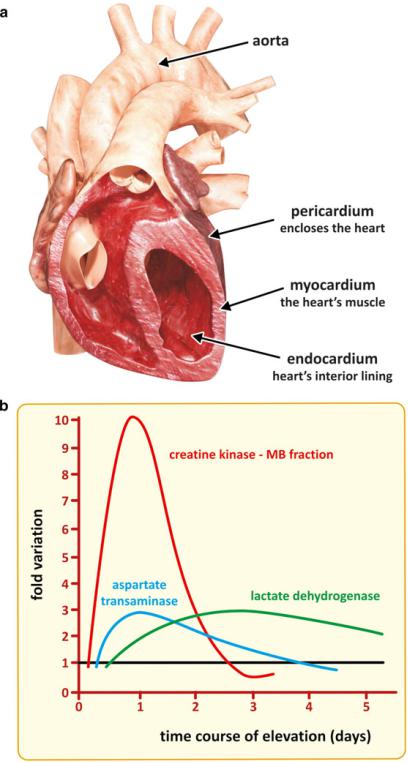
Fig. 3.48 Myocardium is the heart’s muscle (a). Myocardium infarction involves partial tissue death (necrosis) caused by a local deficit of oxygen supply, as a consequence of an obstruction of the tissue’s blood flow. Cardiac muscle enzymes, such as CK-MB, AST, and LDH, appear in the blood after the infarct (b). The combined information of CK-MB and LDH allows to estimate the time of the infarct, which in turn helps devising a therapeutical strategy. CK - creatine kinase, AST - Aspartate transaminase, LDH - Lactate dehydrogenase
124 |
3 The Families of Biological Molecules |
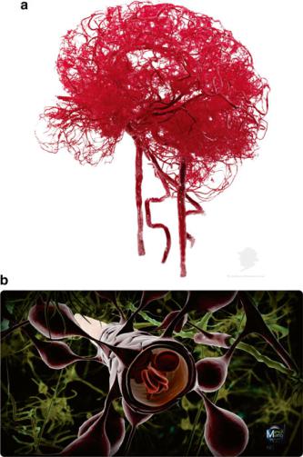
brain enzymes is currently available due to the blood–brain barrier (BBB, Fig. 3.49), the network of capillaries that irrigates the central nervous system. The cells of these capillaries are connected by tight junctions and adhesion molecules that severely restrict the diffusion of hydrophilic macromolecules into the cerebrospinal fluid (CSF). Small gas molecules, such as O2 and CO2, diffuse passively through the barrier, and some nutrients and hormones are actively transported with specific proteins (this will be revisited, for instance, in Box 9.3, which discusses the glucose transport through the BBB).
Fig. 3.49 The blood–brain barrier (BBB) is the network of brain capillaries (top). This photograph shows the result of a technique used to conserve anatomical structures named plastination applied to the human BBB (reprinted with permission of von Hagens Plastination, Germany; © www.vonHagens-Plastination.com). The network of very thin arteries that penetrate the brain forms a very reticulated mesh. The capillaries are associated to a thick basement membrane and astrocytic end feet (brown cells covering the endothelium; bottom panel). Passage of molecules across the endothelial cells of the BBB is highly selective. Enzymes released from nerve cells upon a stroke cannot reach the blood, a fact that makes diagnosis very difficult. Bottom panel is a Ben Brahim Mohammed work reproduced under the Creative Commons license

3.3 Amino Acids and Their Polymers: Peptides and Proteins |
125 |
3.3.4.2 The Nomenclature of Enzymes
Because an enzyme is very specific for the substrate or for a family or related molecules of very similar structure, it is very unique. In the early days of metabolic studies, enzymes were named individually, one by one, with no concern for general rules of nomenclature. With time, the diversity of names and multitude of criteria to indentify newly discovered enzymes was such that the lack of a nomenclature that could be used worldwide was indering to the progress of enzymology (the scientific discipline devoted to study enzymes). The International Union of Pure and Applied Biochemistry (IUPAB), an international organization emerging from the joint efforts of many national societies of biochemists around the globe, appointed a working group to propose general rules that could be used to classify and identify enzymes. The result was a nomenclature based on the kind of reaction catalyzed by the enzyme consisting of:
–A name based on the contraction of “substrate + suffix ase” (e.g., urea + ase = urease, an enzyme that catalyzes a reaction with urea). This name has some flexibility.
–A rigid four-number code preceded by EC (for “Enzyme Commission”), which is unique for each enzyme (or sets of isoenzymes). The numbers refer to a family of enzymes and three successive subfamilies (Box 3.9). EC 5.2.1.3, for instance, identifies an enzyme from family 5 (“isomerases—catalyzes an isomerization reaction), first subfamily 2 (“cis-trans isomerization”), and the total code identifies specifically retinal isomerase, an enzyme involved in vision (see Sect. 2.2). Part of the whole tree of enzyme nomenclatures is presented in Box 3.9.
Box 3.9: Enzyme Classification and Nomenclature
IUBMB, the International Union of Biochemistry and Molecular Biology, is the organization responsible for recommendations on the nomenclature and classification of enzymes. Enzyme classification and strict nomenclature rules allow the unambiguous identification of enzymes. A working group, named Enzyme Commission, was established in 1956 to propose a universal classification and nomenclature system. Nearly 659 enzymes were known by then and the chaos in enzyme naming was clear. Nowadays more than 5500 enzymes are known, and it would be virtually impossible to communicate in enzymology if an official universal classification and nomenclature systems had not been established.
In 1961, the Enzyme Commission presented its report, in which enzymes are divided in six classes according to the reaction they catalyze. Classes and three levels of subclasses are numbered. The Enzyme Commission thus identified enzymes through a four-number code preceded by the letters EC to
(continued)

126 |
3 The Families of Biological Molecules |
Box 3.9 (continued)
clearly identify that the numeric code corresponds to the classification set by the Enzyme Commission. The Enzyme Commission itself has been renamed but the classification system is still the same. The initials EC have remained although the commission they refer to has not.
Besides the numeric EC code, a name is also used because names are more intuitive and immediate than numeric codes. The most commonly used name for the enzyme is preferred, provided that it is unambiguous, but there are alternative systematic names that attempt to describe unambiguously the catalysis. Systematic names consist of two parts. The first contains the name of the substrate or, in the case of a bimolecular reaction, of the two substrates separated by a colon. The second part, ending in -ase, indicates the nature of the reaction, e.g., oxidoreductase, oxygenase, transferase (with a prefix indicating the nature of the group transferred), hydrolase, lyase, racemase, epimerase, isomerase, mutase, and ligase.
In practice, the enzyme classification and nomenclature stem from the classification of enzyme-catalyzed reactions, not from protein structures. A single protein may have two or more EC numbers if it catalyzes two or more reactions. This is the case, for example, for two proteins in Escherichia coli, each of which catalyzes the reactions both of aspartate kinase and of homoserine dehydrogenase. It may also happen that two or more proteins with no detectable evidence of homology catalyze the same reaction. For example, various different proteins catalyze the superoxide dismutase reaction and share a single EC number, EC1.15.1.1. This latter case is relatively rare, but it is almost universal that proteins catalyzing the same reaction in different organisms, or sets of isoenzymes in one organism, are homologous, with easily recognizable similarities in sequence.
Take Class EC 1 of enzymes, oxidoreductases, as example. This class contains the enzymes catalyzing oxidation reactions. Since the oxidation of one group must be accompanied by the reduction of another, they are grouped together as oxidoreductases. The systematic enzyme name is in the form “donor/acceptor oxidoreductase.” The substrate that is being oxidized is regarded as being the hydrogen donor. The name is commonly “donor dehydrogenase.” Although the term reductase is sometimes used as an alternative, it is important to remember that the recommended name does not define the equilibrium position of the reaction or the net direction of flux through the enzyme in vivo. The term “donor oxidase” is used only when O2 is the acceptor.
Enzyme Classes
There are six classes of enzymes:
EC 1: Oxidoreductases catalyze reactions in which a substrate donates one or more electrons to an electron acceptor, becoming oxidized in the process.
(continued)

3.3 Amino Acids and Their Polymers: Peptides and Proteins |
127 |
Box 3.9 (continued)
EC 2: Transferases catalyze reactions in which a chemical group is transferred from a donor substrate to an acceptor substrate.
EC 3: Hydrolases catalyze reactions in which a bond in a substrate is hydrolyzed to produce two fragments.
EC 4: Lyases catalyze non-hydrolytic reactions in which a chemical group is removed from a substrate leaving a double bond.
EC 5: Isomerases catalyze one-substrate one-product reactions that can be regarded as isomerization reactions.
EC 6: Ligases catalyze the joining together of two or more molecules coupled to hydrolysis of ATP or an analogous molecule. These enzymes are also sometimes called synthetases, a name that was already in use before creation of the original Enzyme Commission.
In reality all of the enzymes in classes 1–3 satisfy the definition of transferases. However, as these three classes are all large compared to the other three groups, it is convenient to break them into three classes and to reserve the name transferase for enzymes that are not oxidoreductases or hydrolases.
Enzyme Subclasses
Each of the six classes is divided into subclasses on the basis of the salient differences between the enzymes in the class. In EC 1, for example, the subclasses define the type of substrate acted on:
EC 1.1: Acting on the CH–OH group of donors
EC 1.2: Acting on the aldehyde or oxo group of donors EC 1.19: Acting on reduced flavodoxin as donor
EC 1.97: Other oxidoreductases
This last subclass is numbered EC 1.97 because it is provisional. In due course the enzymes it contains may be reclassified more appropriately. The original report had two subclasses EC 1.99 and EC 1.98 that were removed when sufficient information was available to place the enzymes they contained elsewhere.
Classes EC 3–5 are divided into subclasses on the basis of types of substrate, in much the same way as in EC 1. In EC 2, however, it was more useful to emphasize the nature of the transferred group. So, for example, we have:
EC 2.1: Transferring one-carbon groups
EC 2.2: Transferring aldehyde or ketone residues EC 2.3: Acyltransferases
EC 2.8: CoA-transferases
(continued)

128 3 The Families of Biological Molecules
Box 3.9 (continued)
In EC 6 the division into subclasses is made on the basis of the type of product:
EC 6.1: Forming carbon–oxygen bonds EC 6.2: Forming carbon–sulfur bonds EC 6.3: Forming carbon–nitrogen bonds EC 6.4: Forming carbon–carbon bonds EC 6.5: Forming phosphoric ester bonds
Enzyme Sub-subclasses
The subclasses are divided into sub-subclasses in much the same way as the way the subclasses themselves are defined. For example, EC 1.16 (oxidoreductases oxidizing metal ions) contains two sub-subclasses:
EC 1.16.1: With NAD+ or NADP+ as acceptor EC 1.16.2: With oxygen as acceptor
As with the numbering of subclasses, 99 (or a smaller number if necessary) is used for sub-subclasses containing a miscellaneous group of enzymes. For example, subsection EC 1.6 contains oxidoreductases acting on NADH or NADPH, and within this there is EC 1.6.99 for miscellaneous acceptors.
There are also sub-subsubclasses so that each enzyme is identified by four different numbers. The division of sub-subclasses into sub-subsubclasses follows the same rationale as before. An exhaustive visit of the 4th level of classes is not justified here.
Final note: Text based on “Enzyme Classification and Nomenclature” by S Boyce and K Tipton (Encyclopedia of Life Sciences, 2001) and “Current IUBMB recommendations on enzyme nomenclature and kinetics” by A, Cornish-Bowden (Perspectives in Science, 2014, 1, 74–87)
Selected Bibliography
Boyce S, Tipton KF (2001) Enzyme classification and nomenclature, Encyclopedia of Life Sciences, 1–11. http://www.kois.sk/bioorg/bioorganicka_chemia/BIOORG2/SKLADIK/LITERATURA/ A0000710-Enzyme%20classification%20and%20nomenctature.pdf
Cornish-Bowden A (2014) Current IUBMB recommendations on enzyme nomenclature and kinetics. Perspect Sci 1:74–87
Dobson CM, Gerard JA, Pratt, AJ (2001) Foundations of Chemical Biology, Oxford Chemistry Primers. Oxford University Press
IUPAC-IUB Joint Commission on Biochemical Nomenclature (JCBN) (1985) Nomenclature and symbolism for amino acids and peptides. J Biol Chem 260:14–42
Stevenson J, Brown AJ (2009) How essential is cholesterol? Biochem J 420:e1–e4 Westheimer FH (1987) Why nature chose phosphates. Science 235:1173–1178
Part II
The Interplay and Regulation
of Metabolism
Chapter 4
Introduction to Metabolism
Cells are made up of molecules, but cells are not simple mixtures or assemblies of molecules. If thrown to a test tube separately or in a random mixture, the whole set of molecules of a cell would interact physically and react chemically, but would not spontaneously form a cell. Cells form and exist because the molecular events that create and maintain cells are highly ordered. The sequence and specific place of events and the flows of matter and energy are such that the cell is able to preserve stability and evolve by adaptation to a certain extent, as addressed in Sect. 1.1. In Part II, we will focus on some of the most important chemical reactions occurring in different tissues of the human body and their coordination so that one understands how the changes of matter and transfer of energy enable the human body to exist, move, adapt to external challenges, and reproduce. This ensemble of chemical reactions is called metabolism.
Because the whole metabolism is such a complex array of chemical reactions, biochemists tend to study and concentrate on subsets of reactions separately. The easiest form to understand and explain metabolism is to divide it in parts depending on the chemical nature of the molecules involved, i.e., according to the families of molecules addressed in Part I. So, for practical reasons and for the sake of simplicity, the whole metabolism, which is a single highly complex network of chemical reactions and physical events, is regarded as a sum of “metabolisms”: the metabolism of saccharides, the metabolism of amino acids, the metabolism of lipids, etc.
The part of metabolism more directly related to nutrient absorption and ATP production is the “energy metabolism.” The human body continuously transforms molecules, sometimes forming higher molar mass products, others breaking down molecules into smaller molar mass entities. Typically, these situations correspond to incorporate mass and energy from nutrients, or consuming such mass and energy in the absence of nutrient intake, respectively. The “metabolisms” of the first kind are known as anabolic (construction), the latter being known as catabolic (degrading). So, “amino acid catabolism,” for instance, refers to the breakdown of amino acids into smaller molecules, as opposed to amino acid synthesis (amino acids anabolism).
© Springer Science+Business Media New York 2015 |
131 |
A.T. Da Poian, M.A.R.B. Castanho, Integrative Human Biochemistry,
DOI 10.1007/978-1-4939-3058-6_4
132 |
4 Introduction to Metabolism |
Amino acids, in turn, may polymerize and form proteins: protein anabolism, in contrast to the breakdown of proteins, amino acids resulting therefrom (protein catabolism). Subsets of reactions that are active in both catabolic and anabolic conditions are “amphibolic” (the prefix “amphi” referring to its dual nature). This is the case of the tricarboxylic acid (TCA) cycle, also known as Krebs cycle (see Sect. 7.2).
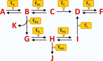
An example of generic metabolism is schematized in Fig. 4.1. No matter how complex the metabolism may seem at first glance, the interpretation of a scheme of consecutive reactions is simple and depends on the identification of five key factors:
1. The reactants that originate the process
2. The end products, regardless of being formed in intermediate reactions or the final reaction
3. The branching points, i.e., steps where the sequence of reactions may follow different courses
4. The irreversible reactions
5.The specific reactions that are catalyzed by enzymes that are finely regulated and so have the ability to highly accelerate segments of the metabolism, or not
Fig. 4.1 Hypothetical schematic metabolism involving metabolites A to K and enzymes Ex (subscript X in Ex represents the substrate, X = A to K). The metabolism is “fed” by A and has K, F, and J as “end products.” An external source of G may also lead to the formation of J and F, but not K. An external source of D can only lead to the formation of F. However, if ED is not present or not operative, F will not be formed in any circumstance. Likewise, if EB2 is not present or not operative, K, G, H, J, and I will not be formed even in the presence of high concentration of B. If EB1 and EB2 are never active or inactive at the same time, F is always formed but not J; the reactions’ scheme assures the permanent formation of F but the selective formation of J when the control of the reaction course is performed by alternate states of activity of EB1 and EB2
These five key factors in the interpretation of a metabolism will help the reader not to look to a metabolism as a chaotic ensemble of chemicals, enzymes, and reaction arrows. Metabolism is an appealing and richly informative text on the organization of life, should we be able to interpret it.
Enzymes are key protagonists of metabolisms together with metabolites (i.e., the intermediate reactants/products of the reactions of metabolisms). Not only enzymes