

новая папка / Integrative Human Biochemistry_ A Textbook for Medical Biochemistry (PDF).pdf
.pdf92 |
3 The Families of Biological Molecules |
Fig. 3.28 Heterocyclic bases are flat. Hydrogen bonding occurs in the plane of the rings. Purines and pyrimidines are able to interact because of the match in the number and orientation of H doning/acceptor groups, forming pairs T-A and C-G (a), which is known as Watson–Crick base pairing. Because bases are so flat, relatively hydrophobic on both sides, and undergo base pairing, nucleic acids may bind complementary sequences of nucleotides in the same polymer or from a different polymer. The entropic effect will cause this arrangement to twist around its long axis forming a double helix in which the polar parts of the molecule, phosphate and pentose residues, are exposed to the aqueous medium shielding the relatively hydrophobic bases. In the center of this helix, the bases stack parallel to each other and are slightly rotated relatively to each other. This kind of organization can be found even in some domains of the transfer RNAs (b; PDB 2TRA). The OH groups present in C2& groups of RNA (asterisk in panel (c) right) but not DNA (panel (c) left) have structural implications in the conformation of nucleic acids as these groups contribute to shield the core of the double strands from the aqueous environment. Panel (c) was reproduced from Goodsell, The Machinery of Life, 2009
3.2 Saccharides and Their Polymers and Derivatives |
93 |
is known as Watson–Crick base pairing. Hydrogen bonding occurs at the edges of heterocyclic bases, in the plane of the rings.
In regions in which the two opposing antiparallel sequences of tRNA have a considerable array of complementary base pairs, both RNA sequences fold into a helical structure to keep the parallel stacked base pairs in the core surrounded by the pentose and phosphate esters backbone. This is the double helix structure frequently associated to DNA but equally present in some regions of the RNA molecule. Ribosomal RNA structure is similar to that of tRNA (Fig. 3.28). It is also worth stressing that DNA polymers may have complementary RNA polymers, associate with them and even fold into helices. However, the alcohol group, OH, at C2& makes the structure of RNA less compact due to its volume and polarity.
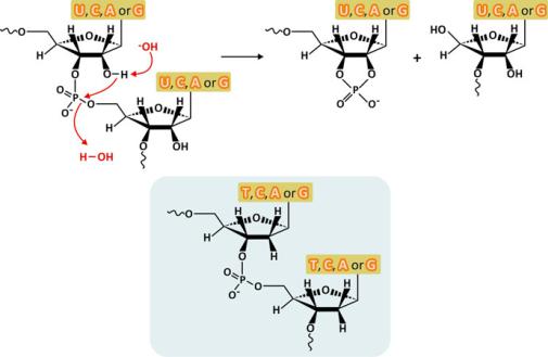
The structure of RNA is not only less compact, but it is also less chemically stable. The OH group at C2& is close to the phosphate diester bond with which it can react to hydrolyze RNA (Fig. 3.29). Because RNA molecules have transient functionalities and are not stored for very long periods in the cells, this limitation of RNA is not a problem. DNA is less prone to hydrolysis because it lacks the OH group in C2&, being the molecule that natural evolution selected to store genetic information for longer periods.
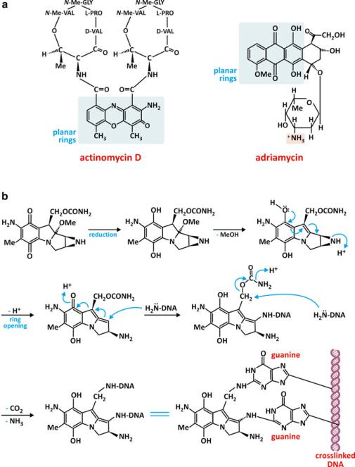
Some drugs target DNA taking advantage from the parallel stacking of heterocyclic bases. Notably, most of these molecules are composed of hydrophobic planar heterocyclic groups able to intercalate the base pairs of DNA (Fig. 3.30). Some of these molecules are used in the treatment of cancer because they prevent cell multiplication.
Fig. 3.29 Intramolecular hydrolysis of the phosphodiester bonds of RNA caused by a base (top). The absence of a hydroxyl group at C2& increases the hydrolytic stability of DNA relative to RNA (bottom shaded structures)
94 |
3 The Families of Biological Molecules |
Fig. 3.30 Actinomycin D is an antibiotic with anticancer activity (a). It binds DNA because it has a flat polycyclic and relatively hydrophobic group able to intercalate the stacked bases of DNA, preventing RNA synthesis. Adriamycin (a) is also an anticancer drug that operates with the same mechanism of action: intercalation of a polycyclic flat hydrophobic group between the base pairs of DNA, preventing cell proliferation. Mitomycin C (b) has a different mechanism of action: it is a DNA cross-linker by covalently linking two guanines. The direct contact with these nucleotide residues is possible because this drug is a polycyclic flat and relatively hydrophobic molecule
3.3 Amino Acids and Their Polymers: Peptides and Proteins |
95 |
3.3Amino Acids and Their Polymers: Peptides and Proteins
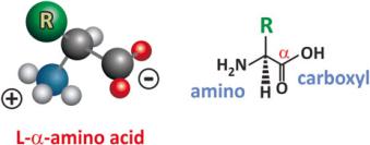
Chemically speaking amino acids are molecules that simultaneously have carboxyl (–COOH) and amine (–NH2) groups. Biochemists focus on !-amino acids, in which these groups are bound to the same terminal carbon (the so-called !-carbon in older organic chemistry nomenclatures), because naturally occurring proteins are polymers of !-amino acids. These amino acids have the structure depicted in Fig. 3.31. Besides the amino and carboxylic acid groups, the !-carbon (also named central carbon) attaches a hydrogen atom and another group, the so-called side chain that is represented by R. In nature R is one of 20 more common groups with few exceptions, which are usually derivatives of these 20 groups.
Fig. 3.31 The structure of !-amino acids. Depending on pH, in aqueous solution the amino group may be protonated and the carboxylic acid deprotonated, which makes amino acids potential zwitterions, i.e., bearing two opposite charges. Being weak bases and acids, amino acids have the ability to constitute buffers themselves (see Sect. 2.1.1)
Another interesting peculiarity of naturally occurring amino acids besides being !-amino acids is that they are almost exclusively L-enantiomers as the !-carbons are chiral centers. The other enantiomer is named D. The L and D nomenclature for the stereochemistry of the amino acids was established by Emil Fisher in analogy with glyceraldehyde, which also has a single chiral center with two possible enantiomers. Another nomenclature, more complex and following modern rules, exists to describe the stereochemistry of amino acids, but the predominance of L-amino acids and the simplicity of the L vs. D system resulted in the long-term longevity and universality of this system.
One simple empirical rule to distinguish L- from D-enantiomers is to adopt the perspective of the chemical structure of the amino acid along the H-!C axis (Fig. 3.32). The groups COOH, NH2, and H appear projected as the vertices of a triangle. You can now recognize “corn” written clockwise in L-enantiomers or counterclockwise in D-enantiomers. This is the CORN rule of thumb.
The chemical nature of the lateral chain, R, is determinant for biochemical processes in which amino acids participate and for the structure that proteins adopt when such amino acids are present. Broadly speaking, amino acids can be grouped in four different categories based on polarity and acidic/basic nature of R (Fig. 3.33): acidic, basic, neutral polar, and neutral nonpolar. Other classification systems are based on the chemical nature of R: hydrocarbons, carboxylic acids, amides (–CONH2), acyclic nitrogen containing, hydroxyl, sulfur containing, and nitrogen heterocycles. Figure 3.33
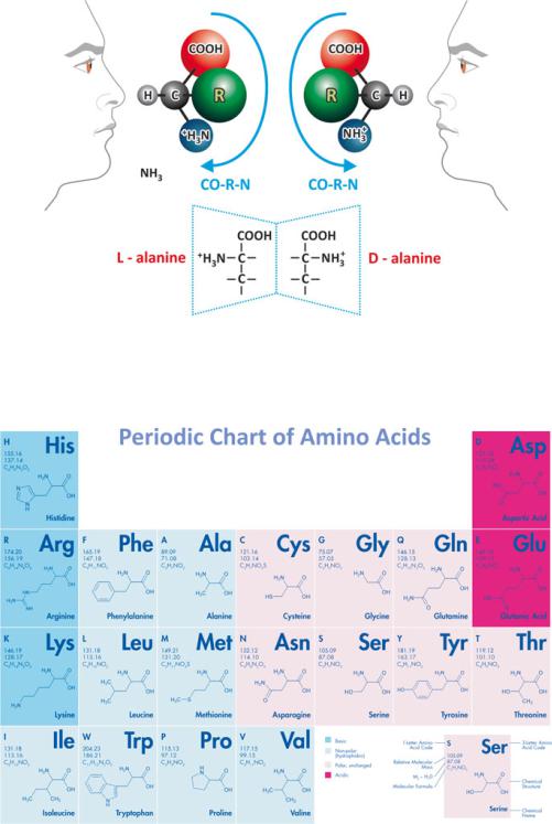
Fig. 3.32 The L vs. D nomenclature revealed by the CORN rule of thumb. When R=H (this happens in glycine, the simplest amino acid), chirality does not exist as two equal substituents (H, in this case) are attached to the central carbon. The example of L- and D-alanine is presented highlighting that they are mirror images
Fig. 3.33 Periodic chart-like arrangement of the natural amino acids. Figure reprinted with the permission of Bachem, Bubendorf, Switzerland
includes grouping of the amino acids according to polarity and charge and shows the chemical nature of the side chains. Table 3.4 clarifies the relationship between the names of amino acids in full and the three-letter and one-letter code abbreviated nomenclature. It also summarizes the most relevant properties of amino acids.
Table 3.4 Natural amino acid nomenclature (three-letter and one-letter code) and main properties. An essential amino acid cannot be synthesized de novo and therefore must be obtained from diet
Nomenclature rationale |
|
Three- |
One-letter |
|
|
for one-letter code |
Amino acid |
letter code |
code |
Main properties |
|
|
|
|
|
|
|
First letter is unique |
Cysteine |
Cys |
C |
Thiol side chain susceptible to oxidization to form disulfides |
|
|
|
|
|
|
|
|
Histidine |
His |
H |
Essential amino acid with imidazole side chain. The imidazole side chain has a pKa |
|
|
|
|
|
of approximately 6.0, which implies that relatively small shifts in most frequent |
|
|
|
|
|
physiologically relevant pH values will change its average charge, which in turn |
|
|
|
|
|
may impact significantly on protein structure |
|
|
|
|
|
|
|
|
Isoleucine |
Ile |
I |
Essential amino acid isomer of leucine. Chiral side chain |
|
|
Methionine |
Met |
M |
Side chain possesses a S-methyl thioether, which may be a source of sulphur for |
|
|
|
|
|
cartilage healing. It has been suggested that methionine is able to strengthen the |
|
|
|
|
|
structure of hair and nails because its side chains may cross react |
|
|
|
|
|
|
|
|
Serine |
Ser |
S |
Residues of serine are found in some phospholipids (besides proteins) |
|
|
|
|
|
|
|
|
Valine |
Val |
V |
Essential amino acid. Like Leu and Ile, has a branched side chain |
|
First letter not unique. |
Alanine |
Ala |
A |
D-Alanine occurs in bacterial cell walls and in some peptide antibiotics. Side chain is |
|
Most frequent amino |
|
|
|
very small (methyl group) |
|
acids have priority |
|
|
|
|
|
Glycine |
Gly |
G |
Side chain consists in H, making Gly the only achiral and the smallest possible |
||
|
|
|
|
amino acid |
|
|
|
|
|
|
|
|
Leucine |
Leu |
L |
Essential branched side-chain amino acid |
|
|
|
|
|
|
|
|
Proline |
Pro |
P |
The amine nitrogen is bound to two alkyl groups forming a cyclic side chain, which |
|
|
|
|
|
gives proline an exceptional conformational rigidity compared to other amino acids. |
|
|
|
|
|
When proline is involved in a peptide bonding, its nitrogen is not bound to any |
|
|
|
|
|
hydrogen, meaning it cannot act as a hydrogen bond donor, causing a disruption of |
|
|
|
|
|
!-helices and "-sheets |
|
|
|
|
|
|
|
|
Threonine |
Thr |
T |
Essential amino acid. Chiral side chain. The hydroxyl group in the side chain is |
|
|
|
|
|
prone to glycosylation and phosphorylation |
|
|
|
|
|
|
|
|
|
|
|
(continued) |
|
Proteins and Peptides Polymers: Their and Acids Amino 3.3
97
Table 3.4 (continued)
Nomenclature rationale |
|
Three- |
One-letter |
|
for one-letter code |
Amino acid |
letter code |
code |
Main properties |
|
|
|
|
|
First letter not unique |
Arginine |
Arg |
R |
The guanidinium group in the side chain is positively charged at physiological pH |
and less frequent: letter |
|
|
|
ranges therefore prone to binding negatively charged groups. This group has also the |
with phonetic similarity |
|
|
|
ability to form multiple H bonds |
or side chain chemical |
|
|
|
|
Asparagine |
Asn |
N |
Its side chain is curiously an amide (like in peptide bonds). Ows its name to |
|
nature |
(side chain |
|
|
asparagus because it was first detected in aspargus juice |
|
contains N) |
|
|
|
|
|
|
|
|
|
Aspartate |
Asp |
D |
Together with glutamic acid, aspartate is an acidic amino acid because of the |
|
|
|
|
carboxylic group in the side chain |
|
|
|
|
|
|
Glutamate |
Glu |
E |
In addition to its role in proteins and amino acid metabolism, in neurosciences |
|
|
|
|
glutamate is a very important neurotransmitter |
|
|
|
|
|
|
Glutamine |
Gln |
Q |
Its side chain is curiously an amide (like in peptide bonds) formed by replacing the |
|
|
|
|
side-chain hydroxyl of Glu with an amine functional group. |
|
|
|
|
|
|
Phenylalanine |
Phe |
F |
Essential amino acid with a benzyl side chain, which makes it fluorescent and |
|
|
|
|
neutral |
|
|
|
|
|
|
Tyrosine |
Tyr |
Y |
Tyrosine has a phenol group in the side chain, which makes it fluorescent. More |
|
|
|
|
importantly, the phenol group functions as a receiver of phosphate mediated by |
|
|
|
|
protein kinases (so-called tyrosine kinases) resulting in alterations in the activity of |
|
|
|
|
the target protein. |
|
|
|
|
|
|
Tryptophan |
Trp |
W |
Essential amino acid having a fluorescent indole functional group in the side chain. |
|
(side chain |
|
|
The indole group is bulky and hydrophobic, so Trp is commonly found in lipid- |
|
with double |
|
|
contacting domains of proteins, such as transmembrane regions of membrane |
|
ring) |
|
|
proteins or fusion domains of viral proteins |
|
|
|
|
|
Nearest first letter |
Lysine |
Lys |
K |
Essential amino acid. Like in Arg, Lys side chain participates in hydrogen bonding |
|
|
|
|
and is cationic at physiological pH range, therefore prone to binding negatively |
|
|
|
|
charged groups |
|
|
|
|
|
Unknown |
(Unknown |
– |
X |
Unidentified amino acids in peptide or protein structure are generically represented |
|
amino acid) |
|
|
by X |
|
|
|
|
|
98
Molecules Biological of Families The 3
3.3 Amino Acids and Their Polymers: Peptides and Proteins |
99 |
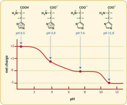
It should be kept in mind that the ionization states of amino acids vary with pH, so depending on pH, amino acids may have different net charges. The example of histidine is presented in Fig. 3.34. Histidine is peculiar as the side chain changes ionization (pKa ~ 6) not far from the range of plasma and cytoplasmic pHs. The intermediate value of the neutrality range (from pH 6 to 9), the so-called isoelectric point, pI, is 7.6, within the range of plasma and cytoplasmic pH range, which happens only for histidine.
Fig. 3.34 Variation of the net charge of His with pH. The molecule has three ionizable groups, one acidic and two basic. Therefore, allowed global charges range from −1 to +2. However, in most common physiological pHs, the global charge is nearly nil
3.3.1From Monomers to Polymers: Peptides and Proteins
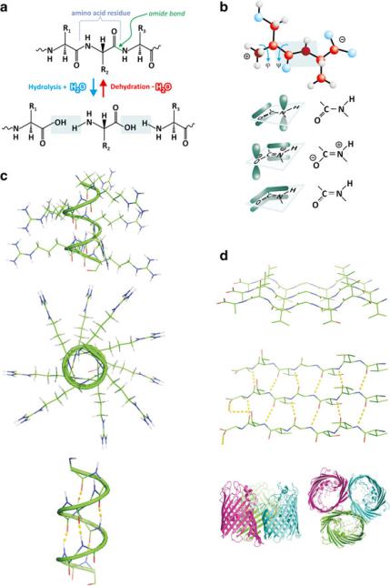
Amine and carboxylic groups may react, forming amide bonds (Fig. 3.35). Amide bonds connecting several amino acids form a peptide. Many amino acids connected through amide bonds form a protein. There is no precise limit to separate the number of amino acid monomers in peptides and proteins although 30 is usually taken as a reference value.
100 |
3 The Families of Biological Molecules |
Fig. 3.35 Dehydration reactions among amino acids lead to polymerization through amide (also known as “peptide”) bonds (a). The CO, CN, and NH bonds are coplanar because of the electronic distribution among the connected OCN set of atoms (b). This implies that when the polymeric chain folds, flexibility is limited and specific arrangements tend to be adopted, which include !-helices (c) and "-sheets (d). Whether a certain sequence of amino acid residues adopts the conformation of !-helix, "-sheet, or any other depends largely on the amino acids involved, their order, and environmental factors such as solvent polarity, pH, and temperature. Both !-helices and "-sheets are conformations that enable the occurrence of frequent intramolecular hydrogen bonding and externalize the location of side chains [(c) and (d)]. Proteins may be formed almost exclusively of !-helices, such as hemoglobin (see figure 3.42), or "-sheets, such as porin (d, bottom) or be a mixture of both. Images of porins are a courtesy of Dr. Claudio Soares, ITQB-UNL, Portugal
3.3 Amino Acids and Their Polymers: Peptides and Proteins |
101 |
Among biochemists, amide bonds forming peptides or proteins are generally referred to as peptide bonds. Because peptide bonds are very planar (C=O, C–N, and N–H bonds are coplanar) due to electron distribution limitations imposed by specific molecular orbitals and have the R groups in close vicinity, the chain of peptide bonds forms a polymer that is not freely flexible. It articulates with spatial constraints, which means that the polymeric chain tends to adopt fixed angles between its amide groups; these angles are the ones that allow accommodating the side chains of the amino acids and adapting the orientation of the amide bonds to each other (Fig. 3.35). As shown in Fig. 3.33, there is a wide diversity of side chains in charge, polarity, and size. All these parameters influence the way a protein folds to cope with the electrostatics, hydrogen bonding, entropic effects (hydrophobicity), and occupation of 3D space. In the end, all these factors determine that amino acid polymers have two different favored kinds of regular conformations: !-helices and "-sheets. Many other folds exist but are not as common because these two are the ones that better accomodate the stabilization of amino acid sequences.
In a !-helix, the peptide bond sequence (i.e., the peptide or protein “backbone”) adopts a helical structure projecting the side chains, R, to the exterior of the helix. It is a very stable structure because there are almost no constraints to spatially accommodate R and because the vast array of C=O and N–H groups in the backbone interact strongly through frequent hydrogen bonds. Many proteins, such as hemoglobin, are composed of several helical segments in their amino acid residues sequence. To facilitate protein representation and reading, helical segments are usually represented as a helical ribbon or a cylinder. This highlights the conformation of the segments, although it overlooks what specific amino acids are involved.
"-sheets are extended conformations that turn in specific points resulting in several linear amino acid residue sequences antiparallel to each other. Like in helices, this enables frequent hydrogen bonding in the protein backbone and projection on the side chain groups to the exterior of this compact arrangement. A certain degree of bending is allowed, and big extensions of "-sheets are usually associated to very stable proteic structures, such as membrane pores. "-sheet representation is usually done with straight ribbons and/or arrows (Fig. 3.35d).
The complete protein structure is described in three or four levels. The primary level is simply the sequence of amino acids that compose the protein, conventionally counted from the free amine terminal to the free carboxyl terminal. This elucidates the chemical nature of the protein but tells us little about what are the domains engaging !-helices, "-sheets, or none, which form the secondary-level structures. !-helices and "-sheets from different parts of the same protein tend to interact with each other toward mutual stabilization by means of electrostatic forces, hydrogen bonding, and entropic effects contributions (Fig. 3.36). The tertiary level arises there from !-helices, "-sheets, and other local arrangements that organize in space to form the protein structure itself. Occasionally, there are different parts of the