

новая папка / Integrative Human Biochemistry_ A Textbook for Medical Biochemistry (PDF).pdf
.pdf112 |
3 The Families of Biological Molecules |
Box 3.7 (continued)
Lysyl hydroxylases are also operative. Hydroxylation of Pro and Lys residues, both exposed in the triple helix of collagen, favors intermolecular adhesion interactions by hydrogen bonding and further chemical modifications, which are strengthened with age (this is one of the reasons why meat from young animals is tenderer than from older animals).
Carnitine is involved in the transport of fatty acids into the mitochondria, where they are oxidized (see Sect. 7.4.3). Ascorbic acid is used by two different enzymes in the carnitine biosynthesis. Without ascorbic acid, production of carnitine declines and fatty acids cannot be used as energy source. This leads to fatigue, which is one of the symptoms of scurvy. Curiously, fatigue appears prior to other symptoms. This may be explained by the fact that the enzymes in carnitine biosynthesis require higher concentrations of ascorbic acid to function (they have “lower affinity” for ascorbic acid) when compared to hydroxylases.
Other pathologies, such as the Ehlers–Danlos syndrome (EDS) and osteogenesis imperfecta (OI), are genetic pathologies associated to deficiencies on protein–protein interactions in collagen. A fraction of the Lys residues in collagen react with one another forming covalent cross-links in collagen fibers. In some forms of EDS, this cross-linking is impaired, rendering the skin less firm and less resistant, hyperelastic. Collagen contributes to the mechanical strength of the skin, joints, muscles, ligaments, blood vessels, and visceral organs. In OI, replacement of Gly residues in the collagen amino acid residue sequence destroys the capacity of the protein to assemble in perfect triple helices because all other residues are bulkier than Gly. This leads to an extremely severe condition that is characterized by alterations in the physical properties of collagen and perturbations in the biochemical processes involving collagen homeostasis. The relationship between the collagen fibrils and hydroxyapatite crystals when bones are formed is altered, causing brittleness. For this reason, OI is also known as “brittle bone disease.”
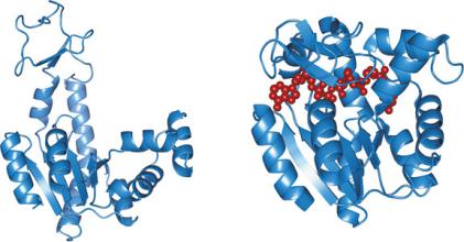
One remarkable property of many globular proteins is the ability to both bind other molecules and change conformation upon binding. Taking adenylate cyclase as example, binding of molecules such as ATP or ADP causes very mobile domains of the protein to change position (Fig. 3.41). This often leads to dynamic distortions of the ligand molecules because of the contact with amino acid residues. The results may be such that covalent bonds are formed or broken, thus chemically transforming the ligand molecule into a product. In practice, the protein action is to increase the rate of transformation of the ligand in the product. If only the ligand, not the protein, is chemically transformed in this process, this can be seen as an enzymatic catalysis, i.e., increase of the velocity of chemical reactions caused by proteins, the enzymes. In these cases, the ligand (reactant) is named substrate.
3.3 Amino Acids and Their Polymers: Peptides and Proteins |
113 |
Fig. 3.41 Adenylate cyclase (or adenylyl cyclase), unbound (PDB 4AKE; left) and bound (PDB 1AKE; right) to a dinucleotide analogue (red). Upon binding to the dinucleotide analogue, the mobile domains adapt by changing position
3.3.3Cooperative Interplay Between Tertiary-Level and Quaternary-Level Structure
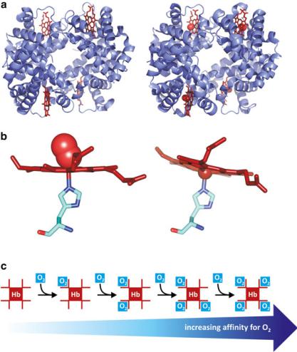
As discussed in the previous section, the tertiary-level structure of a protein is determined and maintained by arrays of sites in which attractive or repulsive forces between groups of atoms exist. This is a relatively delicate balance. When a significant number of such “force spots” are altered, the conformation of the protein adapts by adopting a different tertiary-level structure, which corresponds to the new balance of forces. Likewise, in cases in which a quaternary-level structure exists, the changes in the conformation of one protein monomer at the surface of contact with other monomer may impose alterations in the “force spots” (hydrogen bounds, electrostatic repulsion and attraction, entropic factors, etc.) so that the second monomer changes conformation to adapt. In practice, this means that conformational changes in one protein may be transmitted to a neighboring protein that is in contact with it. In other words, tertiary-level structural changes may be transmitted and amplified to other proteins throughout the quaternary-level structure. Hemoglobin is a good example. It is formed by two subunits, ! and ", forming a dimer that associates to other dimer—a tetramer that is in fact a dimer made of dimers. Dimers are numbered 1 and 2; therefore, hemoglobin is a tetramer of four subunits "1, !1, "2, and !2 (Fig. 3.42). ! − " attractive forces are stronger than 1–2 attractive forces, but both are sufficient to transmit to neighboring monomers conformational changes. This affects the affinity of the hemoglobin monomers to oxygen. Each hemoglobin monomer is covalently associated to a non-proteic
114 |
3 The Families of Biological Molecules |
Fig. 3.42 Hemoglobin is a tetramer (a), each subunit containing a heme group (red). Human deoxyhemoglobin (left; PDB 2HHB) and oxyhemoglobin (right; PDB 1GZX) show subtle but important changes in conformation. Binding of molecular oxygen to the heme group [(b), left; PDB 1HHO] causes a shift in the orientation of the heme relative to the His residue when compared to the heme in deoxyhemoglobin [(b), right; PDB 4HHB). This slight distortion in the position of the heme leads to a variation in the conformation of the protein, which propagates to neighboring monomers in the tetramer. The neighboring monomers acquire higher affinity for O2 (c)
group, i.e., a prosthetic group, of the porphyrin family (Box 3.8). In this case the porphyrin binds in its center an iron ion, forming a heme. The iron ion complexes with the heme through four bonds in the plane of the porphyrin and to a His residue side chain orthogonally to the heme plane. Another orthogonal bond, opposite to His is established with small molecules having electron donor atoms, such as O2 or CO. When an O2 molecule binds to the iron in the heme, the position of the iron slightly shifts, which in turn affects the position of the His residue. When the His

3.3 Amino Acids and Their Polymers: Peptides and Proteins |
115 |
Box 3.8: The Importance of Heme Groups in Proteins.
Many natural proteins are associated to prosthetic groups of similar chemical nature called porphyrins. Porphyrins are macrocyclic compounds related to porphin (see figure). Hemoglobins, for instance, bind porphyrin groups, such as heme b (see figure). Hemes are porphyrins that bind iron ions in the center. The remarkable capacity to bind metallic ions of charge +2 or +3 in the center of the ring may explain the success of these molecules during natural selection and subsequent ubiquity of porphyrins in nature. The porphyrin macrocycle has 26 delocalized (() electrons in total, being classified as aromatic from a chemical point of view. This system of delocalized electrons extends to the nitrogen atoms and is available to bind the cationic metals. It is also responsible for the intense absorption bands in the visible region of electromagnetic radiation. This is the reason why compounds with porphyrins, such as hemoglobin and chlorophyll, are intensely colored. Heme-containing proteins, hemoproteins, and other metal-containing proteins, metalloproteins, due to their unique electronic properties, are adequate for the transient binding of diatomic gases that occurs during their transportation in blood and electron transfer (i.e., electron donation and reception to and from other compounds). It is possible that hemoproteins evolved from ancient proteic forms, whose function was electron transfer in sulfur-based photosynthesis in the ancestors of cyanobacteria before molecular oxygen existed in atmosphere.
In addition to the peculiar intrinsic properties of hemes and other porphy- rin–metal associations, the interaction between the porphyrin and the amino acid residues at the site where it inserts in proteins is of extreme importance. Variations in the shape, volume, and chemical composition of the binding site, in the mode of heme binding, and in the number and nature of heme–protein
(continued)

116 |
3 The Families of Biological Molecules |
Box 3.8 (continued)
interactions result in significantly different heme environments in proteins having different biological roles. The outcome is a fine-tuning of the heme properties. Take the 3D structure of the hemoglobin chain as an example. The position of a His residue is such that its protonation interferes with oxygen release from the iron ion. Acidification of the medium causes the protonation of His, which in turns facilitates the release of oxygen. This is known as Bohr effect (see Fig. 3.44 in main text) and is not a simple curiosity: In the pulmonary vasculature, the pH is higher than in the peripheral tissues because the pH is affected by the local abundance of CO2; therefore, the Bohr effects help in increasing the efficacy of binding oxygen in the lungs and releasing in peripheral tissues.
The porphyrin groups are equally important to stabilize protein structure and resistance to proteolysis, although these properties are frequently overlooked. Even in cases in which the porphyrins are not covalently bound to the protein, the interplay between amino acid residues and the non-proteic groups is very specific.
residue is pulled, the whole structure of the protein changes slightly. This change in conformation induces a change in conformation of the neighboring monomers. As a consequence, the neighboring monomers acquire increased affinity to bind an oxygen molecule. So, binding of O2 to a monomer increases the chances that a second O2 molecule binds to another monomer in the hemoglobin tetramer relative to a monomer in a tetramer without bound O2 molecules. This is called positive cooperativity, i.e. several entities influencing each other favouring a certain event.
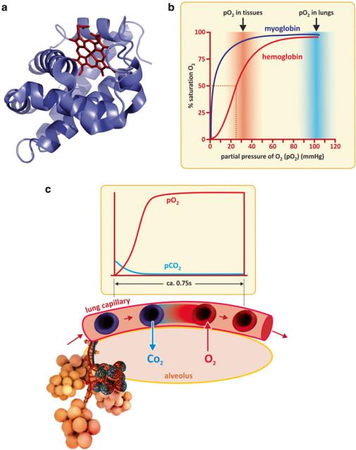
Hemoglobin monomers interact cooperatively to bind up to four oxygen molecules. Myoglobin, a protein abundant in muscles, also binds O2, but this protein occurs as a monomer, in contrast to hemoglobin. Comparing hemoglobin to myoglobin makes the effect of cooperativity clear. The fraction of myoglobin binding O2 relative to total myoglobin increases nearly linearly with partial pressure of oxygen up to saturation. To be more precise, the variation is hyperbolic. In contrast, hemoglobin binds O2 critically at a triggering narrow concentration range, in which it reaches saturation. Hemoglobin changes from highly unsaturated to almost saturated in a narrow O2 partial pressure interval. Interestingly, the narrow interval of transition to near saturation corresponds to the partial pressure of oxygen found in peripheral tissues (Fig. 3.43), away from the lung alveoli. Therefore, cooperativity among the hemoglobin monomers enables hemoglobin to saturates with oxygen in the lungs and delivers its cargo to peripheral tissues. Myoglobin would not be adequate for this function as it is almost saturated in both situations. Myoglobin is fit for oxygen storage in muscle cells. Release of oxygen occurs when the consumption in mitochondria is such that the cell is almost depleted in oxygen (Fig. 3.43).
3.3 Amino Acids and Their Polymers: Peptides and Proteins |
117 |
Fig. 3.43 Myoglobin (a) is a monomeric oxygen-binding protein. Like a hemoglobin monomer, it also binds oxygen through a heme group (red organic structure). The absence of cooperativity in myoglobin when compared to hemoglobin implies distinct binding capacities at different oxygen partial pressures, pO2 (b). Hemoglobin has an abrupt transition from low to high binding when pO2 changes from values typical from peripheral tissues to values typical of the lungs. As the erythrocytes pass adjacent to alveoli, O2 and CO2 diffuse freely across arterial and lung cells driven by partial pressure gradients (c)
118 |
3 The Families of Biological Molecules |
It is worth stressing that hemoglobin transports most, but not all, oxygen used in tissues. Oxygen, like carbon dioxide, is a small, hydrophilic molecule, which easily dissolves in aqueous media. Although being hydrophilic, it is a very small molecule and diffuses freely in tissues. This is the reason why cells do not need oxygen transporters or channels. The same happens with CO2 but it has lower affinity for hemoglobin. In addition, CO2 is converted to HCO3− that equilibrates with H2CO3 (see Sect. 2.1.1). Therefore, plasma CO2 transport is not dependent on a specific protein.
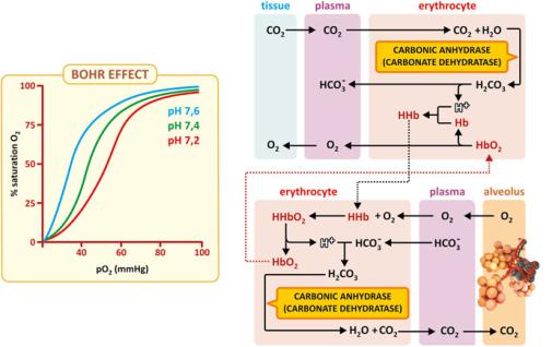
Although the direct binding of CO2 to hemoglobin is not significant, hemoglobin is very important in the chemistry and physiology of CO2 in the human body as the protein itself is a weak base or weak acid depending on pH. In the peripheral tissues, in which CO2 is present at higher partial pressure, CO2 diffuses to plasma and therefore in erythrocytes. Carbonic anhydrase then converts CO2 to H2CO3 that acidifies the medium. Acid pH leads to the protonation of hemoglobin, which has lower affinity for O2. Near the lung alveoli, plasma CO2 diffuses to the alveoli due to the gradient in the partial pressure of CO2 (Fig. 3.43). This drop in plasma partial pressure of CO2 in plasma causes carbonic anhydrase to convert H2CO3 in CO2, therefore shifting the equilibrium HCO3−/H2CO3 toward the consumption of HCO3− and H+. The slight drop in pH causes the deprotonation of hemoglobin, which has higher affinity for oxygen. Thus, there is a coupling between pH and the efficiency of oxygen capture, transport, and release (Fig. 3.44). The coupling of hemoglobin structure with pH is known as the “Bohr effect.”
Fig. 3.44 The Bohr effect associated to hemoglobin. CO2 levels in blood influence the transport of O2 by hemoglobin through plasma pH because protonation/deprotonation of hemoglobin affects cooperativity in O2 binding. Lower pH (found in tissues due to the release of CO2 in plasma) favors hemoglobin protonation and unbinding of oxygen. Hb - Hemoglobin, HHb - protonated hemoglobin
3.3 Amino Acids and Their Polymers: Peptides and Proteins |
119 |
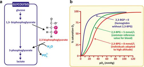
In the same way oxygen binding, transport, and release by hemoglobin are affected by pH, binding of 2,3-bisphosphoglycerate (2,3-BPG) also influences oxygen fixation in a way such that the binding curve of oxygen by hemoglobin is shifted toward higher partial pressures of oxygen (Fig. 3.45). This is far from being a simple curiosity: 2,3-BPG forms from 1,3-BPG, a metabolite of the glycolysis pathway (see Sect. 6.1.3). When glycolysis is highly active, 2,3-BPG is formed and release of O2 from hemoglobin becomes more effective. This is convenient for the cell as higher glycolytic activity implies, in principle, a higher demand for oxygen by the human cells. The fixation of oxygen in lungs remains unaffected.
Fig. 3.45 2,3-BPG forms from 1,3-bisphophoglycerate, an intermediate metabolite of glycolysis; it is therefore, a chemical signal of glycolytic activity (a). 2,3-BPG binding to hemoglobin affects cooperativity in such a way that the O2 binding curves are shifted in a way that O2 release in peripheral tissues is facilitated, but the O2 captured in the lungs is not affected (b)
3.3.4Enzymes
The previous section showed how dynamic the binding of proteins to other molecules may be. In the case of hemoglobin, oxygen binds to the iron of the heme group. However, frequently more complex molecules bind directly to side chains of certain amino acid residues. Specific sets of amino acid residues in a protein may precisely locate and orient in space and have the correct physical properties (charge, polarity, hydrogen donor/acceptor groups, etc.) to specifically bind molecules that establish attractive forces with them (Fig. 3.46). The electronic clouds of these molecules are distorted by the contact with the amino acid residues, which in turn adapt their tertiary-level structure to the presence of the molecules. This mutual adaptation between protein and bound molecules frequently weakens some chemical bounds of the molecules, which may thus be destroyed. Likewise, formation of other bonds is possible. The result is that the molecule that bound to the protein is
120 |
3 The Families of Biological Molecules |
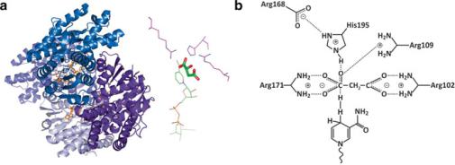
Fig. 3.46 Malate dehydrogenase (PDB 2DFD) is a homodimeric enzyme (A) that catalyzes the oxidation of malate to oxaloacetate concomitantly with the reduction of NAD+ to NADH [highlighted in orange in (a)]. A specific set of amino acids of the enzyme has the right properties (charge, H-binding ability, etc.), the right location, and the right orientation to simultaneously fit the malate molecule (b). This set of residues forms the so-called active site (or active center) of the enzyme. NAD+ binds to other site, specific for it, in close vicinity to the active site and participates in the oxidation of malate facilitated by the action of the amino acids [(b), bottom structure]
converted in a different molecule. If the resulting product dissociates from the protein and the protein returns to the same state as before binding the original molecule, then the protein is an enzyme, i.e., a proteic natural catalyst. The initial molecule that binds the enzyme and undergoes a chemical reaction is said to be a substrate, as previously mentioned in Sect. 3.3.2.
Because the enzymes interact with the substrate and facilitate its conversion to products, they increase the velocity of chemical reactions enormously, typically, above 107-fold. In some cases, the increase may be 1017-fold, which is a figure difficult to conceive intuitively. Considering that a 2 × 10⁸-fold increase in the velocity of a relaxed walk (~1.5 ms−1) would leave us traveling at the speed of light (~3 × 10⁸ ms−1), this intuitive perception becomes clearer. 1017-fold is more than the difference between a relaxed walk and 100 million faster than the speed of light in vacuum. One will see in Chap. 4 that enzymes accelerate reactions but cannot turn impossible reactions in possible ones. However, enzymes turn very slow reactions (so slow that in practice they seem unable to occur) into fast reactions. So, in practice it is almost like if an impossible one was transformed into a possible reaction by the intervention of an enzyme.
3.3.4.1 The Importance of Studying Enzymes
Enzymes are interesting molecules because the dynamics of their tertiary-level structure implies catalytic activity, which has shaped life as it exists today. Even viruses need enzymes to be effective. Because enzymes are so proficient in speeding reactions, controlling the activity of enzymes is, in practice, controlling the course of chemical reactions in a cell. This is an essential piece to impose order in the chemistry of the cells. Also controlling the activity of enzymes ensures that certain
3.3 Amino Acids and Their Polymers: Peptides and Proteins |
121 |
reactions only occur to a significant extent when and where the enzyme is inside the cell. This prevents conflicting reactions in a well regulated cell and permits certain reactions to be coupled. Imagine substrate A and substrate B; now imagine that enzyme EA converts A in B and a second enzyme, EB, converts B in C. The simultaneous presence of both enzymes in the same cell compartment has the practical consequence that A is converted in C. This coupling of reactions may reach considerable complexity, with many substrates, reactions, and enzymes being involved, sometimes with branched and cyclic reaction sequences (recall Figs. 1.3 and 1.4). Such sets of reactions are referred to generally as “metabolisms.” Regulation of metabolism is largely dependent on enzymes. The mechanisms of metabolic regulation are extremely important and will be addressed in Chap. 5. A regulated metabolism is a sine qua non condition for a particular state of organisms we call “health.”
Yet, the structure–activity relationship in enzymes and their significance in metabolic regulation is only part of the importance of studying enzymes. Enzymes can operate outside cells and be used in industrial processes in pharma, food, or detergent processing and manufacturing, for instance. More importantly, in biomedical sciences and clinical practice, they can be used as valuable tools for diagnosis. When enzymes that were supposed to be confined in cellular compartments in specific tissues are found with increased levels in plasma, this is a sign of tissue lesions with rupture of cell membranes (and consequent leakage of enzymes to the plasma). The death of cells in tissues implies a constant flow of intracellular contents to the plasma, but in non-injured tissues this occurs to a very limited extent. A severe lesion in the liver, heart, or other organ leads to unusually high increased levels in the plasma of enzymes that are specific for that organ. Prostatic specific acid phosphatase (PSAP, Fig. 3.47), for instance, is an enzyme produced by the prostate that can be found in
Fig. 3.47 Human prostatic specific acid phosphatase, PSAP [(a); PDB 1cvi), and aspartate aminotransferase, ASP [(b); PDB 3II0], are markers of prostate cancer and hepatitis, respectively. Pyridoxal 5-phosphate is a cofactor that is shown bound to ASP (yellow)