

новая папка / Operative Standards for Cancer Surgery Volume I 1st Edition
.pdf3.Cabioglu N, Hunt KK, Sahin AA, et al. Role for intraoperative margin assessment in patients undergoing breast-conserving surgery. Ann Surg Oncol 2007;14(4):1458-1471.
4.Singh M, Singh G, Hogan KT, et al. The effect of intraoperative specimen inking on lumpectomy re-excision rates. World J Surg Oncol 2010;8:4.
5.Blair SL, Thompson K, Rococco J, et al. Attaining negative margins in breast-conservation operations: is there a consensus among breast surgeons? J Am Coll Surg 2009;209(5):608-613.
6.Bathla L, Harris A, Davey M, et al. High resolution intra-operative two-dimensional specimen mammography and its impact on second operation for re-excision of positive margins at final pathology after breast conservation surgery. Am J Surg
2011;202(4):387-394.
7.Chagpar A, Yen T, Sahin A, et al. Intraoperative margin assessment reduces reexcision rates in patients with ductal carcinoma in situ treated with breast-conserving surgery. Am J Surg 2003;186(4):371-377.
8.Ciccarelli G, Di Virgilio MR, Menna S, et al. Radiography of the surgical specimen in early stage breast lesions: diagnostic reliability in the analysis of the resection margins. Radiol Med 2007;112(3):366-376.
9.Kim SH, Cornacchi SD, Heller B, et al. An evaluation of intraoperative digital specimen mammography versus conventional specimen radiography for the excision of nonpalpable breast lesions. Am J Surg 2013;205(6):703-710.
10.Layfield DM, May DJ, Cutress RI, et al. The effect of introducing an in-theatre intra-operative specimen radiography (IOSR) system on the management of palpable breast cancer within a single unit. Breast 2012;21(4):459-463.
P.26
11.McCormick JT, Keleher AJ, Tikhomirov VB, et al. Analysis of the use of specimen mammography in breast conservation therapy. Am J Surg 2004;188(4):433-436.
12.Carmichael AR, Ninkovic G, Boparai R. The impact of intra-operative specimen radiographs on specimen weights for wide local excision of breast cancer. Breast 2004;13(4):325-328.
13.Hirose M, Kacher DF, Smith DN, et al. Feasibility of MR imaging-guided breast lumpectomy for malignant tumors in a 0.5-T open-configuration MR imaging system. Acad Radiol 2002;9(8):933-941.
14.Tafra L, Fine R, Whitworth P, et al. Prospective randomized study comparing cryo-assisted and needle-wire localization of ultrasound-visible breast tumors. Am J Surg 2006;192(4):462-470.
15.Haka AS, Volynskaya Z, Gardecki JA, et al. In vivo margin assessment during partial mastectomy breast surgery using Raman spectroscopy. Cancer Res 2006;66(6):3317-3322.
16.Fine RE, Schwalke MA, Pellicane JV, et al. A novel ultrasound-guided electrosurgical loop device for intra-operative excision of breast lesions; an improvement in surgical technique. Am J Surg 2009;198(2):283-286.
17.Cortes-Mateos MJ, Martin D, Sandoval S, et al. Automated microscopy to evaluate surgical specimens via touch prep in breast cancer. Ann Surg Oncol 2009;16(3):709-720.
18.Blair SL, Wang-Rodriguez J, Cortes-Mateos MJ, et al. Enhanced touch preps improve the ease of interpretation of intraoperative breast cancer margins. Am Surg 2007;73(10):973-976.
19.Cuntz, MC, Levine EA, O’Dorisio TM, et al. Intraoperative gamma detection of 125I-lanreotide in women with primary breast cancer. Ann Surg Oncol 1999;6(4):367-372.

20.Keller MD, Majumder SK, Kelley MC, et al. Autofluorescence and diffuse reflectance spectroscopy and spectral imaging for breast surgical margin analysis. Lasers Surg Med 2010;42(1): 15-23.
21.Keller MD, Vargis E, de Matos Granja N, et al. Development of a spatially offset Raman spectroscopy probe for breast tumor surgical margin evaluation. J Biomed Opt 2011;16(7):077006.
22.Martin DT, Sandoval S, Ta CN, et al. Quantitative automated image analysis system with automated debris filtering for the detection of breast carcinoma cells. Acta Cytologica 2011;55(3): 271-280.
23.Nguyen FT, Zysk AM, Chaney EJ, et al. Intraoperative evaluation of breast tumor margins with optical coherence tomography. Cancer Res 2009;69(22):8790-8796.
24 Allweis TM, Kaufman Z, Lelcuk S, et al. A prospective, randomized, controlled, multicenter study of a real-time, intraoperative probe for positive margin detection in breast-conserving surgery. Am J Surg 2008;196(4):483-489.
25. Karni T, Pappo I, Sandbank J, et al. A device for real-time, intraoperative margin assessment in breast-conservation surgery.
Am J Surg 2007;194(4):467-473.
26.Rahusen FD, Bremers AJA, Fabry HFJ, et al. Ultrasound-guided lumpectomy of nonpalpable breast cancer versus wireguided resection: a randomized clinical trial. Ann Surg Oncol 2002;9(10):994-998.
27.Krekel NM, Haloua MH, Lopes Cardozo AMF, et al. Intraoperative ultrasound guidance for palpable breast cancer excision (COBALT trial): a multicentre, randomised controlled trial. Lancet Oncol 2013;14(1):48-54.
28.Moore MM, Whitney LA, Cerilli L, et al. Intraoperative ultrasound is associated with clear lumpectomy margins for palpable infiltrating ductal breast cancer. Ann Surg 2001;233(6): 761-768.
29.Davis KM, Hsu CH, Bouton ME, et al. Intraoperative ultrasound can decrease the re-excision lumpectomy rate in patients with palpable breast cancers. Am Surg 2011;77(6):720-725.
30.Olsha O, Shemesh D, Carmon M, et al. Resection margins in ultrasound-guided breast-conserving surgery. Ann Surg Oncol
2011;18(2):447-452.
31.Eichler C, Hübbel A, Zarghooni V, et al. Intraoperative ultrasound: improved resection rates in breast-conserving surgery. Anticancer Res 2012;32(3):1051-1056.
32.Barentsz MW, van Dalen T, Gobardhan PD, et al. Intraoperative ultrasound guidance for excision of non-palpable invasive breast cancer: a hospital-based series and an overview of the literature. Breast Cancer Res Treat 2012;135(1):209-219.
33.Harlow SP, Krag DN, Ames SE, et al. Intraoperative ultrasound localization to guide surgical excision of nonpalpable breast carcinoma. J Am Coll Surg 1999;189(3):241-246.
34.Jakub JW, Gray RJ, Degnim AC, et al. Current status of radioactive seed for localization of non palpable breast lesions. Am J Surg 2010;199(4):522-528.
35.Gray RJ, Salud C, Nguyen K, et al. Randomized prospective evaluation of a novel technique for biopsy or lumpectomy of nonpalpable breast lesions: radioactive seed versus wire localization. Ann Surg Oncol 2001;8(9):711-715.
P.27
36. Lovrics PJ, Goldsmith CH, Hodgson N, et al. A multicentered, randomized, controlled trial comparing radioguided seed localization to standard wire localization for nonpalpable, invasive and in situ breast carcinomas. Ann Surg Oncol
2011;18(12):3407-3414.

37.Ahmed M, Douek M. Radioactive seed localisation (RSL) in the treatment of non-palpable breast cancers: systematic review and meta-analysis. Breast 2013;22(4):383-388.
38.Barentsz MW, van den Bosch MA, Veldhuis WB, et al. Radioactive seed localization for nonpalpable breast cancer. Br J Surg
2013;100(5):582-588.
39.Cox CE, Furman B, Stowell N, et al. Radioactive seed localization breast biopsy and lumpectomy: can specimen radiographs be eliminated? Ann Surg Oncol 2003;10(9):1039-1047.
40.Gobardhan PD, de Wall LL, van der Laan L, et al. The role of radioactive iodine-125 seed localization in breast-conserving therapy following neoadjuvant chemotherapy. Ann Oncol 2013;24(3):668-673.
41.Gray RJ, Pockaj BA, Karstaedt PJ, et al. Radioactive seed localization of nonpalpable breast lesions is better than wire localization. Am J Surg 2004;188(4):377-380.
42.Hughes JH, Mason MC, Gray RJ, et al. A multi-site validation trial of radioactive seed localization as an alternative to wire localization. Breast J 2008;14(2):153-157.
43.Murphy JO, Moo TA, King TA, et al. Radioactive seed localization compared to wire localization in breast-conserving surgery: initial 6-month experience. Ann Surg Oncol 2013;20(13):4121-4127.
44.Donker M, Drukker CA, Valdés Olmos RA, et al. Guiding breast-conserving surgery in patients after neoadjuvant systemic therapy for breast cancer: a comparison of radioactive seed localization with the ROLL technique. Ann Surg Oncol
2013;20(8):2569-2575.
45.Postma EL, Koffijberg H, Verkooijen HM, et al. Cost-effectiveness of radioguided occult lesion localization (ROLL) versus wire-guided localization (WGL) in breast conserving surgery for nonpalpable breast cancer: results from a randomized controlled multicenter trial. Ann Surg Oncol 2013;20(7):2219-2226.
46.Medina-Franco H, Abarca-Pérez L, García-Alvarez MN, et al. Radioguided occult lesion localization (ROLL) versus wireguided lumpectomy for non-palpable breast lesions: a randomized prospective evaluation. J Surg Oncol 2008;97(2):108-111.
47.Sajid MS, Parampalli U, Haider Z, et al. Comparison of radioguided occult lesion localization (ROLL) and wire localization for non-palpable breast cancers: a meta-analysis. J Surg Oncol 2012;105(8):852-858.
48.Audisio RA, Nadeem R, Harris O, et al. Radioguided occult lesion localisation (ROLL) is available in the UK for impalpable breast lesions. Ann R Coll Surg Engl 2005;87(2):92-95.
49.Belloni E, Canevari C, Panizza P, et al. Nonpalpable breast lesions: preoperative radiological guidance in radioguided occult lesion localisation (ROLL). Radiol Med 2011;116(4):564-574.
50.Duarte GM, Cabelloa C, Torresan RZ, et al. Radioguided Intraoperative Margins Evaluation (RIME): preliminary results of a new technique to aid breast cancer resection. Eur J Surg Oncol 2007;33(10):1150-1157.
51.Lavoue V, Nos C, Clough KB, et al. Simplified technique of radioguided occult lesion localization (ROLL) plus sentinel lymph node biopsy (SNOLL) in breast carcinoma. Ann Surg Oncol 2008;15(9):2556-2561.
52.Nadeem R, Chagla LS, Harris O, et al. Occult breast lesions: A comparison between radioguided occult lesion localisation (ROLL) vs. wire-guided lumpectomy (WGL). Breast 2005;14(4):283-289.
53.Zgajnar J, Hocevar M, Frkovic-Grazio S, et al. Radioguided occult lesion localization (ROLL) of the nonpalpable breast lesions. Neoplasma 2004;51(5):385-389.

54.Esbona K, Li Z, Wilke LG. Intraoperative imprint cytology and frozen section pathology for margin assessment in breast conservation surgery: a systematic review. Ann Surg Oncol 2012;19(10):3236-3245.
55.Osborn JB, Keeney GL, Jakub JW, et al. Cost-effectiveness analysis of routine frozen-section analysis of breast margins compared with reoperation for positive margins. Ann Surg Oncol 2011;18(11):3204-3209.
56.Camp ER, McAuliffe PF, Gilroy JS, et al. Minimizing local recurrence after breast conserving therapy using intraoperative shaved margins to determine pathologic tumor clearance. J Am Coll Surg 2005;201(6):855-861.
57.Jorns JM, Visscher D, Sabel M, et al. Intraoperative frozen section analysis of margins in breast conserving surgery significantly decreases reoperative rates: one-year experience at an ambulatory surgical center. Am J Clin Pathol
2012;138(5):657-669.
58. Sabel MS, Jorns JM, Wu A, et al. Development of an intraoperative pathology consultation service at a free-standing ambulatory surgical center: clinical and economic impact for patients undergoing breast cancer surgery. Am J Surg
2012;204(1):66-77.
P.28
59.Olson TP, Harter J, Muñoz A, et al. Frozen section analysis for intraoperative margin assessment during breast-conserving surgery results in low rates of re-excision and local recurrence. Ann Surg Oncol 2007;14(10):2953-2960.
60.Cox CE, Pendas S, Ku NN, et al. Local recurrence of breast cancer after cytological evaluation of lumpectomy margins. Am Surg 1998;64(6):533-537; discussion 537-538.
61.D’Halluin F, Tas P, Rouquette S, et al. Intra-operative touch preparation cytology following lumpectomy for breast cancer: a series of 400 procedures. Breast 2009;18(4):248-253.
62.Weinberg E, Cox C, Dupont E, et al. Local recurrence in lumpectomy patients after imprint cytology margin evaluation. Am J Surg 2004;188(4):349-354.
63.Caruso F, Ferrara M, Castiglione G, et al. Therapeutic mammaplasties: full local control of breast cancer in one surgical stage with frozen section. Eur J Surg Oncol 2011;37(10):871-875.
64.Cendan JC, Coco D, Copeland EM III. Accuracy of intraoperative frozen-section analysis of breast cancer lumpectomy-bed margins. J Am Coll Surg 2005;201(2):194-198.
65.Chen K, Zeng Y, Jia H, et al. Clinical outcomes of breast-conserving surgery in patients using a modified method for cavity margin assessment. Ann Surg Oncol 2012;19(11):3386-3394.
66.Noguchi M, Minami M, Earashi M, et al. Intraoperative histologic assessment of surgical margins and lymph node metastasis in breast-conserving surgery. J Surg Oncol 1995;60(3):185-190.
67.Riedl O, Fitzal F, Mader N, et al. Intraoperative frozen section analysis for breast-conserving therapy in 1016 patients with breast cancer. Eur J Surg Oncol 2009;35(3):264-270.
68.Weber S, Storm FK, Stitt J, et al. The role of frozen section analysis of margins during breast conservation surgery. Cancer J Sci Am 1997;3(5):273-277.
69.Cox CE, Hyacinthe M, Gonzalez RJ, et al. Cytologic evaluation of lumpectomy margins in patients with ductal carcinoma in situ: clinical outcome. Ann Surg Oncol 1997;4(8):644-649.
70.Creager AJ, Shaw JA, Young PR, et al. Intraoperative evaluation of lumpectomy margins by imprint cytology with histologic correlation: a community hospital experience. Arch Pathol Lab Med 2002;126(7):846-848.

71.Mannell A. Breast-conserving therapy in breast cancer patients: a 12-year experience. S Afr J Surg 2005;43(2):28-30.
72.Valdes EK, Boolbol SK, Ali I, et al. Intraoperative touch preparation cytology for margin assessment in breast-conservation surgery: does it work for lobular carcinoma? Ann Surg Oncol 2007;14(10):2940-2945.
73.Fukamachi K, Ishida T, Usami S, et al. Total-circumference intraoperative frozen section analysis reduces margin-positive rate in breast-conservation surgery. Jpn J Clin Oncol 2010;40(6): 513-520.
74.Janes SEJ, Stankheb M, Singha S, et al. Systematic cavity shaves reduces close margins and reexcision rates in breast conserving surgery. Breast 2006;15(3):326-330.
75.Héquet D, Bricou A, Koual M, et al. Systemic cavity shaving: modifications of breast cancer management and long-term local recurrence, a multicentre study. Eur J Surg Oncol 2013;39(8): 899-905.
76.Hewes JC, Imkampe A, Haji A, et al. Importance of routine cavity sampling in breast conservation surgery. Br J Surg
2009;96(1):47-53.
77.Keskek M, Kothari M, Ardehali B, et al. Factors predisposing to cavity margin positivity following conservation surgery for breast cancer. Eur J Surg Oncol 2004;30(10):1058-1064.
78.Kobbermann A, Unzeitig A, Xie XJ, et al. Impact of routine cavity shave margins on breast cancer re-excision rates. Ann Surg Oncol 2011;18(5):1349-1355.
79.Malik HZ, George WD, Mallon EA, et al. Margin assessment by cavity shaving after breast-conserving surgery: analysis and follow-up of 543 patients. Eur J Surg Oncol 1999;25(5):464-469.
80.Rizzo M, Iyengar R, Gabram SG, et al. The effects of additional tumor cavity sampling at the time of breast-conserving surgery on final margin status, volume of resection, and pathologist workload. Ann Surg Oncol 2010;17(1):228-234.
81.Tengher-Barna I, Hequet D, Reboul-Marty J, et al. Prevalence and predictive factors for the detection of carcinoma in cavity margin performed at the time of breast lumpectomy. Mod Pathol 2009;22(2):299-305.
Chapter 2
Total Mastectomy
CRITICAL ELEMENTS
 Incision Placement
 Boundaries of Mastectomy
 Mastectomy Flap Elevation
 Excision of the Pectoralis Major Muscle
 Prevention of Seroma/Drain Placement
1. INCISION PLACEMENT
Recommendation: Incisions for total mastectomy should be placed to facilitate the removal of the preponderance of breast tissue to achieve local disease control and decrease the risk of recurrent breast cancer.
Type of Data: Retrospective case series.
Strength of Recommendation: The consensus of the group supports this guideline based on historic evidence.
Rationale
Incision placement for total mastectomy is dictated by whether immediate reconstruction of the affected breast will be performed. The type of total mastectomy planned—simple total mastectomy, skin-sparing mastectomy, or nipple-sparing mastectomy—also guides incision placement.
Mastectomy without Immediate Breast Reconstruction
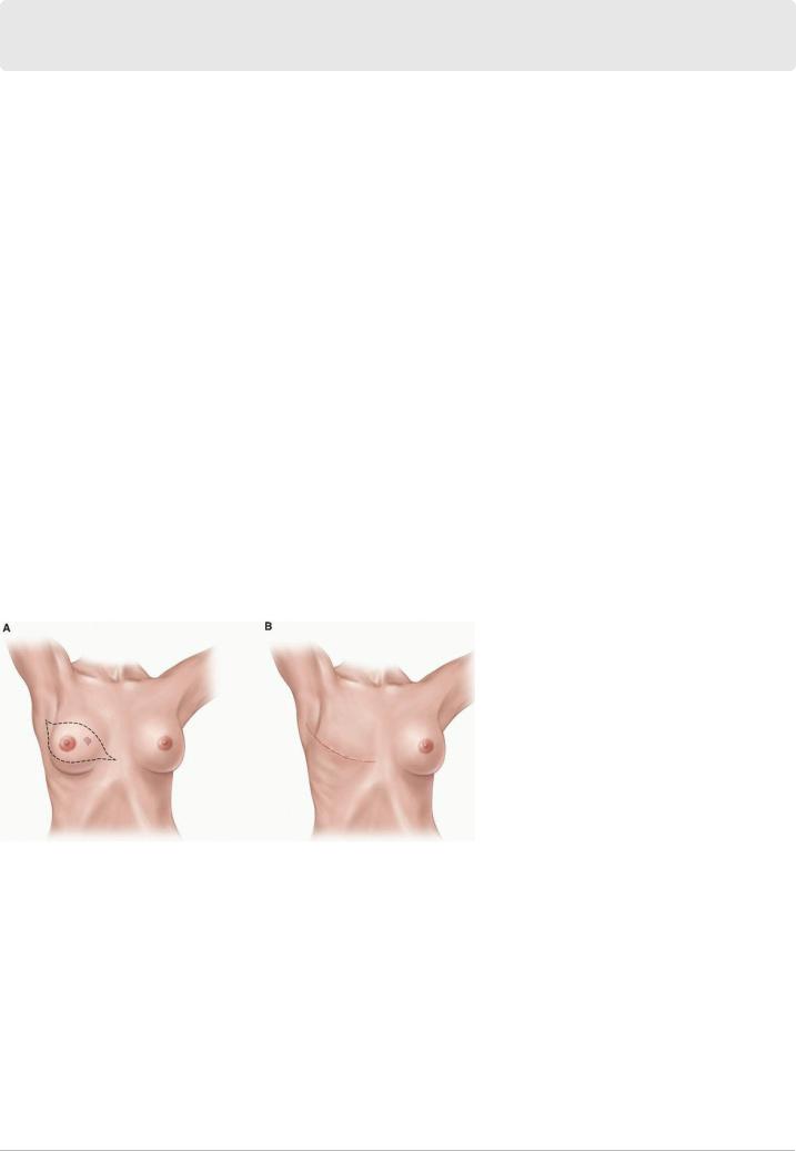
In patients not undergoing immediate breast reconstruction, a simple total mastectomy is performed. In these cases, an elliptical incision that encompasses the nipple-areola complex (NAC) and removes enough skin to achieve a smooth, even chest wall
P.30 without tension or redundant skin (“dog ears”) is made (Fig. 2-1). Although no strong data support the excision of percutaneous core biopsy scars not incorporated within the planned mastectomy incision, the general consensus is that any scar resulting from previous open biopsy or lumpectomy should be removed at the time of mastectomy, ideally within the ellipse of removed skin. The removal of a remote scar caused by previous open biopsy or lumpectomy may necessitate a separate incision.
FIGURE 2-1 Standard mastectomy incisions if no immediate reconstruction is planned. Part A demonstrates removal of skin over tumor and prior core biopsy sites and part B demonstrates the appearance of the chest wall after closure of the skin.
Special consideration must be given to patients with inflammatory breast cancer. These patients have a very high rate of local disease recurrence. They are most successfully treated with neoadjuvant chemotherapy followed by a modified radical mastectomy (i.e., a simple total mastectomy plus axillary lymph node dissection) and subsequently adjuvant therapy with irradiation of the chest wall and regional lymphatics. Modified radical mastectomy involves the resection of all involved skin, confirmation of negative surgical margins, and closure resulting in a flat chest wall without redundancy to facilitate the delivery of adjuvant radiotherapy. Any skin with evidence of residual disease following neoadjuvant chemotherapy must be resected to negative margins. A similar incision is made for modified radical mastectomies as total or simple mastectomies; however, for modified radical mastectomies, the surgeon may extend the incision more laterally to get a complete exposure of the axillary nodes. In simple mastectomies, many surgeons will have a small separate axillary incision for the sentinel node biopsy.
Mastectomy with Immediate Breast Reconstruction
Compared with mastectomy without immediate breast reconstruction, mastectomy with immediate breast reconstruction has more options for incision placement. In patients undergoing immediate breast reconstruction, a skin-sparing mastectomy is performed, and the surgical oncologist and reconstructive surgeon must work together to plan the mastectomy incision(s). In the case of a small or moderately sized breast,
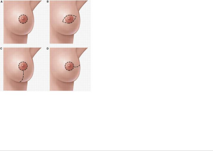
P.31 a circular incision around the edge of the areola with or without lateral extension is employed for skin-sparing mastectomy (Fig. 2-2). In the case of a large breast or mild or moderate breast ptosis, the volume of the skin envelope must be reduced, and this can be achieved by increasing the size of a central elliptical incision or by using a reduction pattern incision according to the plastic surgeon’s preference (see Fig. 2-2).
FIGURE 2-2 Incisions for skin-sparing mastectomy. A: Round periareolar, (B) small elliptical periareolar, (C) perioareolar with inferior extension, (D) perioareolar with lateral extension.
Nipple-sparing mastectomy, a type of skin-sparing mastectomy that preserves not only the breast skin but also the NAC, may also be employed in patients undergoing immediate breast reconstruction (Fig. 2-3). For best results, in nipple-sparing mastectomy, a periareolar incision not exceeding 30% of the circumference of the areola can be used. Incisions that encircle the areola should be avoided, as incisions exceeding 30% of the areolar circumference increase the risk of nipple necrosis.1 Other incisions associated with a low risk of nipple necrosis include the inframammary fold incision and the radial incisions (see Fig. 2-3). The crescent incision is often utilized in women with breast ptosis, as it allows the surgeon good exposure and lifts the NAC to a more cosmetically appropriate position (see Fig. 2-3). At the time of nipple-sparing mastectomy, the mastectomy incision or a separate axillary incision can be accessed to perform nodal staging with either sentinel lymph node surgery or axillary lymph node dissection.
P.32
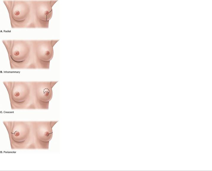
FIGURE 2-3 A-D: Nipple-sparing mastectomy incisions. The four most common nipple-sparing mastectomy incisions are shown. Note that it is important to avoid exceeding 30% of the circumference around the nipple areola complex to prevent necrosis.
P.33
2. BOUNDARIES OF MASTECTOMY
Recommendation: Although the options for incisions and the amount of skin excised for simple total mastectomy, skin-sparing mastectomy, and nipple-sparing mastectomy vary, the anatomical boundaries guiding mastectomy remain uniform in order to remove the entire breast parenchyma.
Type of Data: Retrospective case series.
Strength of Recommendation: The consensus of the group supports this guideline based on historic evidence.
Rationale
The boundaries of the breast include the second rib superiorly, the upper border of the rectus sheath inferiorly, the lateral border of the sternum
medially, and the latissimus dorsi muscle laterally.2 The superior lateral boundary of any mastectomy should include the tail of the breast, which extends into the axilla a variable distance beyond the lateral margin of the pectoralis major muscle. Care should be taken to remove any glandular breast tissue that extends into the axilla. The breast should be dissected off the underlying pectoralis major muscle. Removal of the pectoral fascia with the breast is commonly but not uniformly advised. The fascia overlying the serratus anterior muscle and the rectus sheath should be preserved. Sutures should be placed to provide specimen orientation. Most mastectomies remove more than 95% of the breast; some breast tissue invariably remains. Thus, new primary cancers can, although rarely do, occur even after mastectomy. Therefore, meticulous resection and thorough removal of the breast tissue is essential to achieving optimal outcomes.
3. MASTECTOMY FLAP ELEVATION
Recommendation: Mastectomy flaps should be elevated in a manner that facilitates the removal of essentially all breast tissue to reduce the risk of recurrence and that preserves the overlying subcutaneous tissue and its vascular plexus to minimize the risk of flap necrosis.
Type of Data: Retrospective case series.
Strength of Recommendation: The consensus of the group supports this guideline based on historic evidence.
Rationale
Mastectomy flap thickness varies greatly among patients; the subcutaneous layer in thin patients may be only 2 to 3 mm thick. Persistent glandular

tissue and residual disease have been reported to be more prevalent in patients with mastectomy flaps thicker than 5 mm.3 Anatomic studies have confirmed the interdigitation of the subcutaneous fat and the underlying glandular portion of the breast. Because the border between the subcutaneous tissue and the breast may be poorly defined in patients with excessive adipose tissue in the breast, care must be taken to not extend mastectomy flaps into the
P.34
deeper glandular plane and thus leave breast tissue in the mastectomy flap.4 Thin mastectomy flaps dissected at the level of the dermis are predisposed to necrosis, whereas thick mastectomy flaps may include residual breast tissue that increases the risk of recurrence. Meticulous flap elevation is especially important in cases of skin-sparing or nipple-sparing mastectomy, in which the smaller incision may limit visualization of the upper and lateral borders of dissection depending on which incision is used. All margins at the site of known malignancy should be examined grossly and pathologic analysis of frozen sections performed as indicated to ensure that clear surgical margins are obtained. For patients in whom mammography has revealed diffuse microcalcifications, mammography of the mastectomy specimen with serial sections may be considered; if microcalcifications near a superficial margin are observed, additional skin or subcutaneous tissues can be excised at that site to obtain an adequate
final margin.5 In cases in which the tumor is close to the dermis, a negative margin of skin directly overlying the tumor must be achieved, or the skin must be excised.
Technical Considerations for Flap Elevation
Mastectomy flaps were historically developed using sharp dissection with a scalpel or scissors. Sharp dissection as the primary method of flap
elevation for most surgeons was eventually replaced by electrocautery, which resulted in significantly less blood loss (Fig. 2-4).6 Now, low-energy dissection devices provide an alternative to both sharp dissection and electrocautery for flap elevation. These devices generate temperatures that seal vascular and lymphatic structures, and they divide tissue with less collateral tissue injury and lower seroma volumes compared with sharp
dissection or electrocautery.7,8 To date, however, no specific device has been associated with better oncologic outcomes.
One optional adjunct to flap elevation with sharp dissection is tumescent solution injection, which has been reported to reduce operating room time
without the blood loss or thermal injury associated with sharp dissection or electrocautery alone.9 The tumescent solution is composed of normal saline, lidocaine, and epinephrine. After skin incision, a 20-gauge spinal needle is used to inject the solution into the space between the subcutaneous and glandular tissue to develop a bloodless plane (see Fig. 2-4). The blades of the scissors are opened approximately 1 to 1.5 cm
and the scissors are pushed through the developed plane tissue to elevate the flap.10 Although many breast surgeons have adopted this technique, some groups have reported that tumescent solution injection results in higher rates of flap necrosis than electrocautery does, possibly owing to the
vasoconstriction it causes.10,11,12 Surgeons should select the technique that they are confident will enable them to completely remove the breast with the fewest associated complications.
4. EXCISION OF THE PECTORALIS MAJOR MUSCLE
Recommendation: Although neoadjuvant systemic therapy should minimize the amount of direct muscle invasion encountered during mastectomy, localized excision of the pectoralis major muscle is sometimes necessary to achieve clear margins.
Type of Data: Older prospective trials and more recent case series.
P.35
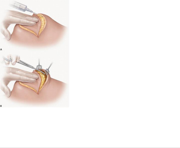
FIGURE 2-4 Flap elevation done sharply after injection of tumescent solution. Part A shows injection of tumescent solution with spinal needle and part B shows flap elevation with scissors.
Strength of Recommendation: The consensus of the group supports this guideline based on historic evidence.
Rationale
Resection of the pectoral muscle, an element of the classic Halstead radical mastectomy described in 1894,13 was routinely performed during mastectomy for more than 70 years. However, the clinical benefit of such radical surgery was questioned once effective adjuvant radiotherapy and systemic therapies came into use. In 1978,
P.36
Baker et al found no difference between the 5-year survival rates of 144 breast cancer patients who underwent radical mastectomy and 188
patients who underwent mastectomy without removal of the pectoralis major muscle at the discretion of the operating surgeon.11 Although radical mastectomy did not result in lower local recurrence rates among patients with stage I or II breast cancer, it did appear to result in lower recurrence rates among patients with stage III disease. On the basis of these findings, the authors concluded that compared with mastectomy without removal of the pectoralis major muscle, radical mastectomy provided a better chance of local control but not survival in patients with stage III breast cancer. The results of the National Surgical Adjuvant Breast and Bowel Project B-04 trial, first published in 1977 and most recently updated with 25-year
follow-up data in 2002,12 demonstrated that there was no significant difference in rates of overall survival or local control in patients who underwent total mastectomy with postmastectomy radiation (PMRT) compared to patients who had radical mastectomy. Local control was inferior in patients with positive nodes without radiation. As a result of this study, the routine en bloc resection of uninvolved pectoralis major muscles was abandoned. However, removal of the pectoralis major muscle during mastectomy may be indicated in those patients with diffuse pectoral muscle involvement at diagnosis.
Removal of the pectoralis major muscle during mastectomy may be indicated in some patients who present with focal or a large area of muscle involvement. Most patients presenting with focal muscle invasion have stage III, locally advanced cancers and should receive neoadjuvant chemotherapy or hormonal therapy to facilitate obtaining clear margins at surgery. In patients whose tumors have limited response to neoadjuvant therapy, muscle excision may have a limited treatment role; it may also have clinical benefit in patients who have local recurrences that are
refractory to treatment.14 In these patients, resection of the pectoralis major muscle at the site of focal invasion to obtain negative margins for improved local control seems prudent. The only aim of this resection should be to achieve a clear margin, and this rarely requires the removal of a large portion of the muscle. In most patients with focal tumor involvement of the pectoralis major muscle, a combination of resection of the area of involved pectoralis muscle plus PMRT are recommended.
Muscle invasion or T4 disease is considered a definite indication for PMRT in patients undergoing mastectomy. Several authors have demonstrated that in the absence of PMRT, close or positive margins and invasion of the pectoralis major muscle or its fascia increase the risk of local recurrence. For example, researchers in the Danish Breast Cancer Cooperative Group 82B and C trials studied a group of more than 1,500 high-
risk patients who received no radiation and found that fascial invasion was an independent predictor of locoregional recurrence.14 Similarly, a group