

новая папка / Operative Standards for Cancer Surgery Volume I 1st Edition
.pdfP.185
these regimens in patients with advanced disease may translate into improvements in the adjuvant and/or neoadjuvant setting. Ongoing trials of both neoadjuvant and adjuvant strategies are necessary to improve results following surgery alone.
Objectives of This Section
Despite the rapidly evolving multidisciplinary care programs that have improved the survival of patients with localized PDAC over the past 35 years, important clinical questions and opportunities for improvement clearly remain. It is interesting and perhaps surprising that many of these questions relate to the technical performance of operations that have not changed significantly for 100 years. Indeed, the specific surgical elements that comprise a pancreatectomy, the only component of care that has ever been proven to be potentially curative,
remain largely nonstandardized within and among surgical centers in the United States.29 For this reason, the potential influence of many of these technical components of surgery on oncologic outcome has not been established. Precisely which lymph node basins should be resected at pancreatoduodenectomy? Within which surgical plane should the deep dissection of distal pancreatectomy be routinely performed? How should the superior mesenteric artery margin of resection be managed surgically, and how should it be evaluated pathologically and reported? Ambiguity with regard to the answers to these and many other technical questions, as well as consequent variability with which both pancreatoduodenectomy and pancreatectomy for cancer are performed, may be responsible for the high rates of margin-positive and node-positive resections and suboptimal
surgical outcomes that are often reported.30,31
Within this section, we present the specific “critical elements” of distal pancreatectomy and pancreatoduodenectomy for cancer that have been catalogued by highvolume pancreatic surgeons and pathologists from both academic and community centers. Recommendations for the performance of each technical element presumed to have a significant association with oncologic outcome, achieved through consensus based on available data, are also presented. Components of surgical therapy believed to represent a “key question” are reviewed in greater depth, with a recommendation generated following a rigorous literature review. Finally, the oncologically significant aspects of surgical care that might be included in a synoptic operative report are proposed. It is hoped that the concepts outlined in this section will be used not only to improve the technical performance of and outcomes associated with pancreatectomy but also to identify opportunities for clinical studies and standardize surgical approaches for clinical protocols.
REFERENCES
1.Fortner JG, Kim DK, Cubilla A, et al. Regional pancreatectomy: en bloc pancreatic, portal vein and lymph node resection. Ann Surg 1977;186(1):42-50.
2.Moossa AR, Lewis MH, Mackie CR. Surgical treatment of pancreatic cancer. Mayo Clin Proc
1979;54(7):468-474.
3.Beall MS, Dyer GA, Stephenson HE Jr. Disappointments in the management of patients with malignancy of pancreas, duodenum, and common bile duct. Arch Surg 1970;101(4):461-465.
4.Bilimoria KY, Bentrem DJ, Ko CY, et al. Multimodality therapy for pancreatic cancer in the U.S.: utilization, outcomes, and the effect of hospital volume. Cancer 2007;110(6):1227-1234.
5.Morrow M, Hilaris B, Brennan MF. Comparison of conventional surgical resection, radioactive implantation,

and bypass procedures for exocrine carcinoma of the pancreas 1975-1980. Ann Surg 1984;199(1):1-5.
P.186
6.Katz MH, Wang H, Fleming JB, et al. Long-term survival after multidisciplinary management of resected pancreatic adenocarcinoma. Ann Surg Oncol 2009;16(4):836-847.
7.Callery MP, Chang KJ, Fishman EK, et al. Pretreatment assessment of resectable and borderline resectable pancreatic cancer: expert consensus statement. Ann Surg Oncol 2009;16(7):1727-1733.
8.Varadhachary GR, Tamm EP, Abbruzzese JL, et al. Borderline resectable pancreatic cancer: definitions, management, and role of preoperative therapy. Ann Surg Oncol 2006;13(8):1035-1046.
9.Exocrine and endocrine pancreas. In: Edge SB, Byrd DR, Compton CC, eds. AJCC Cancer Staging Manual. 7th ed. Chicago: Springer; 2010.
10.Katz MH, Marsh R, Herman JM, et al. Borderline resectable pancreatic cancer: need for standardization and methods for optimal clinical trial design. Ann Surg Oncol 2013;20(8):2787-2795.
11.Kim HJ, Czischke K, Brennan MF, et al. Does neoadjuvant chemoradiation downstage locally advanced pancreatic cancer? J Gastr Surg 2002;6(5):763-769.
12.Tamm EP, Loyer EM, Faria S, et al. Staging of pancreatic cancer with multidetector CT in the setting of preoperative chemoradiation therapy. Abdom Imaging 2006;31(5):568-574.
13.Pawlik TM, Laheru D, Hruban RH, et al. Evaluating the impact of a single-day multidisciplinary clinic on the management of pancreatic cancer. Ann Surg Oncol 2008;15(8):2081-2088.
14.Specht G, Stinshoff K. Walther Kausch (1867-1928) and his significance in pancreatic surgery [article in German]. Zentralblatt fur Chirurgie 2001;126(6):479-481.
15.McClusky DA III, Skandalakis LJ, Colborn GL, et al. Harbinger or hermit? Pancreatic anatomy and surgery through the ages-part 3. World J Surg 2002;26(12):1512-1524.
16.Winter JM, Cameron JL, Campbell KA, et al. 1423 pancreaticoduodenectomies for pancreatic cancer: a single-institution experience. J Gastr Surg 2006;10(9):1199-1210; discussion 1191-1210.
17.Tseng JF, Pisters PW, Lee JE, et al. The learning curve in pancreatic surgery. Surgery 2007;141(5):694-
18.Eppsteiner RW, Csikesz NG, McPhee JT, et al. Surgeon volume impacts hospital mortality for pancreatic resection. Ann Surg 2009;249(4):635-640.
19.Reames BN, Ghaferi AA, Birkmeyer JD, et al. Hospital volume and operative mortality in the modern era.
Ann Surg 2014;260(2):244-251.

20.Gurusamy KS, Kumar S, Davidson BR, et al. Resection versus other treatments for locally advanced pancreatic cancer. Cochrane Database Syst Rev 2014;2:CD010244.
21.Kendrick ML. Laparoscopic and robotic resection for pancreatic cancer. Cancer J 2012;18(6):571-576.
22.Kalser MH, Ellenberg SS. Pancreatic cancer. Adjuvant combined radiation and chemotherapy following curative resection. Arch Surg 1985;120(8):899-903.
23.Simons JP, Ng SC, McDade TP, et al. Progress for resectable pancreatic [corrected] cancer?: a population-based assessment of US practices. Cancer 2010;116(7):1681-1690.
24.Oettle H, Post S, Neuhaus P, et al. Adjuvant chemotherapy with gemcitabine vs observation in patients undergoing curative-intent resection of pancreatic cancer: a randomized controlled trial. JAMA
2007;297(3):267-277.
25.Crane CH, Varadhachary G, Wolff RA, et al. The argument for pre-operative chemoradiation for localized, radiographically resectable pancreatic cancer. Best practice & research. Clin Gastr 2006;20(2):365-382.
26.Evans DB, Farnell MB, Lillemoe KD, et al. Surgical treatment of resectable and borderline resectable pancreas cancer: expert consensus statement. Ann Surg Oncol 2009;16(7):1736-1744.
27.Conroy T, Desseigne F, Ychou M, et al. FOLFIRINOX versus gemcitabine for metastatic pancreatic cancer. N Engl J Med 2011;364(19):1817-1825.
28.Von Hoff DD, Ervin T, Arena FP, et al. Increased survival in pancreatic cancer with nab-paclitaxel plus gemcitabine. N Engl J Med 2013;369(18):1691-1703.
29.Katz MH, Merchant NB, Brower S, et al. Standardization of surgical and pathologic variables is needed in multicenter trials of adjuvant therapy for pancreatic cancer: results from the ACOSOG Z5031 trial. Ann Surg Oncol 2011;18(2):337-344.
30.Merkow RP, Bilimoria KY, Bentrem DJ, et al. National assessment of margin status as a quality indicator after pancreatic cancer surgery. Ann Surg Oncol 2014;21(4):1067-1074.
31.Schwarz RE, Smith DD. Extent of lymph node retrieval and pancreatic cancer survival: information from a large US population database. Ann Surg Oncol 2006;13(9):1189-1200.

Chapter 13
Pancreatic Surgery
CRITICAL ELEMENTS
Evaluation for Extrapancreatic Disease and Assessment of Locoregional Tumor Anatomy
Pathologic Examination of the Surgical Specimen
1. EVALUATION FOR EXTRAPANCREATIC DISEASE AND ASSESSMENT OF LOCOREGIONAL TUMOR ANATOMY
Recommendation: Staging laparoscopy should be performed prior to laparotomy to exclude radiographically occult metastatic disease. If no such disease is identified, a thorough, open exploration to identify local infiltration or metastases not revealed during laparoscopy should be conducted before tumor resection.
Type of Data: Primarily retrospective, low-level evidence.
Strength of Recommendation: Strong.
Rationale
Pancreatic adenocarcinoma has a high malignant potential, and the majority of patients have metastases at diagnosis. Pancreatectomy
is not associated with a survival benefit in these patients.1,2 Pancreatic cancers may also infiltrate locally into the retroperitoneum and root of the mesentery to involve the superior mesenteric artery (SMA), aorta, and/or celiac trunk, and such involvement precludes a safe and effective margin-negative resection. Unfortunately, even the most advanced cross-sectional imaging and echoendoscopy tests do not have the capacity to detect all low-volume metastatic disease. Therefore, for patients with ostensibly localized pancreatic cancer, rigorous operative staging is critical.
P.188
Staging Laparoscopy
The evolution of multidetector row computed tomography and advanced magnetic resonance imaging has increased the accuracy with which the anatomic extent of the primary cancer can be determined. However, the capacity of these modalities to identify small (<1 cm)
metastases to the liver or peritoneum remains limited.3 Therefore, a thorough examination of all visceral and parietal surfaces before tumor resection is mandatory. Historically, this examination was accomplished via laparotomy, and patients found to have radiographically occult metastases or unresectable locoregional disease would undergo surgical biliary and enteric bypass to palliative or prevent symptoms of biliary or gastric outlet obstruction.
Steady advances in endoscopic and laparoscopic techniques have substantially reduced the need for open palliative operations, thereby limiting the role of exploratory surgery to staging alone. Staging laparoscopy was first used as a minimally invasive technique to identify occult metastatic pancreatic ductal adenocarcinoma (PDAC) lesions in 1978, obviating the need for nontherapeutic staging laparotomy
and its associated morbidity, costs, and delay of definitive oncologic therapy (Fig. 13-1).4 When the procedure was first introduced, some voiced concerns about its possible association with trocar site disease and peritoneal dissemination, but subsequent studies did not
support these concerns, so the use of staging laparoscopy became more widespread.5 Many now advocate laparoscopic staging for all
patients with potentially resectable pancreatic cancer prior to open exploration and resection.6,7,8,9 When utilized liberally, staging laparoscopy radiographically may identify occult metastases in 14% to 37% of patients.
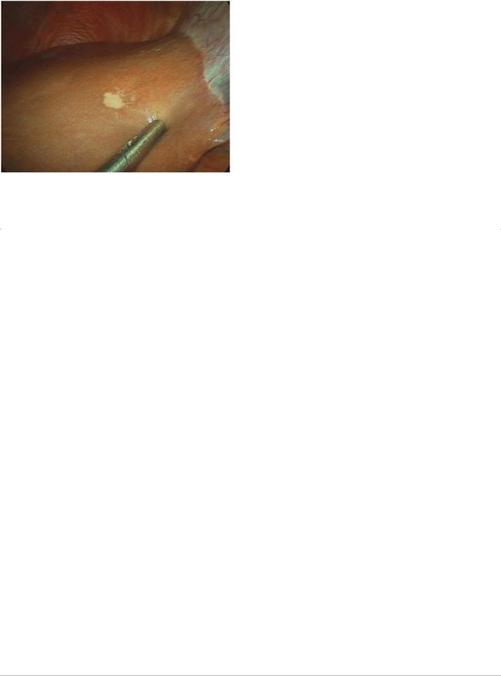
FIGURE 13-1 Radiographically occult lesion on the surface of segment IV of the liver that was identified in a patient with potentially resectable pancreatic cancer upon staging laparoscopy. A tissue biopsy was acquired laparoscopically and immediate histopathologic analysis confirmed metastatic adenocarcinoma. The planned pancreatoduodenectomy was aborted.
P.189
Selective Laparoscopy
Staging laparoscopy is most cost-effective when its findings redirect patients with unresectable disease from unnecessary open operative exploration to nonsurgical palliation. Staging laparoscopy becomes increasingly less cost-effective when yield decreases as preoperative imaging improves or as tumor biology becomes increasingly favorable and the risk of metastatic disease decreases. Thus, staging laparoscopy is most costeffective when it is utilized selectively in patients whose primary cancers are located in the pancreatic body or tail, are large or anatomically extensive, are associated with a high cancer antigen 19-9 level, are associated with equivocal computed tomography findings of metastasis, and/or are associated with clinical findings suggesting advanced disease, such as marked
weight loss.10,11,12,13,14,15,16 In these scenarios, the use of staging laparoscopy is particularly encouraged.17,18
Advanced/Extended Staging Laparoscopy
Although laparoscopy facilitates the detection of small, superficial metastases on the liver and in the peritoneum that are easily missed using radiologic staging techniques, a simple laparoscopic survey of the abdomen may not detect locally advanced disease and vessel
encasement that would also represent a contraindication to resection.19 Modifications to improve the accuracy of laparoscopic staging have included the use of extensive wider laparoscopic dissections and laparoscopic ultrasonography to more thoroughly inspect vascular
structures and evaluate occult intrahepatic lesions, extraanatomic lymph node involvement, and extrapancreatic tumor extension.8,9
Although retrospective analyses have shown that all these techniques have added benefits, their detection yield has diminished as
significant improvements in preoperative imaging have been made.15,16 In general, advanced laparoscopy appears to have limited capacity to detect unresectable locoregional tumor extension in patients who have no radiographic evidence of locally advanced disease.
Peritoneal Lavage
Because pancreatic tumors shed malignant cells into the peritoneum, laparoscopic lavage has been proposed as an additional index of resectability. Several early studies showed that patients with positive peritoneal washings developed metastasis earlier and had shorter
survival durations after resection than did patients with negative peritoneal washings.20,21,22 The results of subsequent investigations have suggested that positive peritoneal cytology alone (without other visible evidence of metastatic disease) should not necessarily preclude resection in pancreatic cancer patients whose tumors would otherwise be considered resectable, as these patients may still
achieve long-term survival.23,24 Regardless, until additional high-level evidence is found to support its widespread use, routine use of peritoneal lavage cannot be recommended at present. It may be used selectively for patients at high risk for surgery to provide support for a nonoperative therapeutic approach.
Timing of Laparoscopy
Staging laparoscopy should typically be performed at the time of the planned resection. Staged procedures to allow time for biopsy specimen analysis, particularly those
P.190
utilizing peritoneal cytology results as a determinant for proceeding to resection, have been described; however, because these procedures carry additional costs and require additional resources, their routine use in the absence of other obvious indications is not recommended.

Technical Aspects
Technique of Staging Laparoscopic Exploration
Camera port incisions can be made along the proposed lines of the laparotomy incision or (most commonly) periumbilically. Following carbon dioxide insufflation, an angled (≥30°) laparoscope is used to visually inspect the abdominal cavity, including the peritoneal, diaphragmatic, and hepatic surfaces and the porta hepatis, for evidence of occult metastases. Additional trocars may be placed to elevate the liver lobes and mobilize viscera to facilitate a more thorough evaluation of the pelvis for drop deposits specifically and the mesenteric root for evidence of tumor extension or matted nodes. Abnormal lesions, which may vary in size and morphology but typically appear firm and have evidence of microvasculature, should be biopsied, and the samples should be sent for frozen section pathologic evaluation.
Beyond simple laparoscopic maneuvers for visual evaluation of the peritoneal and visceral surfaces, additional extensive laparoscopic maneuvers are not considered standard. Such maneuvers include elevating the omentum from the transverse colon to access the omental bursa, performing a partial kocherization and evaluating the aortocaval nodes, and dividing the gastrohepatic ligament and examining the caudate lobe surface and celiac axis. At some centers, laparoscopic exploration is performed in conjunction with laparoscopic ultrasonography. These types of advanced laparoscopic evaluations of the extent of locoregional disease may be best reserved for patients undergoing planned minimally invasive resections, in which laparoscopic palliative procedures can be performed without formally converting to laparotomy.
Technique of Open Exploration for Staging
Formal laparotomy is reserved primarily for potentially curative operations. If no evidence of obvious unresectability is identified at laparoscopy, conversion to open exploration is performed. The incision used for open exploration depends on surgeon preference and abdominal anatomical considerations and is typically a midline, or bilateral, subcostal incision. The surgical field is then thoroughly examined to identify any other occult disease not easily visualized by laparoscopy and to assess extrapancreatic extension and locoregional resectability. The omental bursa is accessed by dividing the gastrocolic ligament and elevating the omentum from the mesocolon. For lesions in the pancreatic body or tail, the dissection is extended sufficiently to the left to facilitate full visualization of the pancreas to its tail and the hilum of the spleen. For proximal lesions, the hepatic flexure is mobilized inferiorly by dissecting in the avascular plane between the hepatic flexure and the duodenum and performing an extended Kocher maneuver to free the third portion of the duodenum from the colonic mesentery. The gastrocolic venous trunk or middle colic vein is used as landmark to identify the superior mesenteric vein (SMV) inferior to the neck of the pancreas, where the SMV is assessed for direct tumor involvement. Tumor involvement of the
P.191
SMA is classically assessed by placing one’s hand posterior to the pancreatic head after kocherization; however, SMA involvement should be readily apparent if highquality preoperative imaging studies have been performed and skillfully interpreted, and any discrepancy between imaging and intraoperative findings should be met with caution. The porta hepatis is assessed for direct tumor infiltration, and the root of the mesentery should be evaluated to rule out nodal disease outside the surgical field. Once locoregional extent has been thoroughly evaluated, formal dissection to resect the tumor and involved tissues begins.
Conclusion
Despite improvements in modern radiographic imaging studies and the use of these studies’ findings to predict locoregional unresectability, cross-sectional imaging studies and echoendoscopy remain relatively nonspecific tests for predicting resectability owing to radiographically occult peritoneal metastases and/or liver metastases in a significant proportion of patients with seemingly localized pancreatic cancer. Minimally invasive procedures and thorough intraoperative evaluation continue to have important roles in staging pancreatic cancers prior to resection with curative intent.
2. PATHOLOGIC EXAMINATION OF THE SURGICAL SPECIMEN
Recommendation: Communication between surgeons and pathologists is essential for the accurate reporting of margin status following pancreatectomy. A standard nomenclature should be used to describe surgical margins. For pancreatoduodenectomy specimens, the status of the pancreatic neck margin, bile duct margin, anterior surface, posterior surface, portal vein/SMV groove, and SMA/uncinate margin should be described. For distal pancreatectomy specimens, the anterior surface, posterior surface, and pancreatic transection margins should be described.
Type of Data: Primarily retrospective, low-level evidence.
Strength of Recommendation: Strong.
Rationale

The oncologic status of the surgical margins (R status) is generally—but not uniformly—regarded as a critical prognostic factor following pancreatectomy and is the most important such factor under the direct influence of the surgeon. For these reasons, the presence or absence of cancer cells at the surgical margins of transection is commonly reported as a primary metric with which the technical success of an individual operation is communicated to patients and other health care providers. The reported association between surgical
margins and survival has led to the use of R status as a measure of surgical quality,25,26 an indicator of technical proficiency,27 a
surrogate marker of the effect associated with preoperative treatment,28 and a key factor used to stratify patients enrolled in clinical trials
of adjuvant therapies.29
Historically, cancer cells have been identified at the inked margins of 20% to 40% of surgical specimens resected from patients treated at
single institutions30,31 and
P.192
within the context of major clinical trials.32 However, recent investigations that have used meticulous histopathologic protocols to critically evaluate pancreatectomy margins have demonstrated that cancer cells can be identified at one or more surgical margins in close to 90% of resected specimens, a rate that can more easily be reconciled with the reality that as many as 80% of patients who undergo curative
resection for pancreatic cancer die with local recurrence.33,34 Therefore, whether a curative operation is reported as microscopically complete (R0) or incomplete (R1) may depend as much on the surgical pathologist as on the surgeon.
Differences in the methods used to handle and process surgical specimens and to interpret and report pathologic findings may contribute
to significant variability in R1 resection rates across populations.35 Because an inaccurate assessment of the surgical margins may alter the purported relationship between margin status and outcome, the utility of R status both as a robust technical metric and as a prognostic variable has been questioned. Furthermore, such variability may adversely influence the accuracy of assessments of patients’ eligibility for clinical trials of adjuvant therapies, the interpretation of the results of such trials, and ultimately oncologic outcomes. A discussion of the precise histopathologic protocols pathologists use to evaluate the status of pancreatectomy margins is beyond the scope of this book. However, surgeons play a critical role in certain aspects of the pathologic analysis of the surgical specimen, and these aspects are reviewed here.
Surgical Margins and Terminology
The seventh edition of the American Joint Committee on Cancer (AJCC) Cancer Staging Manual references the College of American Pathologists (CAP) Checklist for Exocrine Pancreatic Tumors as the standard document that should be used to guide pathologic
evaluation of pancreatic resection specimens in the United States.36,37 This document does not specifically address differences between the handling or processing of pancreatoduodenectomy and distal pancreatectomy specimens. Rather, it lists the following margins and surfaces as important to evaluate when appropriate: the proximal (gastric or duodenal) and distal (jejunal) enteric margins, the pancreatic neck transection and bile duct margins, the deep retroperitoneal posterior surface, and the nonperitonealized surface of the uncinate process that lies adjacent to the SMA.
Among these anatomic components, the nonperitonealized uncinate surface of pancreatoduodenectomy specimens is emphasized by both the AJCC and CAP as having particular oncologic significance, as it is the most commonly positive margin following potentially curative resection and is the site of most local recurrences following the resection of cancers in the pancreatic head. Although the CAP guidelines refer to the corresponding tissue margin as the “uncinate process (retroperitoneal) margin,” the AJCC refers to it as the “SMA margin.” This inconsistency has further complicated the already confusing nomenclature used to describe the margin(s) that lie adjacent to the SMA and SMV, which have also been variably described as the “radial,” “deep,” and “posterior” margin(s), among other terms, by
high-volume pancreatic cancer treatment centers.35
Furthermore, guidelines published by the Royal College of Pathologists promote a different nomenclature that is used by pancreatic surgeons and pathologists in
P.193
Europe.34,38,39 In this system, the anterior and posterior surfaces of the pancreatoduodenectomy specimen flank a medial “circumferential resection margin” (also described as the “vascular” or “superior mesenteric vessel” margin), which is composed of both an SMV groove margin and an SMA margin. As opposed to those published by the CAP, these guidelines describe in greater detail the important anatomic components of distal pancreatectomy specimens (e.g., inking the anterior and posterior surfaces is recommended specifically) and vein segments removed at pancreatectomy (e.g., examination of the proximal and distal ends of the resected vein is recommended). This lexicon and its accompanying analysis protocol were recently utilized in the first multicenter study to prospectively
analyze margin involvement in pancreatectomy specimens.40
A standard terminology should be adopted and utilized in both the operative and pathologic reports to reduce confusion and promote standardization. For pancreatoduodenectomy specimens, the status of the pancreatic neck margin, bile duct margin, anterior surface, posterior surface, portal vein/SMV groove, and SMA/uncinate margin should be described (Fig. 13-2). For distal pancreatectomy
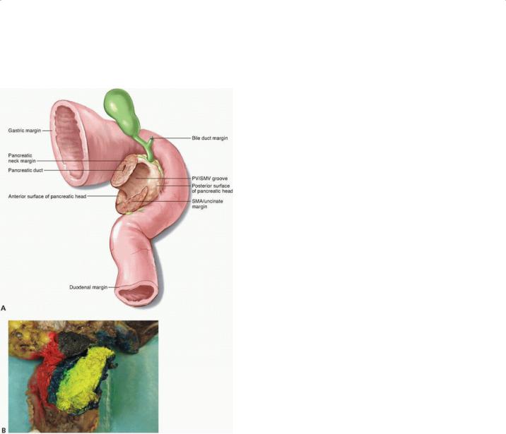
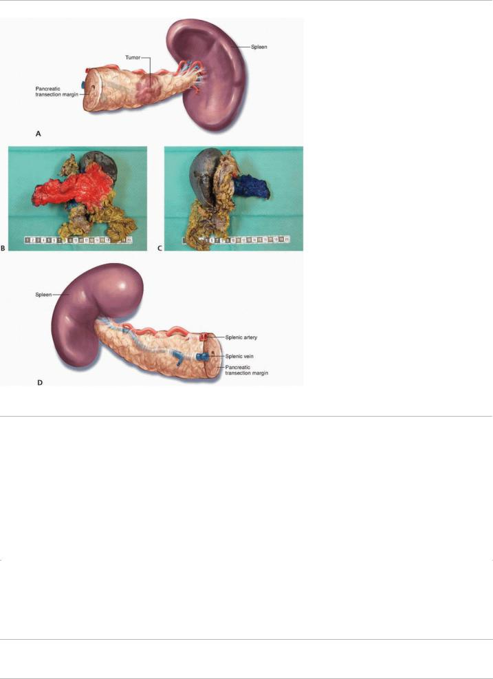
specimens, the anterior surface, posterior surface, and pancreatic transection margins should be described (Fig. 13-3).
Specimen Orientation
Although the histopathologic analysis of surgical margins is ultimately performed by the pathologist, the surgeon plays a crucial role in helping the pathologist accurately assess surgical margins. Several obstacles may prevent the isolated pathologist from accurately assessing surgical margins. First, characterized by multiple sites of transection and multiple surfaces, the anatomy of the pancreatectomy specimen is extraordinarily complex, making orientation of the specimen difficult following its removal, particularly for less experienced
surgical pathologists.41 In particular, the oncologically important uncinate process is less well-defined as a distinct structure ex vivo than it may appear either on cross-sectional images or in the operating room because its anatomy is based on its relationship to the mesenteric vessels. Second, the pathologist cannot reliably determine R status on the basis of the resected specimen alone. Specifically, although the histopathologic techniques the pathologist uses are required to differentiate a microscopically negative (R0) margin from a microscopically positive (R1) margin, only the surgeon, who can view gross residual disease in the abdomen, is in a position to reliably distinguish an R1 margin from a macroscopically positive (R2) margin.
Given these obstacles, the surgeon must be an active participant in the orientation of the surgical specimen. Of particular importance is the surgeon’s identification of structures that may have been resected en bloc, such as a segment of the SMV or portal vein or another organ (e.g., colon, adrenal gland). Although some surgeons prefer to ink or mark the margins in the operating room, surgical pathologists generally prefer to ink the specimen themselves in collaboration with the surgeon. Similarly, although some surgeons cut pancreatic neck or bile duct margins for immediate histopathologic analysis, experienced surgical pathologists generally frown upon this practice because it may introduce confusion or even diagnostic ambiguity if the margin
P.194
proper is artifactually altered. The CAP guidelines recommend that the pathologist ink the posterior surface and the SMA/uncinate margin, bile duct margin, and pancreatic neck margin of pancreatoduodenectomy specimens, working with the surgeon when necessary. However, given the aforementioned recommendations, it would seem reasonable to also ink the anterior surface and portal vein/SMV groove (Fig. 13-2B). For distal pancreatectomy specimens, the anterior and posterior surfaces and the pancreatic transection margin should be inked (Fig. 13-3B,D).
FIGURE 13-2 A: Posterior view of the pancreatoduodenectomy specimen. B: Lateral view of the inked pancreatoduodenectomy specimen depicting the anterior surface (red), pancreatic neck margin (black), portal vein/SMV groove (blue), and SMA/uncinate margin
(yellow).
P.195
FIGURE 13-3 A,B: Anterior and (C,D) posterior surfaces of the distal pancreatectomy with en bloc splenectomy specimen.
P.196 The surgeon and pathologist should begin collaborating immediately after the removal of the surgical specimen. Unfortunately, despite the importance of this practice, it is not common. In a recent evaluation of the operative and pathology reports of patients treated on a prospective, multi-institutional clinical trial of adjuvant therapy following pancreatoduodenectomy, the surgeon clearly participated in the marking of the oncologically important SMA margin in only 25% of cases, and communication regarding specimen orientation between
the surgeon and the pathologist was documented in only 15% of cases.35
REFERENCES
1.De Jong MC, Farnell MB, Sclabas G, et al. Liver-directed therapy for hepatic metastases in patients undergoing pancreaticoduodenectomy: a dual-center analysis. Ann Surg 2010;252(1):142-148.
2.Gleisner AL, Assumpcao L, Cameron JL, et al. Is resection of periampullary or pancreatic adenocarcinoma with synchronous hepatic metastasis justified? Cancer 2007;110(11):2484-2492.
3.Schima W, Fugger R, Schober E, et al. Diagnosis and staging of pancreatic cancer: comparison of mangafodipir trisodiumenhanced MR imaging and contrast-enhanced helical hydro-CT. AJR Am J Roentgenol 2002;179(3):717-724.
4.Warshaw AL, Tepper JE, Shipley WU. Laparoscopy in the staging and planning of therapy for pancreatic cancer. Am J Surg
1986;151(1):76-80.
5. Velanovich V. The effects of staging laparoscopy on trocar site and peritoneal recurrence of pancreatic cancer. Surg Endosc
2004;18(2):310-313.

6. Allen VB, Gurusamy KS, Takwoingi Y, et al. Diagnostic accuracy of laparoscopy following computed tomography (CT) scanning for assessing the resectability with curative intent in pancreatic and periampullary cancer. Cochrane Database Syst Rev
2013;11:CD009323.
7.Contreras CM, Stanelle EJ, Mansour J, et al. Staging laparoscopy enhances the detection of occult metastases in patients with pancreatic adenocarcinoma. J Surg Oncol 2009;100(8):663-669.
8.John TG, Greig JD, Carter DC, et al. Carcinoma of the pancreatic head and periampullary region. Tumor staging with laparoscopy and laparoscopic ultrasonography. Ann Surg 1995;221(2): 156-164.
9.Conlon KC, Dougherty E, Klimstra DS, et al. The value of minimal access surgery in the staging of patients with potentially resectable peripancreatic malignancy. Ann Surg 1996;223(2): 134-140.
10.Karachristos A, Scarmeas N, Hoffman JP. CA 19-9 levels predict results of staging laparoscopy in pancreatic cancer. J Gastrointest Surg 2005;9(9):1286-1292.
11.Satoi S, Yanagimoto H, Toyokawa H, et al. Selective use of staging laparoscopy based on carbohydrate antigen 19-9 level and tumor size in patients with radiographically defined potentially or borderline resectable pancreatic cancer. Pancreas 2011;40(3):426-
12.Maithel SK, Maloney S, Winston C, et al. Preoperative CA 19-9 and the yield of staging laparoscopy in patients with radiographically resectable pancreatic adenocarcinoma. Ann Surg Oncol 2008;15(12):3512-3520.
13.Camacho D, Reichenbach D, Duerr GD, et al. Value of laparoscopy in the staging of pancreatic cancer. JOP 2005;6(6):552-561.
14.Tapper E, Kalb B, Martin DR, et al. Staging laparoscopy for proximal pancreatic cancer in a magnetic resonance imaging-driven practice: what’s it worth? HPB (Oxford) 2011;13(10):732-737.
15.Barabino M, Santambrogio R, Pisani Ceretti A, et al. Is there still a role for laparoscopy combined with laparoscopic ultrasonography in the staging of pancreatic cancer? Surg Endosc 2011;25(1):160-165.
16.Pisters PW, Lee JE, Vauthey JN, et al. Laparoscopy in the staging of pancreatic cancer. Br J Surg 2001;88(3):325-337.
17.Ellsmere J, Mortele K, Sahani D, et al. Does multidetector-row CT eliminate the role of diagnostic laparoscopy in assessing the resectability of pancreatic head adenocarcinoma? Surg Endosc 2005;19(3):369-373.
18.Slaar A, Eshuis WJ, van der Gaag NA, et al. Predicting distant metastasis in patients with suspected pancreatic and periampullary tumors for selective use of staging laparoscopy. World J Surg 2011;35(11):2528-2534.
19.Ahmed SI, Bochkarev V, Oleynikov D, et al. Patients with pancreatic adenocarcinoma benefit from staging laparoscopy. J Laparoendosc Adv Surg Tech A 2006;16(5):458-463.
P.197
20.Warshaw AL. Implications of peritoneal cytology for staging of early pancreatic cancer. Am J Surg 1991;161(1):26-29; discussion 29-30.
21.Leach SD, Rose JA, Lowy AM, et al. Significance of peritoneal cytology in patients with potentially resectable adenocarcinoma of the pancreatic head. Surgery 1995;118(3):472-478.
22.Merchant NB, Conlon KC, Saigo P, et al. Positive peritoneal cytology predicts unresectability of pancreatic adenocarcinoma. J Am Coll Surg 1999;188(4):421-426.
23.Yoshioka R, Saiura A, Koga R, et al. The implications of positive peritoneal lavage cytology in potentially resectable pancreatic cancer. World J Surg 2012;36(9):2187-2191.