

новая папка / Operative Standards for Cancer Surgery Volume I 1st Edition
.pdfrequired for a safe periadventitial dissection, here performed with the ultrasonic dissector, of the right lateral aspect of the artery. This method ensures all tissues to the right of the SMA are removed with the surgical specimen.
If the SMV-PV confluence can be easily mobilized from the uncinate process, the specimen is gently retracted to the right, and the SMV-PV confluence is then swept to the left. This exposes the retropancreatic connective tissue. Meticulous dissection of the SMA proceeds either superiorly from the level of the first jejunal branch of the SMV or inferiorly from the takeoff of the SMA from the aorta (see Fig. 14-3). Although the caudal-to-cranial approach is more commonly described in the literature, a working knowledge of both techniques is necessary to safely deliver the specimen.
P.211 If the tumor is inseparable from the venous confluence, division of the pancreatic neck and leftward retraction of the confluence may be impossible. In this case, the pancreas may be divided to facilitate the exposure of the SMA; ligation of the splenic vein (SV) may also be performed to facilitate the rightward
retraction of the confluence with the surgical specimen.7 Alternatively, dissection of the SMA may be performed using a posterior approach.8 No individual technical maneuver is mandatory; a combination of several maneuvers may be required for a safe, complete resection.
Once the SMA is completely exposed, all lymphatic, nervous, and adipose tissue lateral to the vessel is dissected to the right, and the inferior pancreaticoduodenal artery and branches are individually ligated. The relatively bloodless, periadventitial plane should be used for the dissection; circumferential dissection of the SMA is avoided to preserve the surrounding sympathetic plexus and minimize arterial injury. Skeletonization of the lateral, anterior, and posterior borders of the SMA to the level of the adventitia maximizes the yield of soft tissue adjacent to the SMA and presumably the likelihood of a margin-negative resection at this location.
CONCLUSION
The division of the uncinate process from the SMA is one of the most challenging operative components of pancreatoduodenectomy. A thorough working knowledge of the regional anatomy and its variations is key to performing a safe dissection. Complete mobilization of the SMV-PV confluence and dissection of the SMA margin along its periadventitial plane should maximize uncinate yield and thereby decrease the risk of an R1 resection.
2. RADICAL LYMPHADENECTOMY
Recommendation: A standard lymphadenectomy that includes the resection of nodes along the common bile duct, common hepatic artery, portal vein (PV), posterior and anterior pancreaticoduodenal arcades, superior mesenteric vein (SMV), and right lateral wall of the superior mesenteric artery (SMA) should be performed routinely at pancreatoduodenectomy.
Type of Data: Prospective, randomized (albeit flawed) studies, moderate-level evidence.
Strength of Recommendation: Strong.
Rationale
Pancreatic ductal adenocarcinoma (PDAC) has an extremely high propensity for both locoregional infiltration and systemic metastasis. The majority of tumors that appear localized on current state-of-the-art imaging are found to have regional lymph node involvement at the time of resection. Even among
node-negative tumors, involvement of extrapancreatic lymphatic pathways within the retroperitoneal soft tissues is common.9,10
Lymphatic involvement is the most dominant pathologic prognostic factor for localized PDAC, and the quality of pathologic staging correlates strongly with
the number of lymph nodes evaluated in the pathologic examination.11 Indeed, the primary
P.212 purpose of lymphadenectomy at the time of pancreatoduodenectomy is to obtain staging information. Current American Joint Committee on Cancer staging guidelines recommend that a minimum of 12 lymph nodes be analyzed, but others have argued that the examination of a minimum of 15 nodes is optimal for
staging.12,13 Although higher lymph node counts have been correlated with better survival outcomes11 and one may postulate that lymphadenectomy impacts survival, the likelihood that extensive regional clearance significantly enhances survival is low given the propensity of PDAC toward early systemic
dissemination.14 Nevertheless, it is sensible to perform a standard lymphadenectomy as part of pancreatoduodenectomy both to provide precise staging information and to optimize local control.
Standard Lymphadenectomy
Most peripancreatic lymph nodes are not visible intraoperatively, as they tend to be embedded within retroperitoneal adipose tissue. Thus, lymphadenectomy at the time of pancreatoduodenectomy conceptually is not a resective procedure guided by visible lymphatic anatomy; its extent is instead defined by vascular and visceral anatomic structures. The regional lymph node basins that are included in standard lymphadenectomy at the resection of tumors located in the pancreatic head and neck include nodes along the common bile duct, common hepatic artery, PV, posterior and anterior
pancreaticoduodenal arcades, SMV, and right lateral wall of the SMA.13,15 The anatomy of the basins and their corresponding Japanese nomenclature are reported in Table 14-1 and depicted in Figure 14-4. The anatomic division of regional lymph nodes within the specimen is not necessary, but any nodes the
surgeon separately submits for pathologic analysis should be reported as labeled by the surgeon.13 Completion of a standard lymphadenectomy should lead
to acceptable lymph node counts (median, 13 to 17 nodes),16,17,18,19,20,21 provided that the surgical specimen is subjected to comprehensive pathologic analysis.
TABLE 14-1 Peripancreatic Lymph node stations
Station Number Anatomic Area
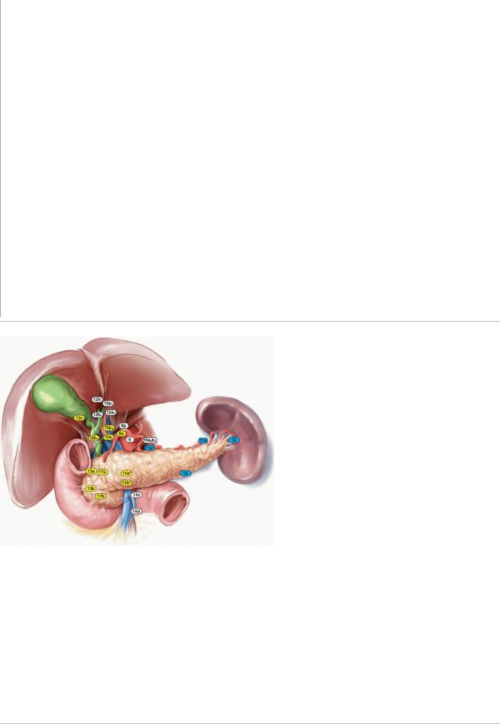
5Suprapyloric
6Infrapyloric
8 (a) Anterior and (p) posterior common hepatic artery
9Celiac artery
10Splenic hilum
11(p) Proximal and (d) distal splenic artery
12(a1, a2) Proper hepatic artery; (b1, b2) common bile duct and hepatic duct; (p1, p2) portal vein; (c) gallbladder, (h) liver hilum
13(a) Posterosuperior and (b) posteroinferior head of pancreas
14(a-d) superior mesenteric artery
16Para-aortic
17(a) Anterosuperior and (b) anteroinferior head of pancreas
18Superior margin of pancreas
Adapted from Japan Pancreas Society as reported in Harisinghani MG, ed. Atlas of Lymph Node Anatomy. New York, NY: Springer Science + Business Media; 2013. doi:10.1007/978-1-4419-9767-8_3.
P.213
FIGURE 14-4 Lymph node basins that should be resected as part of a standard pancreatoduodenectomy (yellow) or distal pancreatectomy with en bloc
splenectomy (blue). •, station 11p should be dissected only for tumors in the proximal body; *, only the right lateral aspect of stations 14a and 14b should be dissected; stations enclosed in dotted lines are posterior.
Standard versus Extended Lymphadenectomy
Five randomized studies have now evaluated the possible benefits associated with extended lymphadenectomy over standard lymphadenectomy. These trials were performed in response to concerns that a more limited resection might leave residual cancer that could lead to local failure and poor survival, as well as in response to several prior retrospective studies that reported that a more extended lymphadenectomy offered a survival advantage over standard lymphatic dissection. These five studies’ designs differed to some degree, and the studies were inconsistent in terms of both the lymph node basins considered to comprise an extended lymphadenectomy and the technical methods used to perform the resections. Furthermore, each of the first four studies that were performed has been individually characterized as being underpowered. These limitations notwithstanding, none of the five studies showed that
extended lymphadenectomy offered a clear benefit over standard lymphadenectomy in terms of long-term survival.16,17,18,19,20,21 Furthermore, associated quality of life analyses revealed that postoperative diarrhea occurred more frequently in patients who underwent extended lymphadenectomy than in patients who underwent standard lymphadenectomy. On the basis of these results, routine extended lymphadenectomy cannot be advocated.

P.214
Technical Aspects
During the Kocher maneuver used to mobilize the duodenum, the retroduodenal and retropancreatic soft tissues anterior to the vena cava are also mobilized. Although the routine resection of the aortocaval nodes between the level of the left renal vein and the lower duodenal border is not advocated, these nodes can be sampled and intraoperatively assessed if positive aortocaval lymphadenopathy is encountered, as survival following resection in this
setting is particularly poor.22 Rather than simply transecting the lymphatic channels in this area, the surgeon should use clips, ligatures, or an energy device to seal these channels to prevent postoperative fluid buildup, which increases the risk for infection and chylous ascites.
After the gallbladder is mobilized en bloc to within the hepatoduodenal ligament, the pericholedochal tissues are resected below the level of the transection site of the common hepatic or bile duct; in addition, all soft tissues adjacent to the proper hepatic artery at this level and those anterior and lateral to the PV are removed en bloc. The removal of retroportal lymph nodes poses a special challenge because these nodes are frequently enlarged in the setting of biliary stenting or cholangitis; these structures posterior to the PV may best be left in place until the final steps of specimen mobilization.
Following the portal dissection, attention is turned to the dissection of the periduodenal and perigastric soft tissues. Depending on the plan for pylorus preservation, the specimen should include all tissue up to the site of gastric or duodenal transection and division of the right gastroepiploic or right gastric vessels. A lymph node is frequently encountered proximal to the origin of the gastroduodenal artery from the common hepatic artery, and this node marks well a transection plane within the soft tissue towards the pancreatic neck. Specific dissection of hepatic artery lymph nodes more proximal to this point is not routinely performed. In select cases, intraoperative sampling of the node may provide important prognostic information, as tumor involvement of this node
is a poor prognostic factor for survival.23
After the pancreas neck is transected anterior to the PV and the distal duodenum and proximal jejunum are transected and mobilized, dissection continues along the SMV-PV confluence. All surrounding soft tissues are dissected, and small vascular branches are divided. This dissection exposes the soft tissues of the mesenteric root that surround the SMA. Unless the presence of tumor extension requires a different approach to be taken, the periarterial tissue is divided anterior to the artery course, and all soft tissues to the right lateral aspect of the SMA are mobilized with the specimen. The goal of this maneuver is to remove 180° of periarterial tissue to the level of the adventitia and to control small branches close to their origin. This facilitates the complete removal of the uncinate process parenchyma, the creation of the best possible margin clearance at the SMA, and the removal of all relevant lymph nodes in this area. This dissection should not be continued beyond the first jejunal branch of the SMA unless specific abnormalities of nodes within the mesenteric root are encountered. Similarly, transverse mesocolon nodes are not routinely resected unless specific findings suggest tumor involvement. The retropancreatic dissection is then completed from the SMA origin towards the liver hilus, and the retroportal lymph nodes can then be removed if necessary. Unless tumor encases it, any replaced right hepatic
P.215 artery originating from the SMA can usually be preserved even after the surrounding lymphoareolar and adipose tissues have been completely dissected. Final hemostatic maneuvers in all divided soft tissue areas complete the regional dissection.
3. RESECTION AND RECONSTRUCTION OF THE SUPERIOR MESENTERIC VEIN/PORTAL VEIN TO OBTAIN NEGATIVE MARGINS
Recommendation: The SMV, PV, or its confluence should be resected and reconstructed at pancreatoduodenectomy if the primary tumor is inseparable from the vessel and vascular involvement is all that impedes the performance of a margin-negative resection.
Type of Data: Primarily retrospective, low-level evidence and meta-analyses thereof.
Strength of Recommendation: Strong.
Rationale
During pancreatoduodenectomy for PDAC, vascular involvement of the tumor poses additional technical challenges to an already complex procedure. Preoperative planning for pancreatoduodenectomy must include high-quality pancreas protocol computed tomography with three-dimensional vascular reconstruction, which enables the surgeon to anticipate the need for venous resection and facilitates adequate preoperative preparation for vascular reconstruction. A review of the imaging should focus on assessing not only the relationship between the tumor and the surrounding major vessels, including the SMV and its first-order tributaries, but also the vascular anatomy itself, including evaluation for the presence of any anatomic variations, such as a replaced right hepatic artery.
The PV courses posterior to the neck of the pancreas and arises from the confluence of the SMV and SV. The SMV originates from the confluence of the jejunal and ileal tributaries, which are adjacent to the uncinate process of the pancreas. Given the proximity of the vessels to tumors in the head, uncinate process, and neck of the pancreas, neoplastic involvement of these vessels is common (Fig. 14-5A,B). Attempts to skeletonize a primary tumor that is densely adherent to the vein may lead to vascular injury and/or macroscopically positive surgical margins (R2 resection). Although pancreatectomy with resection of the major mesenteric arteries carries five times the risk of perioperative mortality and twice the risk of death at 1 year compared with pancreatectomy without arterial resection, pancreatectomy with vein resection, when performed by surgeons experienced with the techniques required,
results in morbidity and mortality equivalent to that of standard pancreatectomy.1,24,25,26 A recent meta-analysis of 19 previous studies estimated 1-, 3-, and 5-year overall survival rates of 61%, 19%, and 12%, respectively, for patients who underwent pancreatoduodenectomy with vein resection and 62%, 27%, and 17%, respectively, for patients who underwent standard pancreatoduodenectomy. This and other meta-analyses included heterogenous, nonrandomized studies; thus, the overall level of evidence is low. Regardless, pancreatoduodenectomy with vein resection and reconstruction should be
P.216 considered standard practice for nonmetastatic PDAC involving the SMV-PV confluence, provided that adequate inflow and outflow veins are present, the
tumor does not involve the SMA or hepatic artery, and an R0/R1 resection can be reasonably expected.27 However, vein resection has no role as part of a “regional pancreatectomy,” initially described in the 1970s and 1980s, in which a greater amount of grossly uninvolved regional soft tissues is cleared in an
attempt to improve outcomes.28,29

FIGURE 14-5 Due to the proximity of the head and uncinate process of the pancreas to the portal and superior mesenteric veins, tumor infiltration into these vessels is common. A: Coronal view, and (B) axial view.
Intraoperative Margin Evaluation
Among patients who undergo venous resection for PDAC, patients with histologic tumor involvement of the resected vein have a poorer prognosis than
patients without tumor involvement.30 However, the clinical significance of a positive venous transection margin (either tangential or segmental) is unclear. Given the logistical difficulties
P.217 associated with performing an intraoperative assessment of the vein margin while the portal system is cross-clamped, routine intraoperative analysis of the vein is therefore not recommended at present. The vein should be resected to grossly clear margins. Resecting clinically uninvolved vein is discouraged because doing so increases the likelihood that a simple lateral venorrhaphy or end-to-end vascular reconstruction must be converted to a more complex reconstruction involving a patch or interposition graft.
Technical Aspects
In cases in which the pancreas can be transected at the neck, the extent of venous involvement may be appreciated as the head and uncinate process are separated from the SMV and PV by dividing the small venous tributaries from the pancreas. If the extent of tumor involvement is more pronounced, transection of the pancreas at the neck may not be possible; in such cases, the pancreas should be divided further to the left of the SMV (superficial to the SV) to prepare for segmental venous resection. The specimen is then separated to fully mobilize the SMV-PV confluence to identify the SMA over its entire proximal course. It may be advantageous to mobilize the specimen from the left side of the vein, separating it from the SMA following acquisition of adequate vascular control proximal and distal to the area of venous involvement. Regardless, it is best to defer final resection and reconstruction of SMV/PV until the entire specimen has been completely resected from all surrounding structures, including the SMA. This allows for better vascular control and minimizes the time needed for venous occlusion.
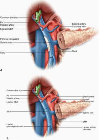
Options for SMV and/or PV resection and reconstruction depend on the site and extent of tumor involvement. If only a small portion of the lateral or posterior aspect of the SMV or PV is involved, the uninvolved uncinate process is separated from the vein, and a Satinsky clamp may be used to gain vascular control of the involved segment; after sharp transection of the involved vein, a primary repair with a 6-0 polypropylene suture can be performed if the lumen of the vessel is not compromised. If a primary repair would create significant narrowing of the venous lumen, a vein patch may be used to repair the SMV or PV (Fig. 14-6A).
If a short segment (≤2 cm) of the PV must be resected, a primary end-to-end anastomosis of the PV should be performed with preservation of the SV and inferior mesenteric vein (IMV) if possible (Fig. 14-6B). Additional PV length for a primary anastomosis can be gained by performing a complete CattellBraasch maneuver with mobilization of the retroperitoneal attachments of the small bowel and right colon mesentery up to the ligament of Treitz, which allows cephalad retraction of the root of the mesentery and the first-order SMV tributaries; division of the middle colic vein may also allow further mobilization of the SMV. In addition, easing the tension of a primary end-to-end PV anastomosis can be achieved by completely dissecting the falciform ligament and right triangular ligament and placing laparotomy pads above the liver, which allows for the caudal displacement of the proximal PV stump. Primary anastomosis of the SMV-PV confluence is usually not possible if the tumor involves the SMV below the level of the SV-PV confluence, as the SV prevents the caudal mobilization of the PV. Therefore, the resection of an SMV segment ≥2 cm with SV preservation generally requires an interposition graft (Fig. 14-
6C). Autologous grafts created from the left internal jugular vein,31 superficial femoral vein,32 and left renal
P.218
P.219
P.220
vein33 have all been described. When the SV is preserved, access to the proximal 3 to 4 cm of the SMA is technically challenging, as the uncinate process attachments to the SMA in this region must be dissected posteriorly.
FIGURE 14-6 Venous reconstruction following resection of a primary cancer of the head of the pancreas with involvement of the portal vein and/or superior mesenteric vein. A: A defect of the right lateral wall of the vein may best be managed by patch venoplasty. Reconstruction of the vessel following removal of a segment of vein may be accomplished without (B) or with (C) an interposition graft. If the splenic vein has been ligated (C) and signs of sinistral portal hypertension develop, implantation into the graft (D) is an option; more straightforwardly, the splenic vein can be implanted into the left renal vein or inferior mesenteric vein to provide venous outflow from the stomach and spleen.
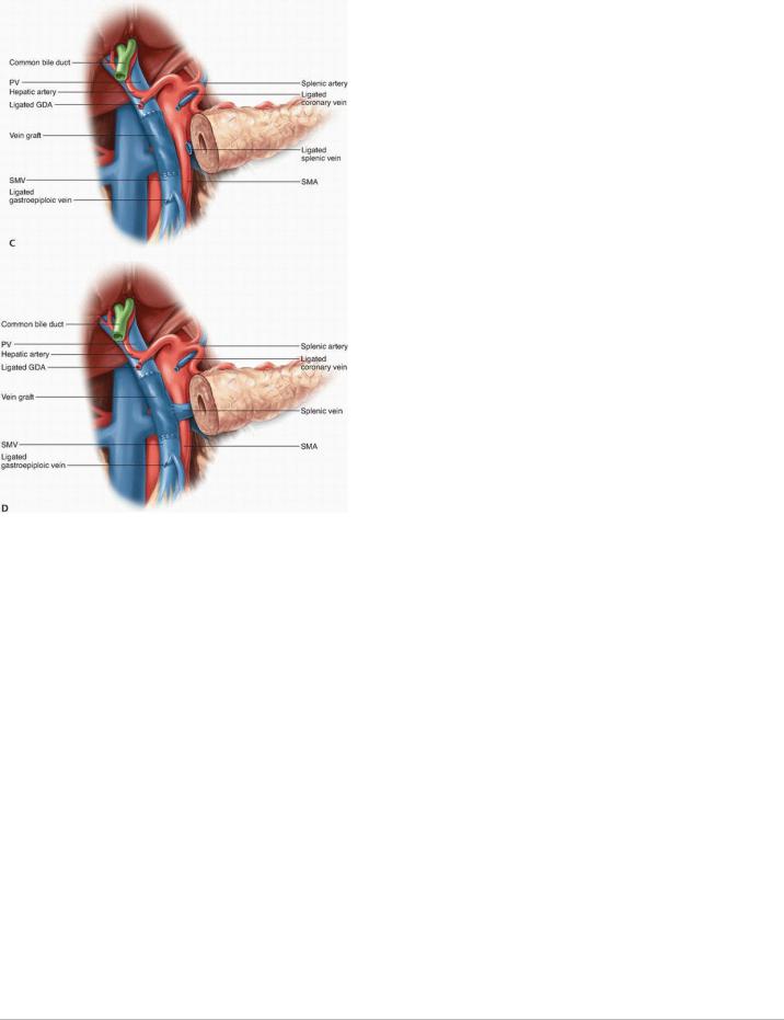
FIGURE 14-6 (Continued).
Management of the Splenic Vein. The SV may have to be divided to gain extra mobility for a direct end-to-end anastomosis between the PV and SMV or if tumor encasement is present at the level of the SV-PV confluence. If the IMV drains into the SV, the SV may be divided at its junction with the PV, as the IMV provides collateral venous drainage of the SV (Fig. 14-6D). However, if the IMV drains into the SMV, ligation of the SV can result in sinistral portal hypertension and increase the risk of future upper gastrointestinal hemorrhage. Various methods to reduce this risk have been reported, including
reimplanting the SV into the side of the interposition vein graft (Fig. 14-6D),32 reimplanting the IMV into the SV,34,35 and performing an end-to-side
splenorenal Warren shunt.36 Mandatory reconstruction of the SV remains a controversial practice, however, because studies suggest that portal
hypertension is not inevitable after SV ligation without reconstruction.35 This has led many surgeons to adopt a selective approach for SV
reconstruction.34,35
Management of the Jejunal and Ileal Tributaries of the Superior Mesenteric Vein
The SMV is formed by the confluence of the ileal and jejunal tributaries in more than 90% of individuals. The jejunal tributary, which drains the proximal small bowel, courses horizontally, typically posterior to the SMA, and enters the right posterolateral aspect of the main trunk of the SMV. The ileal tributary, which drains the distal small bowel, travels vertically through the small bowel mesentery. Anatomic variations in the first-order tributaries of the SMV and the drainage of the IMV are not uncommon, and the operating surgeon must be aware of these.
Isolated tumor involvement of the jejunal tributary requires the resection of this vein to allow for the mobilization of the uncinate process from its retroperitoneal attachments. Reconstruction of the resected jejunal tributary may not be necessary if the ileal tributary is of adequate diameter (i.e., around
1.5 times larger than that of the SMA as revealed by computed tomography37) to provide sufficient venous return to the small bowel. Tumor involvement of the SMV at the confluence of the jejunal and ileal tributaries generally requires resection and reconstruction of the veins. In this setting, the jejunal tributary is typically sacrificed because its thin walls and posterior, horizontal location make reconstructing it to the SMV difficult. Therefore, reconstruction between the ileal tributary and SMV is performed and often requires an interposition graft, especially if the SV has been spared.
Resection and Reconstruction
If possible, the specimen should be completely resected from all surrounding attachments, including the SMA, before resection and reconstruction of the SMV/PV. After the specimen has been removed, systemic anticoagulation is induced with 2,500 to 5,000 U of intravenous heparin. Complete vascular control is obtained by placing vascular clamps proximal and distal to the involved segment of vein and by clamping
P.221
the SV, IMV, and/or jejunal branches if necessary. Subsequent biliary and pancreatic reconstruction can be made less difficult by applying a Rummel tourniquet or soft bulldog clamp to the SMA to prevent inflow to reduce small bowel edema. The anterior walls of the SMV and PV and the interposition graft (when used) are inked proximally and distally with a marker to help orient the vein during reconstruction. The vein is transected, and the tumor is removed en bloc. The vein is then reconstructed using an interrupted or running 5-0 or 6-0 polypropylene suture with care taken to keep the inked walls aligned. Tying the running suture loosely is generally advocated, and when a graft is used, it is typically easier to complete the distal anastomosis first.
4. RESECTION TO NEGATIVE MARGINS AT THE PANCREATIC NECK AND BILE DUCT
Recommendation: The pancreas should be divided at the neck of the gland or, if that is not possible, to the left of the neck. The bile duct should be divided close to the junction of the common bile duct and hepatic duct. In both pancreas division and bile duct division, transection should occur through grossly normal tissue in an attempt to achieve negative margins. Intraoperative evaluation of the status of these margins and re-resection, if positive, may be considered but are not obligatory, particularly if they are likely to result in the sacrifice of a significant amount of normal duct or pancreatic parenchyma.
Type of Data: Primarily retrospective data, low-level evidence.
Strength of Recommendation: Weak.
Rationale
It is reasonably well-established that prognosis following potentially curative pancreatoduodenectomy is associated with the status of the surgical transection margins. In most cases of margin-positive resection, the residual tumor cells are found within the soft tissue adjacent to the SMA. Although they are less commonly found to be positive on histopathologic analysis, the bile duct and pancreatic neck are two other anatomic sites at which residual tumor cells are
commonly found following tissue transection.38,39 In a recent analysis of the pathology reports for patients enrolled on a multi-institutional trial of adjuvant therapy following pancreatoduodenectomy, the SMA, pancreatic neck, and bile duct margins were positive in 38%, 15%, and 3% of evaluated specimens,
respectively.40
Unfortunately, few studies have reported the status of individual surgical margins separately or have evaluated the influence of the status of these individual margins on survival. Furthermore, because specimen analysis is not standardized, comparisons between studies are difficult. Thus, the extent to which residual cells at the bile duct or pancreatic neck margins influence either recurrence or survival is unclear.
Owing to its anatomic location immediately adjacent to the unresectable SMA, the SMA-uncinate margin, if dissected properly, cannot be re-resected in an attempt to clear the margin of malignant tissue if an intraoperative evaluation shows residual
P.222
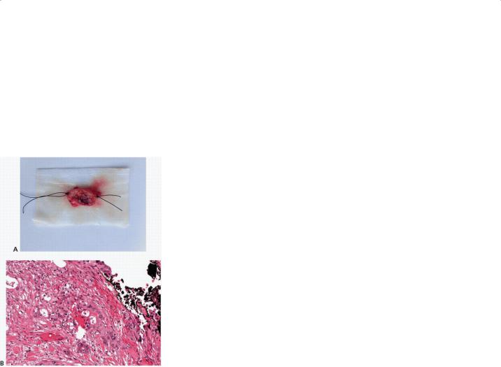
disease. In contrast, the pancreatic neck and bile duct can be re-resected if the initial margin is positive for cancer (Fig. 14-7).41 Surprisingly, however, the data that would substantiate re-resection of the margins in this clinical scenario are conflicting, and so the role of intraoperative evaluation of these margins is the subject of significant debate. One recent single-center study found that the survival of patients in whom an intraoperative extension of the pancreatic resection was performed to convert a microscopically positive pancreatic neck margin was significantly poorer than that of patients who underwent R0
resection initially.42 Similarly, another analysis found that (often multiple) re-resections of the pancreatic parenchyma or bile ducts in patients who initially
had positive margins did not improve survival.43 These studies’ findings imply that a margin-positive resection is a reflection of aggressive tumor biology, not simple anatomy or surgical technique. However, their results must be interpreted cautiously; other studies have found the exact opposite (i.e., that margin-
positive resection reflects anatomy or surgical technique) and suggest that margins should be re-resected until they are negative.44,45 One of these studies even suggested that total
P.223
pancreatectomy, given that it was associated with more favorable overall survival, is preferable to pancreatoduodenectomy with a positive neck margin.45
FIGURE 14-7 Immediate histopathologic analysis of the margin at the pancreatic neck by frozen section should be considered but whether re-resection of a positive margin to a negative one improves survival is unclear. A: If taken, the margin should be properly oriented to direct analysis of the “true” margin. B:
In this example, cancer cells were identified at the pancreatic neck margin so additional pancreatic parenchyma was resected prior to reconstruction.
Given current knowledge, surgeons should clearly attempt to resect to negative margins at the initial transection of the bile duct or pancreatic parenchyma. However, although intraoperative evaluation of those margins should be considered, re-resection should not be pursued to attain negative margins at the expense of significant amounts of otherwise normal-appearing bile duct or pancreatic parenchyma.
Technical Aspects

After a wide Kocher maneuver and cholecystectomy are performed, dissection of the hepatoduodenal ligament can be initiated. Soft tissue anterior to the porta hepatis is divided at approximately the level of the cystic duct stump. Whether arterial (left) or biliary (right) structures are dissected initially is a matter of preference. The clearance of all soft tissues surrounding the portal structures is followed by the isolation of the proper hepatic artery and common hepatic artery and then the ligation of the gastroduodenal artery. Division of the gastroduodenal artery allows for the cephalic retraction of the common hepatic artery and more effective soft tissue clearance, which is achieved by developing a plane of dissection anterior to the PV and extending this plane caudally toward the inferior neck of the pancreas. Lymphadenectomy combined with the clearance of soft tissue directly in contact with the neck of the pancreas is extended to the left as far as needed for transection of the pancreas.
REFERENCES
1.Mollberg N, Rahbari NN, Koch M, et al. Arterial resection during pancreatectomy for pancreatic cancer: a systematic review and meta-analysis. Ann Surg 2011;254(6):882-893.
2.Verbeke CS, Menon KV. Redefining the R1 resection in pancreatic cancer. Br J Surg 2006; 93(10):1232-1237.
3.Gaedcke J, Gunawan B, Grade M, et al. The mesopancreas is the primary site for R1 resection in pancreatic head cancer: relevance for clinical trials. Langenbecks Arch Surg 2010;395(4):451-458.
4.Katz MH, Wang H, Balachandran A, et al. Effect of neoadjuvant chemoradiation and surgical technique on recurrence of localized pancreatic cancer. J Gastrointest Surg 2012;16(1):68-78; discussion 78-79.
5.Baqué P, Iannelli A, Delotte J, et al. Division of the right posterior attachments of the head of the pancreas with a linear stapler during pancreaticoduodenectomy: vascular and oncological considerations based on an anatomical cadaver-based study. Surg Radiol Anat 2009;31(1):13-17.
6.D’Souza MA, Singh K, Hawaldar RV, et al. The vascular stapler in uncinate process division during pancreaticoduodenectomy: technical considerations and results. Dig Surg 2010;27(3):175-181.
7.Katz MH, Lee JE, Pisters PW, et al. Retroperitoneal dissection in patients with borderline resectable pancreatic cancer: operative principles and techniques. J Am Coll Surg 2012;215(2): e11-e18.
8.Dumitrascu T, David L, Popescu I. Posterior versus standard approach in pancreatoduodenectomy: a case-match study. Langenbecks Arch Surg
2010;395(6):677-684.
9. Hatzaras I, George N, Muscarella P, et al. Predictors of survival in periampullary cancers following pancreaticoduodenectomy. Ann Surg Oncol
2010;17(4):991-997.
10.Chen JW, Bhandari M, Astill DS, et al. Predicting patient survival after pancreaticoduodenectomy for malignancy: histopathological criteria based on perineural infiltration and lymphovascular invasion. HPB (Oxford) 2010;12(2):101-108.
11.Schwarz RE, Smith DD. Extent of lymph node retrieval and pancreatic cancer survival: information from a large US population database. Ann Surg Oncol 2006;13(9):1189-1200.
12.Tomlinson JS, Jain S, Bentrem DJ, et al. Accuracy of staging node-negative pancreas cancer: a potential quality measure. Arch Surg
2007;142(8):767-723; discussion 773-774.
13. Edge SB, Byrd DR, Compton CC, et al, eds; American Joint Committee on Cancer. AJCC Cancer Staging Manual. 7th ed. New York, NY: Springer; 2010.
P.224
14. Pawlik TM, Abdalla EK, Barnett CC, et al. Feasibility of a randomized trial of extended lymphadenectomy for pancreatic cancer. Arch Surg
2005;140(6):584-589; discussion 589-591.
15.Pedrazzoli S, Günther Beger H, Obertop H, et al. A surgical and pathological based classification of resective treatment of pancreatic cancer. Summary of an international workshop on surgical procedures in pancreatic cancer. Dig Surg 1999;16(4):337-345.
16.Farnell MB, Aranha GV, Nimura Y, et al. The role of extended lymphadenectomy for adenocarcinoma of the head of the pancreas: strength of the evidence. J Gastrointest Surg 2008;12(4): 651-656.
17.Michalski CW, Kleeff J, Wente MN, et al. Systematic review and meta-analysis of standard and extended lymphadenectomy in pancreaticoduodenectomy for pancreatic cancer. Br J Surg 2007;94(3):265-273.
18.Pedrazzoli S, DiCarlo V, Dionigi R, et al. Standard versus extended lymphadenectomy associated with pancreatoduodenectomy in the surgical treatment of adenocarcinoma of the head of the pancreas: a multicenter, prospective, randomized study. Lymphadenectomy Study Group. Ann Surg

1998;228(4):508-517.
19. Yeo CJ, Cameron JL, Lillemoe KD, et al. Pancreaticoduodenectomy with or without distal gastrectomy and extended retroperitoneal lymphadenectomy for periampullary adenocarcinoma, part 2: randomized controlled trial evaluating survival, morbidity, and mortality. Ann Surg
2002;236(3):355-366; discussion 366-368.
20.Farnell MB, Pearson RK, Sarr MG, et al. A prospective randomized trial comparing standard pancreatoduodenectomy with pancreatoduodenectomy with extended lymphadenectomy in resectable pancreatic head adenocarcinoma. Surgery 2005;138(4):618-628; discussion 628-630.
21.Jang JY, Kang MJ, Heo JS, et al. A prospective randomized controlled study comparing outcomes of standard resection and extended resection, including dissection of the nerve plexus and various lymph nodes, in patients with pancreatic head cancer. Ann Surg 2014;259(4):656-664.
22.Shimada K, Sakamoto Y, Sano T, et al. The role of paraaortic lymph node involvement on early recurrence and survival after macroscopic curative resection with extended lymphadenectomy for pancreatic carcinoma. J Am Coll Surg 2006;203(3):345-352.
23.LaFemina J, Chou JF, Gönen M, et al. Hepatic arterial nodal metastases in pancreatic cancer: is this the node of importance? J Gastrointest Surg
2013;17(6):1092-1097.
24. Chua TC, Saxena A. Extended pancreaticoduodenectomy with vascular resection for pancreatic cancer: a systematic review. J Gastrointest Surg
2010;14(9):1442-1452.
25.Yu XZ, Li LJ, Fu DL, et al. Benefit from synchronous portal-superior mesenteric vein resection during pancreaticoduodenectomy for cancer: a metaanalysis. Eur J Surg Oncol 2014;40(4):371-378.
26.Zhou Y, Zhang Z, Liu Y, et al. Pancreatectomy combined with superior mesenteric vein-portal vein resection for pancreatic cancer: a meta-analysis. World J Surg 2012;36(4):884-891.
27.Callery MP, Chang KJ, Fishman EK, et al. Pretreatment assessment of resectable and borderline resectable pancreatic cancer: expert consensus statement. Ann Surg Oncol 2009;16(7): 1727-1733.
28.Fortner JG. Regional resection of cancer of the pancreas: a new surgical approach. Surgery 1973;73(2):307-320.
29.Fortner JG, Kim DK, Cubilla A, et al. Regional pancreatectomy: en bloc pancreatic, portal vein and lymph node resection. Ann Surg 1977;186(1):42-
30.Wang J, Estrella JS, Peng L, et al. Histologic tumor involvement of superior mesenteric vein/portal vein predicts poor prognosis in patients with stage II pancreatic adenocarcinoma treated with neoadjuvant chemoradiation. Cancer 2012;118(15):3801-3811.
31.Tseng JF, Raut CP, Lee JE, et al. Pancreaticoduodenectomy with vascular resection: margin status and survival duration. J Gastrointest Surg
2004;8(8):935-949; discussion 949-950.
32. Fleming JB, Barnett CC, Clagett GP. Superficial femoral vein as a conduit for portal vein reconstruction during pancreaticoduodenectomy. Arch Surg
2005;140(7):698-701.
33.Smoot RL, Christein JD, Farnell MB. An innovative option for venous reconstruction after pancreaticoduodenectomy: the left renal vein. J Gastrointest Surg 2007;11(4):425-431.
34.Ferreira N, Oussoultzoglou E, Fuchshuber P, et al. Splenic vein-inferior mesenteric vein anastomosis to lessen left-sided portal hypertension after pancreaticoduodenectomy with concomitant vascular resection. Arch Surg 2011;146(12):1375-1381.
35.Strasberg SM, Bhalla S, Sanchez LA, et al. Pattern of venous collateral development after splenic vein occlusion in an extended Whipple procedure: comparison with collateral vein pattern in cases of sinistral portal hypertension. J Gastrointest Surg 2011;15(11):2070-2079.
36.Christians KK, Tsai S, Tolat PP, et al. Critical steps for pancreaticoduodenectomy in the setting of pancreatic adenocarcinoma. J Surg Oncol
2013;107(1):33-38.
P.225
37.Katz MH, Fleming JB, Pisters PWT, et al. Anatomy of the superior mesenteric vein with special reference to the surgical management of first-order branch involvement at pancreaticoduodenectomy. Ann Surg 2008;248(6):1098-1102.
38.Gnerlich JL, Luka SR, Deshpande AD, et al. Microscopic margins and patterns of treatment failure in resected pancreatic adenocarcinoma. Arch Surg 2012;147(8):753-760.

39.Raut CP, Tseng JF, Sun CC, et al. Impact of resection status on pattern of failure and survival after pancreaticoduodenectomy for pancreatic adenocarcinoma. Ann Surg 2007;246(1):52-60.
40.Katz MH, Merchant NB, Brower S, et al. Standardization of surgical and pathologic variables is needed in multicenter trials of adjuvant therapy for pancreatic cancer: results from the ACOSOG Z5031 trial. Ann Surg Oncol 2011;18(2):337-344.
41.Dillhoff M, Yates R, Wall K, et al. Intraoperative assessment of pancreatic neck margin at the time of pancreaticoduodenectomy increases likelihood of margin-negative resection in patients with pancreatic cancer. J Gastrointest Surg 2009;13(5):825-830.
42.Lad NL, Squires MH, Maithel SK, et al. Is it time to stop checking frozen section neck margins during pancreaticoduodenectomy? Ann Surg Oncol
2013;20(11):3626-3633.
43.Hernandez J, Mullinax J, Clark W, et al. Survival after pancreaticoduodenectomy is not improved by extending resections to achieve negative margins. Ann Surg 2009;250(1):76-80.
44.Fatima J, Schnelldorfer T, Barton J, et al. Pancreatoduodenectomy for ductal adenocarcinoma: implications of positive margin on survival. Arch Surg
2010;145(2):167-172.
45. Schmidt CM, Glant J, Winter JM, et al. Total pancreatectomy (R0 resection) improves survival over subtotal pancreatectomy in isolated neck margin positive pancreatic adenocarcinoma. Surgery 2007;142(4):572-578; discussion 578-580.
P.226
Pancreatoduodenectomy: Key Question
In patients with resectable adenocarcinoma of the pancreatic head, should a periadventitial dissection of the superior mesenteric artery (i.e.,
total mesopancreas excision) be performed at the time of pancreatoduodenectomy?
INTRODUCTION
Pancreatic ductal adenocarcinoma (PDAC) is the fourth leading cause of cancer-related death in the United States and the eighth worldwide. Despite advances in medical therapy, the median survival duration of patients diagnosed with PDAC is only 4 to 6 months. However, for the 10% to 20% of PDAC patients who are candidates for pancreatoduodenectomy (PD) at the time of diagnosis, the 5-year overall survival rate approaches 25%, and the median survival duration is 20 to 22 months. Among patients who undergo resection, multiple clinical variables have been determined to be associated with postoperative prognosis. Of these, margin status appears to be one of the most important.
The American Joint Committee on Cancer staging guidelines have attempted to standardize the pathologic evaluation of PD specimens to facilitate margin
assessment.1 According to these guidelines, PD specimen margins that should be evaluated by the pathologist include the pancreatic neck, bile duct, duodenum, and stomach margins, as well as the superior mesenteric artery (SMA) margin. The last of these margins, referred to in this book as the SMA/uncinate margin, is specifically emphasized. The SMA/uncinate margin comprises the tissue that connects the uncinate process to the right lateral border of the proximal 3 to 4 cm of the SMA. PDAC has a propensity to spread through this tissue along the perineural autonomic plexus that surrounds the artery. However, the SMA cannot be removed and reconstructed at surgery in the absence of considerable morbidity. Many surgeons therefore strongly recommend that a periadventitial dissection of the SMA be performed at PD to skeletonize the right lateral aspect of the vessel from the uncinate process and adjacent tissues to maximize the likelihood of obtaining a negative margin in the retroperitoneum.
Although this recommendation is commonly made, the association between the status of the SMA/uncinate margin and oncologic outcomes is unclear. Indeed, the incidence of a positive SMA/uncinate margin and any association between margin status and outcome may reflect “tumor biology” rather than
surgical approach or technical skill.2,3 The contribution of this specific surgical technique to postoperative outcome is therefore incompletely understood. Herein, we describe our review of the literature to determine the effect of performing a periadventitial dissection of the SMA during PD.
METHODOLOGY
We searched Medline and PubMed for English language articles published from January 1990 through January 2014 that addressed the effect of margin status on
P.227 survival and recurrence following PD. Combinations of the following keywords were used: “pancreas,” “retroperitoneal margin,” “pancreaticoduodenectomy,” “Whipple,” “margin,” “SMA dissection,” “mesopancreas,” “retroperitoneal dissection,” “morbidity,” and “uncinate dissection.” Our initial search yielded 520 unique articles. Of these articles, 66 were selected for further review following review of the abstract. The articles selected for analysis focused specifically on pancreatic cancer, commented on margin status following PD, and reported outcomes related to PD and/or margin status. Directed searches of embedded references from the selected articles yielded an additional 16 articles, providing a total of 82 articles for review. Among these, 43 were selected for further analysis.
Of the 43 articles selected, 5 were prospective trials; the others were retrospective reviews of institutional databases (37 articles) or national registry data (1 article). Articles selected for the analysis were specifically scrutinized for standardization of the surgical technique for SMA dissection, pathologic evaluation of the retroperitoneal margin, and margin status-related outcomes, including local recurrence and overall survival. All articles were then reviewed by two members of the group and assigned a strength of recommendation using the GRADE (Grades of Recommendation, Assessment, Development, and Evaluation) system.