

новая папка / Operative Standards for Cancer Surgery Volume I 1st Edition
.pdfduring EUS. When rapid on-site cytologic evaluation (ROSE) is available, a sample from one lymph node station should be confirmed as adequate for evaluation before the next station is sampled. When ROSE is not available, at least three passes of one lymph node station should be
obtained unless a tissue block that is adequate for evaluation is grossly obtained during the first or second pass.3
2. NODE STATION ASSESSMENT UTILIZING ENDOBRONCHIAL ULTRASONOGRAPHY/ENDOSCOPIC ULTRASONOGRAPHY
Recommendation: Morphology ultrasonography should be used to assess mediastinal lymph node stations 2R, 4R, 2L, 4L, and 7 during EBUS and lymph node stations 8 and 9 during EUS.
Type of Data: Retrospective.
Strength of Recommendation: Weak.
Rationale
The recommendations for the extent of lymph node analysis are based on published guidelines. 4 Convex probe EBUS can access the upper paratracheal (stations 2R and 2L), lower paratracheal (stations 4R and 4L), subcarinal (station 7) and retrotracheal (station 3p) lymph nodes, as well as the hilar (station 10), interlobar (station 11), and lobar (station 12) nodes in the lower lobes. However, convex probe EBUS cannot access the prevascular (station 3a), subaortic (station 5), para-aortic (station 6), paraesophageal (station 8), pulmonary ligament (station 9), or lobar lymph nodes in the upper and middle lobes (Table 6-1).
EUS can access the pulmonary ligament (station 9), paraesophageal (station 8), subcarinal (station 7), retrotracheal (station 3p), and paratracheal (stations 2 and 4) lymph nodes. EUS usually cannot access the prevascular (station 3a), subaortic (station 5), para-aortic (station 6), or N1 lymph nodes. Some authors have reported that EUS can access stations 5 and 6, but the assessment of these nodes with EUS is
P.100
more difficult than that of other stations with EUS and requires significant expertise and experience to accomplish successfully.5
TABLE 6-1 Lymph Nodes Accessible by EBUS/EUS
EBUS |
EUS |
2 |
2 |
3p |
3p |
4 |
4 |
7 |
7 |
10 |
8 |
11 |
9 |
12* |
|
EBUS, endobronchial ultrasonography; EUS, endoscopic ultrasonography.
*Lower lobe lobar lymph nodes.
Technical Aspects
Although the instrumentation used for EUS and EBUS have some similarities, they also have distinct characteristics that limit the way in which each can be utilized. EBUS-TBNA is performed using a convex probe. A saline-filled balloon at the tip of the probe helps improve image quality. Samples are obtained using either a 21or 22-gauge needle with real-time EBUS visualization. The needle has a number of safety features, including a mechanism that prevents the needle from protruding more than 40 mm and an internal sheath that prevents the needle from contaminating the working channel of the scope. In contrast, EUS-FNA is performed using a side-viewing videogastroscope with a dedicated curved linear array transducer attached to the tip of the probe. A number of different scopes from different companies are available for EUS-FNA, and all have minor differences in their overall diameter and size of the ultrasonography probe. A dedicated 22-gauge needle is usually used for EUS-FNA, but smaller (25-gauge) and larger (19-gauge) needles are also available. Similar to that performed using EBUS, tissue sampling using EUS is performed with the aid of real-time ultrasonography.

Both EBUS-TBNA and EUS-FNA can be performed on an outpatient basis utilizing either conscious sedation or general anesthesia. (When general anesthesia is used, the cough reflex is minimal, which may be an advantage during the procedure.) An endotracheal tube (size 8 minimum) or laryngeal mask airway is used to accommodate the scope. The size of the scope limits nasal insertion.
EBUS-TBNA should include an examination of the airway using regular flexible bronchoscopy. Ultrasonically visible vascular landmarks should be
used to identify the specific lymph node stations according to the International Association for the Study of Lung Cancer lymph node map.2
Doppler ultrasonography is used to identify surrounding vessels as well as the blood flow within lymph nodes. The needle is introduced into the lymph node with a sharp stab. The node is aspirated by removing the internal stylet and using a Vac-Lok syringe to apply negative pressure. (Because negative pressure can cause bloody samples of hypervascular lymph nodes, EBUSTBNA of such nodes can be done without suction.) The needle is moved back and forth inside the lymph node to obtain samples. Finally, the needle is retracted into its sheath and removed from the bronchoscope.
The technique for EUS-FNA is similar to that for EBUS-TBNA. Anatomical landmarks such as the inferior vena cava, right and left atrium, azygos vein, main pulmonary artery, and aorta are identified. Any lymph nodes encountered are described and numbered according to the International Association for the Study of Lung Cancer lymph node map (see Fig. I-3 in the Introduction to Section II). Lymph nodes are then biopsied, usually with a 22-gauge needle, using real-time ultrasonography guidance. Finally, one cannot stress enough the importance of proper specimen handling during EBUS-TBNA and/or EUS-FNA. Institutional standards differ, and close collaboration between the surgeon and cytopathologist is essential. However, when ROSE is available, both EBUSTBNA and EUS-FNA can be performed without the aid of a cytopathologist.
P.101
REFERENCES
1. Fujiwara T, Yasufuku K, Nakajima T, et al. The utility of sonographic features during endobronchial ultrasound-guided transbronchial needle aspiration for lymph node staging in patients with lung cancer: a standard endobronchial ultrasound image classification system. Chest
2010;138:641-647.
2.Rusch VW, Asamura H, Watanabe H, et al. The IASLC Lung Cancer Staging Project. A proposal for a new international lymph node map in the forthcoming seventh edition of the TNM classification for lung cancer. J Thorac Oncol 2009;4:568-577.
3.Lee HS, Lee GK, Lee HS, et al. Real-time endobronchial ultrasound-guided transbronchi al needle aspiration in mediastinal staging of nonsmall cell lung cancer: how many aspirations per target lymph node station? Chest 2008;134(2):368-374.
4.Howington JA, Blum MG, Chang AC, et al. Treatment of stage I and II non-small cell lung cancer: diagnosis and management of lung cancer, 3rd ed: American College of Chest Physicians Evidence-Based Clinical Practice Guidelines. Chest 2013;143(5)(suppl):e278S-e313S.
5.Liberman M, Durnceau A, Grunenwald E, et al. Initial experience with a new technique of endoscopic and ultrasonographic access for biopsy of para-aortic (station 6) mediastinal lymph nodes without traversing the aorta. J Thorac Cardiovasc Surg 2012;144(4):787-792; discussion 792-793.
P.102
Endobronchial Ultrasonography/Endoscopic Ultrasonography: Key Question
When should negative endobronchial ultrasonography findings be confirmed by a more invasive procedure?
INTRODUCTION
Accurate staging is of paramount importance in the management of non-small cell lung cancer (NSCLC). The treatment of NSCLC, whether surgery, chemotherapy, radiotherapy, or a multimodality approach, depends on the stage of the disease. In NSCLC patients without metastatic disease, the status of the mediastinum is the most important determinant of candidacy for curative-intent treatment.
The first-line tests in the diagnosis and staging of NSCLC are imaging studies, including computed tomography (CT) and positron emission tomography (PET). Based on the most recent American College of Chest Physicians (ACCP) guidelines, the sensitivity and specificity of CT in
identifying mediastinal lymph node metastasis are 55% and 81%, respectively, and those of PET are 77% and 86%, respectively.1 These imaging studies have substantial false positive and false negative rates, and PET findings in particular must be confirmed with lymph node biopsy.
Traditionally, the gold standard for sampling lymph nodes in the mediastinum to confirm imaging studies’ findings has been mediastinoscopy. Recently, however, endobronchial ultrasonography (EBUS)- or endoscopic ultrasonography (EUS)-guided needle aspiration techniques have also emerged as options for staging the mediastinum. In many institutions, EBUS-guided transbronchial needle aspiration (EBUSTBNA) has replaced mediastinoscopy as the first-line means of sampling mediastinal lymph nodes, and mediastinoscopy is reserved to confirm the absence of N2
disease.1,2
As surgeons gain experience using EBUS-TBNA for lung cancer staging, the role of confirmatory mediastinoscopy and other invasive approaches to confirm EBUS-TNBA findings continues to diminish. Despite the impressive results obtained with EBUSTBNA overall, confirmatory mediastinoscopy is still useful in certain situations. These situations are discussed in the following texts.
LITERATURE REVIEW
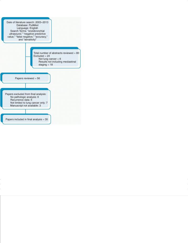
We conducted a Medline search of English language studies published from 2003 to 2013. We used the Medical Subject Heading term “endobronchial ultrasound”; the terms “negative predictive value,” “false negative,” “accuracy,” and “sensitivity” were introduced to limit the number of studies identified. A total of 80 articles were found. Of the 80 abstracts we reviewed, 24 were excluded because the studies included patients who did not have lung cancer or examined EBUS in a role other than its use in invasive mediastinal staging. A careful review of the remaining 56 articles
P.103 identified 35 articles that included data about the false negative rate, sensitivity, or negative predictive value of EBUS in the assessment of N2 disease in patients with NSCLC. This is summarized in Figure 6-2.
FIGURE 6-2 CONSORT diagram summarizing the literature search used for review of endobronchial ultrasound performance.
FINDINGS
Factors that affect the negative predictive value of EBUS
Multiple studies have demonstrated the utility of EBUS-TBNA as a staging modality in patients with NSCLC (Table 6-
2).3,4,5,6,7,8,9,10,11,12,13,14,15,16,17,18,19,20,21,22,23,24,25,26,27,28,29,30 The most important parameter defining the role of a staging modality in the preoperative staging of NSCLC is its ability to rule out mediastinal lymph node disease. In other words, a test for mediastinal disease must have a high sensitivity and high negative predictive value to prevent surgeons from performing a futile thoracotomy or thoracoscopic resection. However, both sensitivity and negative predictive value can be influenced by a number of factors.
P.104
P.105
P.106
P.107
TABLE 6-2 Studies Reporting Sensitivity and/or Negative Predictive Value of Real-Time Endobronchial Ultrasonography in the Mediastinal Staging of Confirmed or Highly Suspected Lung Cancer
|
|
|
Prevalence |
|
|
|
|
Potential |
|
|
|
of N2 |
|
|
|
|
for |
Author, |
Study |
No. of |
Disease |
|
|
|
|
Significant |
Year |
Design |
Patients |
(%) |
NPV |
Sensitivity |
Accuracy |
Key Findings |
Bias |
Cornwell |
Retrospective |
62 |
5 |
93 |
67 |
94 |
In patients with |
Yes |

et al, |
|
|
|
|
|
|
clinical stage I |
|
20133 |
|
|
|
|
|
|
NSCLC, EBUS |
|
|
|
|
|
|
|
|
results in a lower |
|
|
|
|
|
|
|
|
incidence of |
|
|
|
|
|
|
|
|
nontherapeutic |
|
|
|
|
|
|
|
|
thoracotomy than |
|
|
|
|
|
|
|
|
noninvasive staging |
|
|
|
|
|
|
|
|
does, but this |
|
|
|
|
|
|
|
|
difference is not |
|
|
|
|
|
|
|
|
significant. |
|
Herth et al, |
Prospective |
97 |
10 |
98.9 |
89 |
NA |
EBUS is accurate in |
Yes |
20084 |
observational |
|
|
|
|
|
NSCLC staging for |
|
|
|
|
|
|
|
|
patients with clinical |
|
|
|
|
|
|
|
|
stage I NSCLC |
|
|
|
|
|
|
|
|
determined by CT |
|
|
|
|
|
|
|
|
and PET findings. |
|
Herth et al, |
Prospective |
100 |
121 |
96.3 |
92.3 |
NA |
EBUS is beneficial |
Yes |
20065 |
observational |
|
|
|
|
|
in patients with |
|
|
|
|
|
|
|
|
clinical stage I |
|
|
|
|
|
|
|
|
NSCLC. It prevents |
|
|
|
|
|
|
|
|
nontherapeutic |
|
|
|
|
|
|
|
|
thoracotomy in 1 of |
|
|
|
|
|
|
|
|
6 patients. PET was |
|
|
|
|
|
|
|
|
not used routinely. |
|
Hwangbo |
Prospective |
126 |
26 |
96.7 |
90 |
97.4 |
EBUS is useful for |
Yes |
et al, |
observational |
|
|
|
|
|
confirming N2 |
|
20096 |
|
|
|
|
|
|
disease detected by |
|
|
|
|
|
|
|
|
PET. It is also |
|
|
|
|
|
|
|
|
useful for detecting |
|
|
|
|
|
|
|
|
N2 disease in |
|
|
|
|
|
|
|
|
patients with |
|
|
|
|
|
|
|
|
radiographic N0 |
|
|
|
|
|
|
|
|
disease. |
|
Lee et al, |
Retrospective |
102 |
30 |
96.9 |
93.8 |
97.9 |
Optimal results with |
Yes |
20087 |
|
|
|
|
|
|
EBUS are obtained |
|
|
|
|
|
|
|
|
when at least 3 |
|
|
|
|
|
|
|
|
aspirations of each |
|
|
|
|
|
|
|
|
lymph node are |
|
|
|
|
|
|
|
|
performed. |
|
Yasufuku |
Prospective |
153 |
35 |
91 |
81 |
93 |
EBUS is equivalent |
No |
et al, |
controlled |
|
|
|
|
|
to mediastinoscopy |
|
20118* |
trial |
|
|
|
|
|
in the mediastinal |
|
|
|
|
|
|
|
|
staging of NSCLC. |
|
Feller- |
Retrospective |
131 |
35 |
89.7 |
85 |
NA |
EBUS is an |
Yes |
Kopman et |
|
|
|
|
|
|
accurate and |
|
al, 20099* |
|
|
|
|
|
|
sensitive method for |
|
|
|
|
|
|
|
|
diagnosing and |
|
|
|
|
|
|
|
|
staging NSCLC. |
|
Petersen |
Retrospective |
157 |
43 |
90 |
85 |
NA |
EBUS is accurate in |
Yes |
et al, |
|
|
|
|
|
|
staging the |
|
200910 |
|
|
|
|
|
|
mediastinum in |
|
|
|
|
|
|
|
|
NSCLC patients. |
|
|
|
|
|
|
|
|
The routine |
|
|
|
|
|
|
|
|
confirmation of |
|
|
|
|
|
|
|
|
negative EBUS |
|
|
|
|
|
|
|
|
findings with |
|
|
|
|
|
|
|
|
|
|

|
|
|
|
|
|
|
mediastinoscopy |
|
|
|
|
|
|
|
|
has a minor role in |
|
|
|
|
|
|
|
|
NSCLC staging. |
|
Sanz- |
Retrospective |
296 |
51 |
93.6 |
NA |
NA |
EBUS can be used |
Yes |
Santos et |
|
|
|
|
|
|
to sample lymph |
|
al, 201211 |
|
|
|
|
|
|
node regions 4R, |
|
|
|
|
|
|
|
|
4L, and 7 in more |
|
|
|
|
|
|
|
|
than 80% of |
|
|
|
|
|
|
|
|
patients. In such |
|
|
|
|
|
|
|
|
patients, EBUS has |
|
|
|
|
|
|
|
|
an NPV of >90% for |
|
|
|
|
|
|
|
|
mediastinal |
|
|
|
|
|
|
|
|
malignancy. |
|
Nakajima |
Retrospective |
438 |
52 |
90 |
97 |
98 |
ROSE during EBUS |
Yes |
et al, |
|
|
|
|
|
|
results in a low |
|
201312* |
|
|
|
|
|
|
incidence of |
|
|
|
|
|
|
|
|
nondiagnostic |
|
|
|
|
|
|
|
|
samples. |
|
Jhun et al, |
Retrospective |
151 |
55 |
84.3 |
91.6 |
93.8 |
The diagnostic yield |
Yes |
201213 |
|
|
|
|
|
|
of EBUS is lower for |
|
|
|
|
|
|
|
|
left paratracheal |
|
|
|
|
|
|
|
|
lymph nodes. The |
|
|
|
|
|
|
|
|
diagnostic yield is |
|
|
|
|
|
|
|
|
not related to lymph |
|
|
|
|
|
|
|
|
node size. |
|
Szlubowski |
Retrospective |
226 |
57 |
89 |
83.5 |
92.9 |
EBUS is an |
Yes |
et al, |
|
|
|
|
|
|
effective and safe |
|
200914 |
|
|
|
|
|
|
technique for |
|
|
|
|
|
|
|
|
mediastinal staging |
|
|
|
|
|
|
|
|
in NSCLC patients. |
|
|
|
|
|
|
|
|
In patients with |
|
|
|
|
|
|
|
|
negative EBUS |
|
|
|
|
|
|
|
|
results, surgical |
|
|
|
|
|
|
|
|
exploration of the |
|
|
|
|
|
|
|
|
mediastinum should |
|
|
|
|
|
|
|
|
be performed. |
|
Bauwens |
Retrospective |
106 |
58 |
91 |
95 |
97 |
EBUS is a |
Yes |
et al, |
|
|
|
|
|
|
reasonable first |
|
200815 |
|
|
|
|
|
|
step in the |
|
|
|
|
|
|
|
|
confirmation of N2 |
|
|
|
|
|
|
|
|
disease in NSCLC |
|
|
|
|
|
|
|
|
patients. Surgical |
|
|
|
|
|
|
|
|
mediastinal staging |
|
|
|
|
|
|
|
|
should be used to |
|
|
|
|
|
|
|
|
confirm negative |
|
|
|
|
|
|
|
|
EBUS findings. |
|
Joesph et |
Retrospective |
131 |
58 |
90 |
92 |
NA |
ROSE does not |
Yes |
al, 201316* |
|
|
|
|
|
|
affect clinical |
|
|
|
|
|
|
|
|
decisions made |
|
|
|
|
|
|
|
|
during staging |
|
|
|
|
|
|
|
|
EBUS. |
|
Lee et al, |
Retrospective |
73 |
62 |
94 |
95 |
97 |
EBUS can be used |
Yes |
201217 |
|
|
|
|
|
|
to accurately |
|
|
|
|
|
|
|
|
assess the |
|
|
|
|
|
|
|
|
mediastinum in |
|
|
|
|
|
|
|
|
patients with |
|
|
|
|
|
|
|
|
NSCLC and |
|

|
|
|
|
|
|
|
radiographic N2 |
|
|
|
|
|
|
|
|
disease. |
|
Cerfolio et |
Retrospective |
72 |
63 |
79 |
57 |
83 |
EBUS and EUS |
Yes |
al, 201018 |
|
|
|
|
|
|
have high false |
|
|
|
|
|
|
|
|
negative rates, and |
|
|
|
|
|
|
|
|
negative results |
|
|
|
|
|
|
|
|
should be confirmed |
|
|
|
|
|
|
|
|
prior to |
|
|
|
|
|
|
|
|
thoracotomy. |
|
Navani et |
Retrospective |
774 |
65 |
88 |
72 |
NA |
EBUS samples are |
Yes |
al, 201219 |
|
|
|
|
|
|
suitable for use in |
|
|
|
|
|
|
|
|
NSCLC subtyping |
|
|
|
|
|
|
|
|
and EGFR mutation |
|
|
|
|
|
|
|
|
analysis. |
|
Kuo et al, |
Retrospective |
43 |
65 |
85.7 |
80.6 |
91 |
The diagnostic |
Yes |
201120 |
|
|
|
|
|
|
accuracy of EBUS |
|
|
|
|
|
|
|
|
is higher than that |
|
|
|
|
|
|
|
|
of PET in a |
|
|
|
|
|
|
|
|
tuberculosis- |
|
|
|
|
|
|
|
|
endemic population. |
|
Hu et al, |
Retrospective |
231 |
67‡ |
92 |
88 |
87 |
Proficiency using |
Yes |
201321 |
|
|
|
|
|
|
EBUS requires 22 |
|
|
|
|
|
|
|
|
cases. Lymph node |
|
|
|
|
|
|
|
|
size is a predictor of |
|
|
|
|
|
|
|
|
success. |
|
Yasufuku |
Prospective |
105 |
67 |
89.5 |
94.6 |
96.3 |
EBUS is an |
Yes |
et al, |
observational |
|
|
|
|
|
accurate staging |
|
200522* |
|
|
|
|
|
|
procedure in |
|
|
|
|
|
|
|
|
patients with |
|
|
|
|
|
|
|
|
NSCLC. |
|
Rintoul et |
Retrospective |
109 |
71 |
60 |
91 |
92 |
EBUS can be used |
Yes |
al, 200923 |
|
|
|
|
|
|
to accurately |
|
|
|
|
|
|
|
|
evaluate PET- |
|
|
|
|
|
|
|
|
positive hilar and |
|
|
|
|
|
|
|
|
mediastinal lymph |
|
|
|
|
|
|
|
|
nodes. Negative |
|
|
|
|
|
|
|
|
findings should be |
|
|
|
|
|
|
|
|
confirmed by |
|
|
|
|
|
|
|
|
surgical means. |
|
Cetinkaya |
Retrospective |
52 |
80 |
83 |
95 |
96 |
EBUS is safe and |
Yes |
et al, |
|
|
|
|
|
|
accurate in NSCLC |
|
201124 |
|
|
|
|
|
|
staging. |
|
Ernst et al, |
Prospective |
60 |
89 |
78 |
87 |
NA |
The difference |
Yes |
200825 |
cross-over |
|
|
|
|
|
between EBUS and |
|
|
|
|
|
|
|
|
mediastinoscopy in |
|
|
|
|
|
|
|
|
determining the N |
|
|
|
|
|
|
|
|
status of patients |
|
|
|
|
|
|
|
|
with NSCLC is not |
|
|
|
|
|
|
|
|
statistically |
|
|
|
|
|
|
|
|
significant. |
|
Gu et al, |
Meta- |
1,299 |
NA |
93 |
NA |
NA |
EBUS has a high |
|
200926 |
analysis |
|
|
|
|
|
NPV and is |
|
|
|
|
|
|
|
|
costeffective in the |
|
|
|
|
|
|
|
|
|
|

|
|
|
|
|
|
|
mediastinal staging |
|
|
|
|
|
|
|
of NSCLC. |
Adams et |
Meta- |
782 |
NA |
NA |
88 |
NA |
EBUS has high |
al, 200927 |
analysis |
|
|
|
|
|
sensitivity in the |
|
|
|
|
|
|
|
mediastinal staging |
|
|
|
|
|
|
|
of NSCLC. |
Abu-Hijleh |
Retrospective |
200 |
NA |
75 |
87 |
91 |
EBUS is similar to |
et al, |
|
|
|
|
|
|
surgical staging in |
201328* |
|
|
|
|
|
|
patients with |
|
|
|
|
|
|
|
NSCLC. The NPV |
|
|
|
|
|
|
|
of EBUS is highest |
|
|
|
|
|
|
|
after the initial 25- |
|
|
|
|
|
|
|
50 cases. The |
|
|
|
|
|
|
|
accuracy of EBUS |
|
|
|
|
|
|
|
is independent of |
|
|
|
|
|
|
|
lymph node size or |
|
|
|
|
|
|
|
location and |
|
|
|
|
|
|
|
number of passes. |
Dong et al, |
Meta- |
1,066 |
NA |
93 |
90 |
96 |
EBUS is accurate |
201329* |
analysis |
|
|
|
|
|
and safe in staging |
|
|
|
|
|
|
|
NSCLC. |
Whitson et |
Retrospective |
120 |
NA |
66† |
83† |
87† |
The inclusion of |
al, 201330* |
|
|
|
85 |
93 |
95 |
nondiagnostic |
results yields a lower NPV, sensitivity, and accuracy.
*ROSE was used.
†For the Whitson study, the first set of numbers includes nondiagnostic specimens. The values for when nondiagnostic studies are included are shown below in the same field.
‡Incidence of N1 and N2 disease.
CT, computed tomography; EBUS, endobronchial ultrasonography; EGFR, epidermal growth factor receptor; NA, not available; NPV, negative predictive value; NSCLC, non-small cell lung cancer; PET, positron emission tomography; ROSE, rapid on-site evaluation.
P.108 When examining the negative predictive values of EBUS reported in the literature, one must ask a number of questions to arrive at appropriate conclusions regarding the accuracy of staging.
What is the definition of false negative in the study?
There is no uniform definition of what constitutes a false negative EBUS result, and this has contributed to confusion regarding the applicability of the EBUS for mediastinal staging. For example, several reports have suggested that the false negative rate should include nondiagnostic
results.17,30 Although we agree that it is important to note the incidence of nondiagnostic results, we do not believe that such results should be included in the calculation of false negative rates. A nondiagnostic test is neither positive nor negative, and either a repeat EBUS or some other invasive staging modality should be performed to obtain diagnostic information.
The location of the malignant lymph nodes at the time of thoracotomy or thoracoscopic resection also presents an issue when defining a false negative EBUS result. One may ask, for example, whether a negative EBUS-directed biopsy of level 4L and 7 lymph nodes in a patient with a left upper lobe lesion constitutes a false negative result if metastases are found in level 5 or 6 lymph nodes at resection. Although such a finding signifies the inability of the test to prevent futile thoracotomy or thoracoscopic resection, it is not a false negative result per se, as level 5 and 6 lymph nodes cannot be evaluated using EBUS. This is not a failure of EBUS but rather a failure of the staging approach as a whole—CT, PET/CT, and EBUS may be insufficient for staging in this patient. Such situations are precisely why the most recent ACCP guidelines state that patients with tumors in the left upper lobe and an indication for invasive mediastinal staging should undergo evaluation with thoracoscopy, anterior mediastinotomy, or extended mediastinoscopy in the event that nodes outside the reach of standard cervical mediastinoscopy may be involved.

We and others have used endoscopic transesophageal ultrasonography to assess these lymph nodes.31,32
Given the above considerations, we believe that a true false negative value includes an adequate sample of negative lymph nodes with abundant lymphocytes for nodal stations assessable to EBUS but not positive lymph nodes not assessable using EBUS (level 5, 6, 8, and 9 lymph nodes). The diagnostic yield (includes nondiagnostic samples) and the ability of EBUS to prevent futile resection (includes positive results in level 5, 6, 8, and 9 stations) are important results but are different than the false negative rate.
What is the pretest probability of the disease?
The impact of prevalence on negative predictive value is well known; the smaller the prevalence of mediastinal lymph node involvement, the better the negative predictive value. In our practice, there are two types of EBUS performed for mediastinal assessment of lung cancer patients; a staging EBUS and diagnostic or confirmatory EBUS. The technique is similar; however, the indications are different. In a staging EBUS, the patient has an intermediate suspicion of N2 or N3 involvement, a radiographically normal mediastinum (by CT and PET) and a central tumor or N1
lymph node enlargement, and no distant metastases (category C radiographic disease by ACCP guidelines).1
P.109 These patients receive a staging EBUS with systemic evaluation of all accessible mediastinal lymph nodes and biopsy of any lymph nodes with specific ultrasonographic criteria, such as lymph node size, shape, and echogenicity (see Fig. 6.2). The prevalence of mediastinal disease is low,
and as specific reports have shown, the performance of EBUS in this setting yields a very high negative predictive value.4,5 In patients with radiographically abnormal mediastinum (category B), especially if disease is bulky (category A), the prevalence of disease is higher, and the
negative predictive value of EBUS tends to be lower.23,24,25
WHAT ADJUNCTS TO EBUS HAVE BEEN USED?
As mentioned earlier, most studies investigating EBUS for NSCLC staging focus on the modality’s false negative rate and sensitivity. Ultimately, however, the important question is whether performing EBUS prevents futile resection in patients with N2 disease, and multiple recent studies have evaluated the addition of EUS-guided fine needle aspiration (EUS-FNA) to EBUS to increase the ability to prevent futile resection. Compared with EBUS alone, the combination of EBUS plus EUS-FNA has a greater sensitivity, negative predictive value, and diagnostic yield and
heightened ability to reduce futile thoracotomy (Table 6-3).33,34,35,36,37,38,39,40 Although the addition of EUS-FNA to EBUS enables access to additional lymph node stations, the assessment of level 5 and 6 lymph nodes using the combined technique remains difficult and is best done with thoracoscopy. (Anterior mediastinotomy and extended mediastinoscopy can also be used to assess these nodes but is not employed by the majority of surgeons owing to the ease with which thoracoscopy can be performed.)
Whether rapid on-site evaluation (ROSE) of the specimens has been used must also be considered. The usefulness of ROSE in conjunction with EBUS to improve accuracy is controversial, with studies reporting mixed results. Some reports note that the addition of ROSE to EBUS reduces the number of biopsies performed and potentially increases diagnostic yield; however, these findings have not been validated. One recent randomized trial demonstrated that although the addition of ROSE to EBUS elicited no improvement in the diagnostic yield of adequate specimens, patients who underwent EBUS with ROSE, possibly because fewer biopsies were attempted in these patients, had a lower
complication rate than patients who underwent EBUS without ROSE.41 Other studies have demonstrated the reduced utility of ROSE during EUS-
FNA.16
SITUATIONS IN WHICH NEGATIVE EBUS FINDINGS SHOULD BE CONFIRMED WITH ADDITIONAL INVASIVE MEANS
Absence of lymphocytes
All patients with nondiagnostic EBUS results (i.e., an absence of lymphocytes) should undergo confirmatory mediastinoscopy (grade of recommendation: 1B). Some studies have reported a substantial incidence of malignancy in N2 nodes considered to be nondiagnostic specimens
on EBUS.19,30 Although nondiagnostic samples technically are not a false negative result, patients in whom EBUS yields nondiagnostic samples should be re-evaluated using another invasive test if no other samples yielded
P.110
P.111 adequate information for staging purposes. For example, a patient with a right upper lobe tumor in whom EBUS yields a nondiagnostic level 7 sample but a positive 4R sample need not undergo additional tests to confirm the nondiagnostic result because the patient has N2 disease by definition; however, an additional procedure would be warranted if all the other samples were negative. The most likely next procedure would be mediastinoscopy.
TABLE 6-3 Studies Reporting Sensitivity and/or Negative Predictive Value of Real-Time Endobronchial Ultrasonography plus Endoscopic Ultrasonography in the Mediastinal Staging of Confirmed or Highly Suspected Lung Cancer
|
|
|
Prevalence |
|
|
|
|
Potential |
|
|
|
of N2 |
|
|
|
|
for |
Author, |
Study |
No. of |
Disease |
|
|
|
|
Significant |
Year |
Design |
Patients |
(%) |
NPV |
Sensitivity |
Accuracy |
Key Findings |
Bias |
|
|
|
|
|
|
|
|
|

Szlubowski |
Prospective |
120 |
22 |
88 for |
52 for |
89 for |
In the |
Yes |
et al, |
observational |
|
|
EBUS, |
EBUS, 76 |
EBUS, 93 |
radiographically |
|
201033 |
|
|
|
94 for |
for EBUS- |
for EBUS- |
normal |
|
|
|
|
|
EBUS- |
EUS |
EUS |
mediastinum, |
|
|
|
|
|
EUS |
|
|
EBUS-EUS has a |
|
|
|
|
|
|
|
|
higher sensitivity |
|
|
|
|
|
|
|
|
and NPV than |
|
|
|
|
|
|
|
|
EBUS alone |
|
|
|
|
|
|
|
|
does. |
|
|
|
|
|
|
|
|
Confirmatory |
|
|
|
|
|
|
|
|
mediastinoscopy |
|
|
|
|
|
|
|
|
is not necessary if |
|
|
|
|
|
|
|
|
EBUS-EUS |
|
|
|
|
|
|
|
|
results are |
|
|
|
|
|
|
|
|
negative. |
|
Wallace et |
Prospective |
138 |
30 |
97 |
93 |
NA |
EBUS-EUS has |
Yes |
al, 200834 |
observational |
|
|
|
|
|
higher sensitivity |
|
|
|
|
|
|
|
|
and NPV than |
|
|
|
|
|
|
|
|
EBUS alone does |
|
|
|
|
|
|
|
|
in NSCLC |
|
|
|
|
|
|
|
|
staging. |
|
Hwangbo |
Retrospective |
143 |
31 |
93 for |
84 for |
95 for |
EBUS-EUS has a |
Yes |
et al, |
|
|
|
EBUS, |
EBUS, 91 |
EBUS, 97 |
higher NPV, |
|
201035 |
|
|
|
96 for |
for EBUS- |
for EUS |
sensitivity, and |
|
|
|
|
|
EBUS- |
EUS |
|
accuracy than |
|
|
|
|
|
EUS |
|
|
EBUS alone |
|
|
|
|
|
|
|
|
does, but this |
|
|
|
|
|
|
|
|
difference is not |
|
|
|
|
|
|
|
|
statistically |
|
|
|
|
|
|
|
|
significant. |
|
Block et al, |
Retrospective |
42 |
36 |
87 |
84 |
NA |
EBUS-EUS |
Yes |
201036 |
|
|
|
|
|
|
reduces the need |
|
|
|
|
|
|
|
|
for |
|
|
|
|
|
|
|
|
mediastinoscopy. |
|
|
|
|
|
|
|
|
Accuracy |
|
|
|
|
|
|
|
|
increases with |
|
|
|
|
|
|
|
|
user experience. |
|
Annema et |
Randomized |
123 |
49 |
85 |
85 |
NA |
EBUS-EUS with |
No |
al, 201037 |
controlled |
|
|
|
|
|
confirmatory |
|
|
trial |
|
|
|
|
|
surgical staging in |
|
|
|
|
|
|
|
|
the event of |
|
|
|
|
|
|
|
|
negative results is |
|
|
|
|
|
|
|
|
more accurate |
|
|
|
|
|
|
|
|
than surgical |
|
|
|
|
|
|
|
|
staging alone. |
|
|
|
|
|
|
|
|
EBUS-EUS |
|
|
|
|
|
|
|
|
results in fewer |
|
|
|
|
|
|
|
|
nontherapeutic |
|
|
|
|
|
|
|
|
thoracotomies. |
|
Herth et al, |
Retrospective |
139 |
52 |
92 for |
91 for |
NA |
EBUS-EUS is |
Yes |
201038 |
|
|
|
EBUS, |
EBUS, 96 |
|
more accurate |
|
|
|
|
|
96 for |
for EBUS- |
|
than either EBUS |
|
|
|
|
|
EBUS- |
EUS |
|
or EUS alone. |
|
|
|
|
|
EUS |
|
|
|
|
Vilmann et |
Prospective |
33 |
71 |
72 for |
85 for |
89 for |
EBUS-EUS is |
Yes |
al, 200540 |
observational |
|
|
EBUS, |
EBUS, 100 |
EBUS, |
accurate and may |
|

|
series |
|
|
100 |
for EBUS- |
100 for |
replace surgical |
|
|
|
|
|
for |
EUS |
EBUS- |
mediastinal |
|
|
|
|
|
EBUS- |
|
EUS |
staging methods. |
|
|
|
|
|
EUS |
|
|
|
|
Zielinski et |
Retrospective |
632 |
64 |
87.8 |
82.8 |
NA |
Transcervical |
Yes |
al, 201339 |
observational |
|
|
|
|
|
extended |
|
mediastinal lymphadenectomy has a higher sensitivity and NPV than EBUSEUS.
EBUS, endobronchial ultrasonography; EBUS-EUS, combined endobronchial ultrasound-endoscopic ultrasound; EUS, endoscopic ultrasonography; NA, not available; NPV, negative predictive value; NSCLC, non-small cell lung cancer.
Highly suspicious computed tomography or positron emission tomography findings
Patients with negative EBUS findings and highly suspicious CT or PET findings (category A or B radiographic disease by ACCP guidelines) should undergo additional invasive tests for mediastinal staging (grade of recommendation: 2B). The negative predictive value of EBUS in this setting is lower owing to the higher prevalence of disease, but this is also true of any other test, including mediastinoscopy. Multiple prospective
comparative trials have reported that EBUS and mediastinoscopy have very similar results in this setting.8,25 The addition of mediastinoscopy to EBUS has been reported to slightly improve the procedure’s negative predictive value and sensitivity; however, this difference was statistically
insignificant in most studies.25 One study from the Mayo Clinic investigated the use of mediastinoscopy to confirm negative EBUS findings in
patients with a high suspicion for N2 disease.42 Of the more than 400 patients who underwent EBUS for lung cancer staging over a 2-year period, 29 with negative EBUS findings underwent confirmatory mediastinoscopy; of these 29 patients, eight (29%) were found to have N2 disease. Of the patients with negative EBUS and mediastinoscopy findings who proceeded to surgery, four were found to have N2 disease missed by both procedures. In all four of these patients, the disease was in lymph nodes that had been assessed by both EBUS and mediastinoscopy: two patients had disease in the level 4 lymph nodes, and two patients had disease in the level 7 lymph nodes.
Annema et al37 showed that the combination of EBUS-EUS and mediastinoscopy has the highest sensitivity and negative predictive value in detecting disease involvement in the mediastinum. In this study, the disease prevalence was 44%. Surgical staging identified positive mediastinal lymph nodes that had not been detected by EBUS-EUS in an additional 9% of patients. The sensitivity and negative predictive value of EBUSEUS alone were both 85%. The addition of surgical staging to endosonography increased the sensitivity to 94% and the negative predictive value to 93%.
Suspicious mediastinal lymph nodes not assessable using endobronchial ultrasonography
Patients in whom CT or PET reveals suspicious mediastinal lymph nodes that cannot be assessed with EBUS (level 5, 6, 8, and 9 lymph nodes) should undergo an additional procedure to sample those nodes (grade of recommendation: 1A). Disease within lymph node stations that cannot
be assessed using EBUS is a common cause of false negative results.6,8,10,11,14,25,37 Nevertheless, EBUS is a reasonable first-line study, as it can be used to document disease in the paratracheal lymph nodes and establish a diagnosis of N2 disease. However, in the event of negative EBUS findings, further testing should be undertaken. Options depend on the position of the lymph node.
P.112 For level 5 and 6 lymph nodes, thoracoscopy, anterior mediastinotomy, or extended mediastinoscopy have been used. Although they are rarely positive, level 8 and 9 lymph nodes can be sampled via EUS.
In addition, patients with a tumor in the left upper lobe in whom invasive mediastinal staging is indicated should undergo separate sampling of the level 5 and 6 lymph nodes if EBUS reveals other mediastinal node stations to be uninvolved (grade of recommendation: 2B). The relatively higher incidence of metastatic disease in the level 5 and 6 lymph nodes in patients with left upper lobe tumors, even in the absence of metastases in other mediastinal lymph nodes, presents a special situation in the staging of NSCLC. In these patients, staging EBUS can be performed; however, if EBUS findings are negative, additional assessment of the level 5 and 6 lymph nodes by thoracoscopy, anterior mediastinotomy, or extended mediastinoscopy is warranted.
CONCLUSIONS
Additional invasive staging should be considered if no lymphocytes were obtained in the EBUS specimen or if the pretest probability of cancer involvement was high. One should also take into consideration the ability of EBUS to assess the suspicious lymph node station. Some nodal stations are best assessed by other invasive means.