

новая папка / Operative Standards for Cancer Surgery Volume I 1st Edition
.pdf15.Donker M, van Tienhoven G, Straver ME, et al. Radiotherapy or surgery of the axilla after a positive sentinel node in breast cancer (EORTC 10981-22023 AMAROS): a randomised, multicentre, open-label, phase 3 non-inferiority trial. Lancet Oncol 2014;15(12):13031310.
16.Overgaard M, Nielsen HM, Overgaard J. Is the benefit of postmastectomy irradiation limited to patients with four or more positive nodes, as recommended in international consensus reports? A subgroup analysis of the DBCG 82 b&c randomized trials. Radiother Oncol 2007;82(3): 247-253.
17.Ragaz J, Olivotto IA, Spinelli JJ, et al. Locoregional radiation therapy in patients with high-risk breast cancer receiving adjuvant chemotherapy: 20-year results of the British Columbia randomized trial. J Natl Cancer Inst 2005;97(2):116-126.
18.Clarke M, Collins R, Darby S, et al. Effects of radiotherapy and of differences in the extent of surgery for early breast cancer on local recurrence and 15-year survival: an overview of the randomised trials. Lancet. 2005;366(9503):2087-2106.
19.Clarke M, Collins R, Darby S, et al. Effects of radiotherapy and of differences in the extent of surgery for early breast cancer on local recurrence and 15-year survival: an overview of the randomised trials. Lancet 2005;366(9503):2087-2106.
20.Whelan T, Olivotto I, Ackerman I, et al. NCIC-CTG MA. 20: an intergroup trial of regional nodal irradiation in early breast cancer. J Clin Oncol 2011;29(18)(suppl 1):LBA1003.
21.Galimberti V, Cole BF, Zurrida S, et al. Axillary dissection versus no axillary dissection in patients with sentinel-node micrometastases (IBCSG 23-01): a phase 3 randomised controlled trial. Lancet Oncol 2013;14(4):297-305.
22.Milgrom S, Cody H, Tan L, et al. Characteristics and outcomes of sentinel node-positive breast cancer patients after total mastectomy without axillary-specific treatment. Ann Surg Oncol 2012;19(12):3762-3770.
23.Yegiyants S, Romero LM, Haigh PI, et al. Completion axillary lymph node dissection not required for regional control in patients with breast cancer who have micrometastases in a sentinel node. Arch Surg 2010;145(6):564-569.
24.Schrenk P, Konstantiniuk P, Wolfl S, et al. Prediction of non-sentinel lymph node status in breast cancer with a micrometastatic sentinel node. Brit J Surg 2005;92(6):707-713.
P.66
25.Guenther JM, Hansen NM, DiFronzo LA, et al. Axillary dissection is not required for all patients with breast cancer and positive sentinel nodes. Arch Surg 2003;138(1):52-56.
26.Fant JS, Grant MD, Knox SM, et al. Preliminary outcome analysis in patients with breast cancer and a positive sentinel lymph node who declined axillary dissection. Ann Surg Oncol 2003;10(2):126-130.
27.Mansel RE, Fallowfield L, Kissin M, et al. Randomized multicenter trial of sentinel node biopsy versus standard axillary treatment in operable breast cancer: the ALMANAC Trial. J Natl Cancer Inst 2006;98(9):599-609.
28.Zavagno G, De Salvo GL, Scalco G, et al. A randomized clinical trial on sentinel lymph node biopsy versus axillary lymph node dissection in breast cancer: results of the Sentinella/GIVOM trial. Ann Surg 2008;247(2):207-213.
29.Mansel RE, Fallowfield L, Kissin M, et al. Randomized multicenter trial of sentinel node biopsy versus standard axillary treatment in operable breast cancer: the ALMANAC Trial. J Natl Cancer Inst 2006;98(9):599-609.
30.Veronesi U, Paganelli G, Viale G, et al. A randomized comparison of sentinel-node biopsy with routine axillary dissection in breast cancer. N Engl J Med 2003;349(6):546-553.
31.Canavese G, Catturich A, Vecchio C, et al. Sentinel node biopsy compared with complete axillary dissection for staging early breast cancer with clinically negative lymph nodes: results of randomized trial. Ann Oncol 2009;20(6):1001-1007.

Chapter 4
Sentinel Lymphadenectomy
CRITICAL ELEMENTS
 Identification of All Sentinel Nodes
 Technique for Injecting Localizing Tracer or Dye
 Preincision Evaluation of Drainage Pattern
 Node Removal Technique to Limit Seroma Formation
1. IDENTIFICATION OF ALL SENTINEL NODES
Recommendation: All sentinel nodes must be identified, removed, and subjected to pathologic analysis to ensure that sentinel lymph node mapping and sentinel lymphadenectomy provide accurate information for breast cancer staging. Sentinel nodes are defined by the presence of a tracer (radioactive tracer and/or colored dye) that has been previously injected into the affected breast or by the presence of a dominant palpable lymph node identified by the operating surgeon.
Type of Data: Randomized multicenter prospective trials.
Strength of Recommendation: The group strongly endorses this recommendation based on strong evidence.
Rationale
The original definition of a sentinel lymph node was “the first draining lymph node on the direct pathway from the
primary tumor site.”1 According to the sentinel node hypothesis, tumor cells migrate from a primary tumor focus to the first draining lymph node(s) before involving distal lymph nodes. Sentinel lymph nodes are variably located
but are usually within the level I or II axilla near the lateral thoracic vein.2,3 The median number of sentinel nodes removed during a sentinel lymphadenectomy is between two and three; in the two largest randomized clinical trials comparing sentinel
P.68 lymphadenectomy to axillary node dissection, the mean numbers of sentinel nodes removed per procedure were
2.8 in the National Surgical Adjuvant Breast and Bowel Project B32 trial3 and 2.2 in the ALMANAC (Axillary
Lymphatic Mapping Against Nodal Axillary Clearance) trial.4 For cases in which only one sentinel lymph node is removed, the reported false negative rate of the procedure is greater than 10%, potentially leading to the
assignment of lower than actual disease stages to some breast cancers.5
Identification Using a Radioactive Tracer
Most commonly technetium sulfur colloid is injected in the breast an hour prior to the planned sentinel node biopsy, but the tracer can be injected the day before if more convenient. After the patient is under general anesthesia, a handheld gamma detection probe is held over the axilla to identify the area of greatest radioactivity. A 3- to 6-cm incision is then made near the area of greatest radioactivity within the region at the base of the axillary hairline. The clavipectoral fascia is opened to the level I axilla, and the area around the lateral thoracic vein and second intercostobrachial nerve is evaluated using the gamma detection probe. The first sentinel node is the node with the highest absolute radioactivity count. The nodes are excised using clip, tie, or sealing device closure of the indwelling lymphatics and vessels, and blunt dissection of the surrounding fat is performed to prevent the removal of multiple nonsentinel nodes. After the first sentinel node is excised, its ex vivo
highest or 10-second radioactive count is obtained and recorded, and the radioactivity of the axillary basin is reassessed. All nodes whose radioactive count is at least 10% that of the most radioactive node are considered sentinel nodes and are removed in a similar fashion, and ex vivo radioactive counts are recorded for each node. Confirmation of an elevated ex vivo count of the node ensures that it is indeed a hot node and not that the count was falsely elevated in vivo due to scatter from the primary injection in the breast.
Identification Using Vital Blue (or Colored) Dye
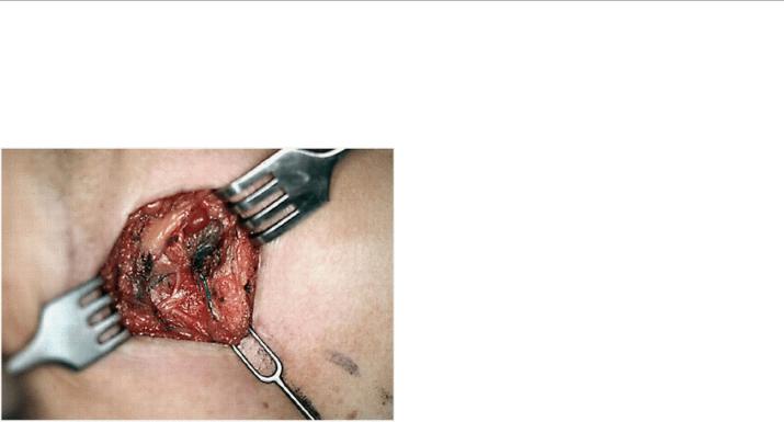
For sentinel node identification with a blue dye, isosulfan or methylene blue are most commonly used. The dye is injected in the breast and massaged; subsequently, the axilla is incised and opened as described for sentinel node identification with a radioactive tracer. Blunt dissection is performed to identify the dye-filled lymphatic tract. This tract is then followed proximally and distally until a blue-stained sentinel node is identified (Fig. 4-1). If more than one dye-filled lymphatic tract is identified, each is followed until a blue node is identified. Blue-stained sentinel nodes are removed in a fashion similar to that used to remove sentinel nodes identified using a radioactive tracer. Another colored tracer in current use is indocyanine green; in cases in which this tracer is used, all nodes with fluorescent tracer uptake must be identified.
Identification Using a Dual-Tracer Approach
The majority of sentinel lymphadenectomy procedures utilize a dual-tracer technique.6 If both a radioactive tracer and blue dye have been injected, nodes that are
P.69 radioactive and/or stained blue are considered sentinel nodes. All blue-stained nodes should be assessed with a gamma detection probe for radioactivity, and all radioactive nodes that are removed should be assessed for the presence of blue dye. Some nodes may only be identified by one modality, as studies show that the procedure is
the most accurate when dual tracer technique is utilized.6
FIGURE 4-1 Sentinel node procedure demonstrating blue lymphatic leading to blue sentinel node. (Photo courtesy of Sarah Blair, MD, and Marek Dobke, MD.)
Identification Using Palpation of the Axilla
As a component of sentinel lymphadenectomy, careful palpation of the level I and II axilla is essential to guiding the complete removal of all sentinel nodes. Nodes that feel abnormal on palpation should be categorized as sentinel nodes and removed regardless of whether they are radioactive or stained blue.7
2. TECHNIQUE FOR INJECTING LOCALIZING TRACER OR DYE
Recommendation: The site of localizing tracer or dye injection within the affected breast and/or subareolar plexus does not influence the identification of the axillary sentinel node(s).
Type of Data: Multiple single institutional series, small prospective randomized study, and systematic review.
Strength of Recommendation: Consensus of the group is that the evidence is strong.
Rationale
Over the past 15 years, several different techniques and combinations of techniques have been employed for the
injection of radioactive tracer and/or dye for sentinel node identification. Pesek et al5 published the most comprehensive and systematic
P.70
review of injection techniques for sentinel node identification. They concluded that of the seven different techniques and combinations of techniques they studied, including peritumoral (intraparenchymal) injection, subor (peri)areolar injection (Figs. 4-2 and 4-3), subdermal but nonintratumoral injection, intratumoral injection, peritumoral/subareolar injection, peritumoral/subdermal injection, and subareolar/subdermal injection, none had a significantly lower false negative rate than another; therefore, they could not justify recommending one
technique over another.5 A multicenter prospective randomized study found that, compared with peritumoral injection
P.71 of a radioactive tracer and blue dye, periareolar injection of a tracer and dye had a higher sentinel lymph node
detection rate and higher within-node dye-tracer concordance.7 One small study investigating radioactive tracer injection in both subareolar and peritumoral locations in individual patients (27 patients total) noted no difference in the number of axillary sentinel nodes identified using subareolar or peritumoral radioactive tracer injection; however, the study did find that peritumoral injection resulted in the localization of extra-axillary drainage sites
(e.g., internal mammary [IM] nodes, paraclavicular nodes).8,9 If extra-axillary sites are of interest, the radioactive tracer should be injected peritumorally. In developing quality metrics for sentinel node mapping (other than false
negative rate, which requires a full axillary node dissection), Quan et al6 did not include the tracer or dye injection site as one of the quality indicators because they found no data supporting one injection location over another. The findings of these studies demonstrate that no specific injection site ensures the lowest false negative rate and highest sentinel node identification rate. However, surgeons should monitor their performance of dye and radioactive tracer injection to ensure that the injection sites they use result in the identification of all sentinel nodes, an average number of removed sentinel nodes greater than 1.9, and a sentinel node positivity
rate of 20% to 30%.6
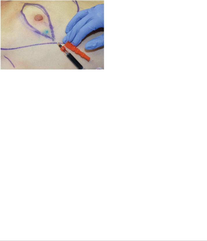
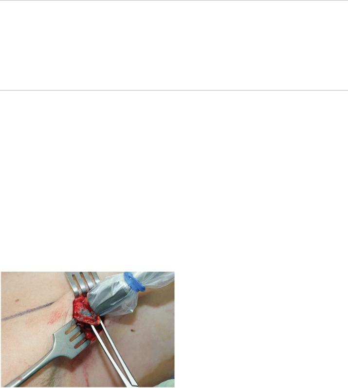
FIGURE 4-2 In cases of dual tracer, all blue nodes should be assessed for radioactivity and all radioactive nodes should be assessed for presence of blue dye. This illustrates checking for radioactivity in a blue node.
FIGURE 4-3 This demonstrates subdermal and perioareolar injection of blue dye in a patient undergoing mastectomy for clinically T2N0 breast cancer (Image courtesy of Sarah Blair MD and Marek Dobke MD)
Radioactive Tracer
Technetium-99m (99mTc)-labeled sulfur colloid is the radioactive tracer most commonly utilized for sentinel node identification; however, newer agents such as 99mTctilmanocept, whose use in sentinel lymphadenectomy has
been approved by the U.S. Food and Drug Administration, are at least equally effective.10 Usually, a nuclear medicine specialist injects the nuclear tracer the day of or the day before surgery, but it has also been injected
by surgeons intraoperatively.11,12 With either approach, the expected average number of sentinel nodes identified is two. The 99mTc-labeled sulfur colloid tracer, which can be filtered or unfiltered, is administered in volumes of 0.1 to 1.0 mL. The tracer can be injected at one site or multiple sites. In the National Surgical Adjuvant Breast and Bowel Project B-32 clinical trial, it was injected into multiple sites around the tumor and in
the dermis above the tumor.13 The tracer’s radioactivity, which typically ranges from 0.25 mCi to 2.5 mCi at injection, depends on the time between the injection and operative intervention; higher doses are given when longer times between injection and surgery are expected.
Vital Blue (or Colored) Dye
Isosulfan blue dye is the most commonly used colored dye for sentinel node identification (Fig. 4-3). At the time of surgery, 1 to 5 mL of undiluted dye is injected into the site or sites of choice, and the breast is massaged for 2 to 5 minutes to travel through the lymphatic vessels. In 0.7% to 1.1% of cases, isosulfan blue dye has been
associated with severe anaphylactic reactions requiring patient resuscitation.13 Methylene blue dye, which has been used as a substitute for isosulfan blue dye, is less expensive and has a lower risk of allergic reaction but has been reported to have more frequent side effects such as skin necrosis, induration, and erythema (14).
Methylene blue dye used for sentinel node identification is commonly diluted in normal saline at a 1:2 ratio.14
P.72
Documentation
All removed sentinel lymph nodes must be accurately labeled in the operating room. Detailed information must be given for each sentinel lymph node, including its description as a radioactive, blue-stained, or both radioactive and blue-stained (“hot and blue”) node; a palpable hard node; or an unstained node found at the termination of a blue-stained lymphatic channel. The location of the node—axillary level I, II, or III or IM—should also be

described. If a radioactive tracer was used for sentinel node identification, documentation for each removed node must include either a 10-second ex vivo radioactive count or the highest radioactive count recorded with the resected node placed on the tip of the gamma detector probe. Accurate documentation of the characteristics of the sentinel node(s) is an important indicator of the quality of sentinel lymphadenectomy (15).
3. PREINCISION EVALUATION OF DRAINAGE PATTERN
Recommendation: For sentinel node identification using a radioactive tracer, preincision skin localization of the area of highest radioactivity facilitates a minimally invasive approach to exposure in the axilla and the identification of any extra-axillary sites of nodal drainage (see Fig. 4-4). Lymphoscintigraphy is not required for sentinel node localization unless an extra-axillary site of drainage is suspected.
Type of Data: Retrospective review.
Strength of Recommendation: No strong data to recommend but low risk with potential benefit associated with
adding this step.
Rationale
In cases involving nodal mapping using a radioactive colloid, surgeons should confirm an area of radioactivity concentration within the axilla and localize the area of highest radioactivity before making the incision for axillary sentinel node biopsy. These preincision steps are important regardless of whether preoperative lymphoscintigraphy is performed or whether the radioactive tracer is used with or without a colored dye. Routine lymphoscintigraphy to identify areas of high radioactivity is not recommended on the basis of a retrospective analysis of prospective data indicating that the procedure does not improve the success rate of sentinel node
identification.15,16,17 However, lymphoscintigraphy should be considered if (1) a surgeon or institution is in the early phases of learning sentinel lymphadenectomy or (2) there is concern that extra-axillary drainage is likely (for example, in a patient undergoing repeat sentinel node mapping for recurrent cancer) (20).
Identification of the Axillary “Hot Spot”
The majority of sentinel lymph nodes are encountered in the level I axilla within a 5-cm diameter circle centered midway between the pectoralis major and latissimus dorsi muscles at the inferior border of the axillary hairline (16). The surgeon should initiate cutaneous scanning prior to incision. Scanning should progress in either
P.73 increasingly larger concentric circles or radially until an area of elevated radioactivity (the “hot spot”) is identified. Marking the skin overlying the hot spot (Fig. 4-4) and confirming a diminution in the radioactive counts moving from the axillary hot spot medially towards the injection site in the breast helps identify the best location for the skin incision in the axilla, minimizing unnecessary blunt dissection of the axilla and disruption of nonsentinel nodes. In cases in which an axillary hot spot cannot be identified and lymphoscintigraphy has not been performed, lymphatic mapping with a radioactive tracer alone may not facilitate the successful removal of all sentinel nodes, and blue dye should be injected for intraoperative lymphatic mapping. The operative record should indicate whether an axillary hot spot was identified before incision.
Internal Mammary Node Removal
For patients in whom preoperative lymphoscintigraphy reveals a radioactive IM lymph node, surgeons may consider pursuing IM node removal, particularly if an axillary sentinel node is not identified or if pathologic analysis reveals the axillary sentinel node to be negative. Preincision scanning for the hot spot in such patients should be performed along the IM node chain, particularly in the second and third intercostal spaces along the lateral border of the sternum. If preoperative lymphoscintigraphy was not performed and no axillary hot spot is present, scanning along the parasternal border, particularly in the second and third intercostal spaces, is useful
to determine whether an IM hot spot is present. Although the prognosis of patients with IM drainage and/or
metastasis18 is worse than that of patients without these conditions, no studies have shown that removing IM
nodes conveys a survival advantage.19 Furthermore, because isolated IM metastasis is rare, IM node removal is
seldom performed.20 Because adjuvant treatment decisions are more likely to be made from molecular panels and not necessarily low-volume regional metastasis, the
P.74
rarity of IM node removal seldom affects adjuvant treatment decisions.21 In addition, IM node removal is
associated with an increased complication rate compared to sentinel node biopsy in the axilla.19
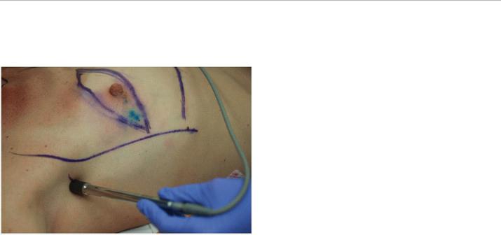
FIGURE 4-4 Demonstrates preincision localization of the area of highest count with the gamma probe in order to plan the axillary incision closest to the in-vivo sentinel nodes for resection. (Image courtesy of Sarah Blair, MD, and Marek Dobke, MD.).
4. NODE REMOVAL TECHNIQUE TO LIMIT SEROMA FORMATION IN ORDER TO PREVENT DELAY TO ADJUVANT TREATMENT
Recommendation: Because no one method or device has been found to result in a lower incidence of seroma formation following sentinel lymphadenectomy, surgeons should use the approach that their experience indicates will have optimal outcomes.
Type of Data: Retrospective or small prospective studies, one systematic review.
Strength of Recommendation: No strong data for specific method to avoid seromas.
Rationale
The morbidity that may result from sentinel node biopsy is minimal compared with that which may result from conventional axillary lymph node dissection. The most frequently observed complication of sentinel lymph node biopsy is seroma, an accumulation of serous fluid under the skin. Persistent seroma can lead to mild or severe
infections and/or delays in adjuvant therapy, thus potentially affecting oncologic outcomes.22,23 In a randomized
controlled trial by McCaul et al,24 sentinel node biopsy had a significantly lower rate of seroma than conventional axillary lymph node dissection did. In their analysis of the outcomes following sentinel node surgery in the
ACOSOG Z0011 trial, Lucci et al reported a seroma rate of only 6% after sentinel lymph node biopsy alone.25
Although seroma is not a serious complication, it can lead to adverse events (including wound infections and dehiscence), prolong recovery time, increase treatment costs, and/or delay the initiation of adjuvant therapy.
Various surgical methods have been utilized in an effort to reduce seroma formation following sentinel node

biopsy. However, whether one or another method results in better outcomes is unclear and no standard surgical technique for seroma prevention or treatment exists. One area of focus for reducing seroma following sentinel lymphadenectomy is lymph vessel sealing and axillary hemostasis, which traditionally have been performed using electrocautery, suture ligation, or vascular clip placement. Each of these techniques, whose use is based on surgeon preference, has its own potential complications. Suture ligation is time-consuming, and the knots used to secure the sutures may slip, causing the ligation to fail. Vascular clips may become dislodged. Electrocautery ligation may cause thermal damage to adjacent tissues. Given these techniques’ potential complications, the use of electrothermal bipolar vessel sealing systems during axillary procedures to prevent seroma formation is becoming increasingly popular, although the method has not been found to significantly improve seroma rates. In a recent Italian study, 116 women were randomly assigned to conventional node dissection performed using a scalpel and monopolar cautery or to node dissection performed using an electrothermal bipolar vessel sealing system. The use
P.75 of a bipolar vessel sealing system offered only marginal advantages over the conventional technique, and the
groups had similar incidences of seroma.26 Furthermore, no study has shown that the use or nonuse of additional devices (e.g., laser scalpels, argon diathermy electrodes, ultrasonic scalpels) during conventional node dissection using a scalpel and monopolar cautery significantly affects seroma incidence. Therefore, surgeons should use the technique with which they have had the most success in obtaining optimal surgical outcomes.
REFERENCES
1.Morton DL, Wen DR, Wong JH, et al. Technical details of intraoperative lymphatic mapping for early stage melanoma. Arch Surg 1992;127(4):392-399.
2.Clough KB, Nasr R, Nos C, et al. New anatomical classification of the axilla with implications for sentinel node biopsy. Br J Surg 2010;97(11):1659-1665.
3.Krag DN, Anderson SJ, Julian TB, et al. Sentinel-lymph-node resection compared with conventional axillary-lymph-node dissection in clinically node-negative patients with breast cancer: overall survival findings from the NSABP B-32 randomised phase 3 trial. Lancet Oncol 2010; 11(10):927-933.
4.Goyal A, Newcombe R, Mansel R. Clinical relevance of multiple sentinel nodes in patients with breast cancer. Brit J Surg 2005;92(4):438-442.
5.Pesek S, Ashikaga T, Krag LE, et al. The false-negative rate of sentinel node biopsy in patients with breast cancer: a meta-analysis. World J Surg 2012;36(9):2239-2251.
6.Quan ML, Wells BJ, McCready D, et al. Beyond the false negative rate: development of quality indicators for sentinel lymph node biopsy in breast cancer. Ann Surg Oncol 2010;17(2):579-591.
7.Cody HS III. Clinical aspects of sentinel node biopsy. Breast Cancer Res 2001;3:104-108.
8.Rodier J-F, Velten M, Wilt M, et al. Prospective multicentric randomized study comparing periareolar and peritumoral injection of radiotracer and blue dye for the detection of sentinel lymph node in breast sparing procedures: FRANSENODE trial. J Clin Oncol 2007;25(24):3664-3669.

9.Fearmonti RM, Gayed IW, Kim E, et al. Intra-individual comparison of lymphatic drainage patterns using subareolar and peritumoral isotope injection for breast cancer. Ann Surg Oncol 2010;17(1):220-227.
10.Wallace AM, Han LK, Povoski SP, et al. Comparative evaluation of [99mTc] tilmanocept for sentinel lymph node mapping in breast cancer patients: results of two phase 3 trials. Ann Surg Oncol
2013;20(8):2590-2599.
11.Johnson CB, Boneti C, Korourian S, et al. Intraoperative injection of subareolar or dermal radioisotope results in predictable identification of sentinel lymph nodes in breast cancer. Ann Surg 2011;254(4):612-618.
12.Stell VH, Flippo-Morton TS, Norton HJ, et al. Effect of intraoperative radiocolloid injection on sentinel lymph node biopsy in patients with breast cancer. Ann Surg Oncol 2009;16(8):2300-2304.
13.Krag DN, Anderson SJ, Julian TB, et al. Technical outcomes of sentinel-lymph-node resection and conventional axillary-lymph-node dissection in patients with clinically node-negative breast cancer: results from the NSABP B-32 randomised phase III trial. Lancet Oncol 2007;8(10):881-888.
14.Zakaria S, Hoskin TL, Degnim AC. Safety and technical success of methylene blue dye for lymphatic mapping in breast cancer. Am J Surg 2008;196(2):228-233.
15.Burak WE Jr, Waler MJ, Yee LD, et al. Routine preoperative lymphoscintigraphy is not necessary prior to sentinel node biopsy for breast cancer. Am J Surg 1999;177(6):445-449.
16.McMasters KM, Wong SL, Tuttle TM, et al. Preoperative lymphoscintigraphy for breast cancer does not improve the ability to identify axillary sentinel lymph nodes. Ann Surg 2000;231(5): 724-731.
17.Goyal A, Newcombe RG, Mansel RE. Role of routine preoperative lymphoscintigraphy in sentinel node biopsy for breast cancer. Eur J Cancer 2005;41(2):238-243.
18.Kong AL, Tereffe W, Hunt KK, et al. Impact of internal mammary lymph node drainage identified by preoperative lymphoscintigraphy on outcomes in patients with stage I to III breast cancer. Cancer
2012;118(24):6287-6296.
19. Lacour J, Bucalossi P, Caceres E, et al. Radical mastectomy versus radical mastectomy plus internal mammary dissection. Ten year results of an international cooperative trial in breast cancer. Cancer
1983;51(10):1941-1943.
P.76
20.Chagpar AB, Kehdy F, Scoggins CR, et al. Effect of lymphoscintigraphy drainage patterns on sentinel lymph node biopsy in patients with breast cancer. Am J Surg 2005;190(4):557-562.
21.Jain S, Gradishar WJ. The application of oncotype DX in early-stage lymph-node-positive disease. Curr Oncol Rep 2014;16(1):1-6.

22.Huang J, Barbera L, Brouwers M, et al. Does delay in starting treatment affect the outcomes of radiotherapy? A systematic review. J Clin Oncol 2003;21(3):555-563.
23.Downing A, Twelves C, Forman D, et al. Time to begin adjuvant chemotherapy and survival in breast cancer patients: a retrospective observational study using latent class analysis. Breast J 2014;20(1):29-36.
24.McCaul J, Aslaam A, Spooner RJ, et al. Etiology of seroma formation in patients undergoing surgery for breast cancer. Breast 2000;9(3):144-148.
25.Lucci A, McCall LM, Beitsch PD, et al. Surgical complications associated with sentinel lymph node dissection (SLND) plus axillary lymph node dissection compared with SLND alone in the American College of Surgeons Oncology Group Trial Z0011. J Clin Oncol 2007;25(24):3657-3663.
26.Nespoli L, Antolini L, Stucchi C, et al. Axillary lymphadenectomy for breast cancer. A randomized controlled trial comparing a bipolar vessel sealing system to the conventional technique. Breast
2012;21(6):739-745.