

- •Preface
- •Contents
- •1: The Eye Scanning
- •1 An Overview of Anatomy
- •2.1 Scanning Method
- •2.2 Section Structure
- •2.4 Clinical Value
- •2.5 Notice
- •3.1 Scanning Method
- •3.2 Sectional Structure
- •3.4 Clinical Value
- •4.1 Scanning Method
- •4.2 Sectional Structure
- •4.4 Clinical Value
- •5.1 Scanning Method
- •5.2 Sectional Structure
- •5.4 Clinical Value
- •6.1 Scanning Method
- •6.2 Sectional Structure
- •6.4 Clinical Value
- •2: Transcranial Ultrasonography
- •1.1 Scanning Method
- •1.2 Section Structure
- •1.4 Clinical Application Value
- •2.1 Scanning Method
- •2.2 Section Structure
- •2.4 Clinical Application Value
- •3.1 Scanning Method
- •3.2 Section Structure
- •3.4 The Clinical Application Value
- •4.1 Scanning Method
- •4.2 Section Structure
- •4.4 The Clinical Application Value
- •5.1 Scanning Method
- •5.2 Section Structure
- •5.4 Clinical Application Value
- •6.1 Scanning Method
- •6.2 Section Structure
- •6.4 Clinical Application Value
- •1.1 Scanning Method
- •1.2 Section Structure
- •1.4 Clinical Application Value
- •1.5 Notice
- •2.1 Scanning Method
- •2.2 Section Structure
- •2.3 Clinical Application Value
- •3 Para-parotid Gland (Transverse Scan)
- •3.1 Scanning Method
- •3.2 Section Structure
- •3.3 Clinical Application Value
- •4 Longitudinal Scan of the Submandibular Gland
- •4.1 Scanning Method
- •4.2 Section Structure
- •4.4 Clinical Application Value
- •5.1 Scanning Method
- •5.2 Section Structure
- •5.3 Clinical Application Value
- •4: Thyroid Scanning
- •1.1 Scanning Method
- •1.2 Section Structure
- •2 The Longitudinal View of the Thyroid
- •2.1 Scanning Method
- •2.2 Section Structure
- •2.4 The Clinical Application Value
- •3.1 Scanning Method
- •3.2 Section Structure
- •3.3 The Clinical Application Value
- •4 Color Doppler Flow Image of the Thyroid
- •4.1 Scanning Method
- •4.2 Section Structure
- •4.3 The Clinical Application Value
- •5.1 Scanning Method
- •5.2 Section Structure
- •5.4 The Clinical Application Value
- •6.1 Scanning Method
- •6.2 Section Structure
- •6.4 The Clinical Application Value
- •7 Doppler Spectrum Flow Imaging of the Inferior Thyroid Artery
- •7.1 Scanning Method
- •7.2 Section Structure
- •7.4 The Clinical Application Value
- •5: The Breast Scanning
- •1.1 Scanning Method
- •1.2 Section Structures
- •1.4 Clinical Application Value
- •2.1 Scanning Method
- •2.2 Section Structures
- •2.3 Clinical Application Value
- •3.1 Scanning Method
- •3.2 Section Structures
- •3.4 Clinical Section Value
- •4.1 Scanning Method
- •4.2 Section Structures
- •4.3 Clinical Application Value
- •5.1 Scanning Method
- •5.2 Section Structures
- •5.3 Clinical Application Value
- •6.1 Scanning Method
- •6.2 Section Structures
- •6.3 Clinical Application Value
- •7 Color Doppler Imaging of Normal Breast in Lactating Women
- •7.1 Scanning Method
- •7.2 Section Structures
- •7.3 Clinical Application Value
- •8 Doppler Spectrum of Normal Breast in Lactating Women
- •8.1 Scanning Method
- •8.2 Section Structures
- •8.3 Clinical Application Value
- •9.1 Scanning Method
- •9.2 Section Structures
- •9.3 Clinical Application Value
- •10.1 Scanning Method
- •10.2 Section Structures
- •10.3 Clinical Application Value
- •11.1 Scanning Method
- •11.2 Section Structures
- •11.3 Clinical Value
- •Bibliography
- •6: Echocardiography
- •1.1 Parasternal Left Ventricular Long-Axis View: M-Mode, Echo Pattern of Heart Base (Aortic Root)
- •1.1.1 Scanning Method
- •1.1.2 Section Structure
- •1.1.3 Measuring Method and Normal Value
- •1.1.4 The Clinical Application
- •1.2 Parasternal Left Ventricular Long-Axis-View M-Mode, Echo Pattern of Mitral Valve
- •1.2.1 Scanning Method
- •1.2.2 Section Structure
- •1.2.3 Measuring Method and Normal Value
- •1.2.4 The Clinical Application
- •1.3 The Parasternal Left Ventricular Long-Axis-View M-Mode, Echo Pattern of Left Ventricle
- •1.3.1 Scanning Method
- •1.3.2 Section Structure
- •1.3.3 Measuring Method and Normal Value
- •1.3.4 The Clinical Application
- •1.4 Great Artery Short-Axis View (M-Mode Scan at Pulmonary Valve Level)
- •1.4.1 Scanning Method
- •1.4.2 Section Structure
- •1.4.3 Measuring Method and Normal Value
- •1.4.4 The Clinical Application
- •2.1.1 Scanning Method
- •2.1.2 Section Structure
- •2.1.3 Measuring Method and Normal Value
- •2.1.4 The Clinical Application
- •2.2.1 Scanning Method
- •2.2.2 Section Structure
- •2.2.3 Measuring Method and Normal Value
- •2.2.4 The Clinical Application
- •2.3.1 Scanning Method
- •2.3.2 Section Structure
- •2.3.3 Measuring Method and Normal Value
- •2.3.4 The Clinical Application
- •2.4.1 Scanning Method
- •2.4.2 Section Structure
- •2.4.3 The Clinical Application
- •2.5.1 Scanning Method
- •2.5.2 Section Structure
- •2.5.3 Measuring Method and Normal Value
- •2.5.4 The Clinical Application
- •2.6.1 Scanning Method
- •2.6.2 Section Structure
- •2.6.3 Measuring Method and Normal Value
- •2.6.4 The Clinical Application
- •2.7.1 Scanning Method
- •2.7.2 Section Structure
- •2.7.3 The Clinical Application
- •2.8.1 Scanning Method
- •2.8.2 Section Structure
- •2.8.3 The Clinical Application
- •2.9.1 Scanning Method
- •2.9.2 Section Structure
- •2.9.3 Measuring Method and Normal Value
- •2.9.4 The Clinical Application
- •2.10 Apical Four-Chamber View
- •2.10.1 Scanning Method
- •2.10.2 Section Structure
- •2.10.3 Measuring Method and Normal Value
- •2.10.4 The Clinical Application
- •2.11.1 Scanning Method
- •2.11.2 Section Structure
- •2.11.3 Measuring Method and Normal Value
- •2.11.4 The Clinical Application
- •2.12.1 Scanning Method
- •2.12.2 Section Structure
- •2.12.3 The Clinical Application
- •2.13.1 Scanning Method
- •2.13.2 Section Structure
- •2.13.3 The Clinical Application
- •2.14.1 Scanning Method
- •2.14.2 Scanning Section Structure
- •2.14.3 Measuring Method and Normal Value
- •2.14.4 The Clinical Application
- •2.15 Coronary Sinus View
- •2.15.1 Scanning Method
- •2.15.2 Section Structure
- •2.15.3 Measuring Method and Normal Value
- •2.15.4 The Clinical Application
- •2.16.1 Scanning Method
- •2.16.2 Section Structure
- •2.16.3 Measuring Method and Normal Value
- •2.16.4 The Clinical Application
- •2.17.1 Scanning Method
- •2.17.2 Section Structure
- •2.17.3 The Clinical Application
- •2.18 Apical Five-Chamber View
- •2.18.1 Scanning Method
- •2.18.2 Section Structure
- •2.18.3 Measuring Method and Normal Value
- •2.18.4 The Clinical Application
- •2.19 Short-Axis View of the Inferior Vena Cava
- •2.20.1 Scanning Method
- •2.20.2 Section Structure
- •2.20.3 Measuring Method and Normal Value
- •2.20.4 The Clinical Application
- •2.21.1 Scanning Method
- •2.21.2 Section Structure
- •2.21.3 Measuring Method and Normal Value
- •2.21.4 The Clinical Application
- •2.22.1 Scanning Method
- •2.22.2 Section Structure
- •2.22.3 The Clinical Application
- •2.23 Subxiphoid Four-Chamber View
- •2.23.1 Scanning Method
- •2.23.2 Section Structure
- •2.23.3 Measuring Method and Normal Value
- •2.23.4 The Clinical Application Value
- •2.24 Subxiphoid Two-Atrium View
- •2.24.1 Scanning Methods
- •2.24.2 Section Structure
- •2.24.3 Measuring Method and Normal Value
- •2.24.4 The Clinical Application Value
- •2.25 Subxiphoid Aortic Ventricular View
- •2.25.1 Scanning Methods
- •2.25.2 Section Structure
- •2.25.3 Measuring Method and Normal Value
- •2.25.4 The Clinical Application Value
- •2.26 Subxiphoid Inferior Vena Cava (Long-Axis) View
- •2.26.1 Scanning Methods
- •2.26.2 Section Structure
- •2.26.3 Measuring Method and Normal Value
- •2.26.4 The Clinical Application Value
- •2.27 Subxiphoid Ventricular Short-Axis View
- •2.27.1 Scanning Methods
- •2.27.2 Section Structure
- •2.27.3 Measuring Method and Normal Value
- •2.27.4 The Clinical Application Value
- •2.28 Suprasternal Aortic Arch Short-Axis View
- •2.28.1 Scanning Methods
- •2.28.2 Section Structure
- •2.28.3 Measuring Method and Normal Value
- •2.28.4 The Clinical Application Value
- •2.29 Suprasternal Superior Vena Cava Long-Axis View
- •2.29.1 Scanning Methods
- •2.29.2 Section Structure
- •2.29.3 Measuring Method and Normal Value
- •2.29.4 The Clinical Application Value
- •2.30 Suprasternal Aortic Arch Short-Axis View
- •2.30.1 Scanning Methods
- •2.30.2 Section Structure
- •2.30.3 Measuring Method and Normal Value
- •2.30.4 The Clinical Application Value
- •2.31 Main Pulmonary Artery Long-Axis View by Subxiphoid
- •2.31.1 Scanning Methods
- •2.31.2 Section Structure
- •2.31.3 Measuring Method and Normal Value
- •2.31.4 The Clinical Application Value
- •2.32 Transesophageal Two-Atrium View
- •2.32.1 Scanning Method
- •2.32.2 Section Structure
- •2.32.3 Measuring Method and Normal Value
- •2.32.4 The Clinical Application Value
- •2.33 Transesophageal Left Atrial Appendage View
- •2.33.1 Scanning Methods
- •2.33.2 Section Structure
- •2.33.3 Measuring Method and Normal Value
- •2.33.4 The Clinical Application Value
- •2.34 Transesophageal Aortic Root Long-Axis View
- •2.34.1 Scanning Methods
- •2.34.2 Section Structure
- •2.34.3 Measuring Method and Normal Value
- •2.34.4 The Clinical Application Value
- •3.1.1 Scanning Methods
- •3.1.2 Section Structure
- •3.1.3 Measuring Method and Normal Value
- •3.1.4 The Clinical Application Value
- •3.1.5 Notice
- •3.2.1 Scanning Methods
- •3.2.3 The Clinical Application Value
- •3.3.1 Scanning Method
- •3.3.2 Section Structure
- •3.3.3 Measuring Method and Normal Value
- •3.3.4 The Clinical Application Value
- •3.4.1 Scanning Method
- •3.4.2 Section Structure
- •3.4.3 The Clinical Application Value
- •3.4.4 Notice
- •3.5.1 Scanning Method
- •3.5.2 Section Structure
- •3.5.4 The Clinical Application Value
- •3.5.5 Notice
- •3.6.1 Scanning Method
- •3.6.2 Section Structures
- •3.6.3 The Clinical Application Value
- •3.6.4 Notice
- •3.7.1 Method
- •3.7.2 Section Structure
- •3.7.3 Measuring Method and Normal Value
- •3.7.4 The Clinical Application Value
- •3.7.5 Notice
- •3.8.1 Scanning Method
- •3.8.2 Section Structure
- •3.8.3 The Clinical Application Value
- •3.8.4 Notice
- •7: Vascular System Scanning
- •1 Artery System
- •1.1.1 Scanning Method
- •1.1.2 Sectional Structure
- •1.1.4 Clinical Significance
- •1.2.1 Scanning Method
- •1.2.2 Sectional Structure
- •1.2.3 Measurement Methods
- •1.2.4 Clinical Significance
- •1.3.1 Scanning Method
- •1.3.2 Sectional Structure
- •1.3.4 Clinical Significance
- •1.4.1 Scanning Method
- •1.4.2 Sectional Structure
- •1.4.3 Measurement Methods
- •1.4.4 Clinical Significance
- •1.4.5 Note
- •1.5.1 Scanning Method
- •1.5.2 Sectional Structure
- •1.5.3 Measurement Methods
- •1.5.4 Clinical Significance
- •1.5.5 Note
- •1.6.1 Scanning Method
- •1.6.2 Section Structure
- •1.6.3 Measurement Methods
- •1.6.4 Clinical Significance
- •1.7 Doppler Spectrum of the External Carotid Artery
- •1.7.1 Scanning Method
- •1.7.2 Section Structure
- •1.7.3 Measurement Methods
- •1.7.4 Clinical Significance
- •1.8.1 Scanning Method
- •1.8.2 Sectional Structure
- •1.8.3 Measurement Methods
- •1.8.4 Clinical Significance
- •1.9.1 Scanning Method
- •1.9.2 Sectional Structure
- •1.9.3 Measurement Methods
- •1.9.4 Clinical Significance
- •1.10.1 Scanning Method
- •1.10.2 Sectional Structure
- •1.10.3 Measurement Methods
- •1.10.4 Clinical Significance
- •1.11.1 Scanning Method
- •1.11.2 Sectional Structure
- •1.11.3 Clinical Significance
- •1.12.1 Scanning Method
- •1.12.2 Sectional Structure
- •1.12.3 Measurement Methods
- •1.12.4 Clinical Significance
- •1.12.5 Note
- •1.13.1 Scanning Method
- •1.13.2 Sectional Structure
- •1.13.3 Measurement Methods
- •1.13.4 Clinical Significance
- •1.14.1 Scanning Method
- •1.14.2 Sectional Structure
- •1.14.3 Measurement Methods
- •1.14.4 Clinical Significance
- •2.1.1 Scanning Method
- •2.1.2 Sectional Structure
- •2.1.3 Measurement Methods
- •2.1.4 Clinical Significance
- •2.1.5 Note
- •2.2.1 Scanning Method
- •2.2.2 Sectional Structure
- •2.2.3 Measurement Methods
- •2.2.4 Clinical Significance
- •2.2.5 Note
- •2.3.1 Scanning Method
- •2.3.2 Sectional Structure
- •2.3.3 Measurement Methods
- •2.3.4 Clinical Significance
- •2.4.1 Scanning Method
- •2.4.2 Sectional Structure
- •2.4.3 Measurement Methods
- •2.4.4 Clinical Significance
- •2.5.1 Scanning Method
- •2.5.2 Section Structure
- •2.5.3 Measuring Method
- •2.5.4 Clinical significance
- •2.6.1 Scanning Method
- •2.6.2 Sectional Structure
- •2.6.3 Measurement Methods
- •2.6.4 Clinical Significance
- •2.7.1 Scanning Method
- •2.7.2 Sectional Structure
- •2.7.3 Measurement Methods
- •2.7.4 Clinical significance.
- •2.8.1 Scanning Method
- •2.8.2 Sectional Structure
- •2.8.3 Measurement Methods
- •2.8.4 Clinical Significance
- •2.9.1 Scanning Method
- •2.9.2 Sectional Structure
- •2.9.3 Measurement Methods
- •2.9.4 Clinical Significance
- •2.10.1 Scanning Method
- •2.10.2 Sectional Structure
- •2.10.3 Measurement Methods
- •2.10.4 Clinical Significance
- •2.11.1 Scanning Method
- •2.11.2 Sectional Structure
- •2.11.3 Measurement Methods
- •2.11.4 Clinical Significance
- •2.12.1 Sectional Structure
- •2.12.2 Measurement Methods
- •2.12.3 Clinical Significance
- •2.13.1 Sectional Structure
- •2.13.2 Measurement Methods
- •2.13.3 Clinical Significance
- •2.14.1 Scanning Method
- •2.14.2 Sectional Structure
- •2.14.3 Measurement Methods
- •2.14.4 Clinical Significance
- •2.15.1 Scanning Method
- •2.15.2 Sectional Structure
- •2.15.3 Measurement Methods
- •2.15.4 Clinical Significance
- •2.16.1 Scanning Method
- •2.16.2 Sectional Structure
- •2.16.3 Measurement Methods
- •2.16.4 Clinical Significance
- •2.17.1 Scanning Method
- •2.17.2 Sectional Structure
- •2.17.3 Measurement Methods
- •2.17.4 Clinical Significance
- •2.18.1 Scanning Method
- •2.18.2 Sectional Structure
- •2.18.3 Measurement Methods
- •2.18.4 Clinical Significance
- •2.18.5 Note
- •2.19.1 Scanning Method
- •2.19.2 Sectional Structure
- •2.19.3 Measurement Methods
- •2.19.4 Clinical Significance
- •2.19.5 Note
- •3 Vein System
- •3.1.1 Scanning Method
- •3.1.2 Sectional Structure
- •3.1.3 Measurement Methods
- •3.1.4 Clinical Significance
- •3.2 Transverse scannng of the internal jugular vein
- •3.2.1 Scanning Method
- •3.2.2 Sectional Structure
- •3.2.3 Measurement Methods
- •3.2.4 Clinical Significance
- •3.3.1 Scanning Method
- •3.3.2 Sectional Structure
- •3.3.3 Measurement Methods
- •3.3.4 Clinical Significance
- •3.3.5 Note
- •3.4.1 Scanning Method
- •3.4.2 Sectional Structure
- •3.4.3 Measurement Methods
- •3.4.4 Clinical Significance
- •3.5.1 Scanning Method
- •3.5.2 Sectional Structure
- •3.5.3 Measurement Methods
- •3.5.4 Clinical Significance
- •3.6.1 Scanning Method
- •3.6.2 Sectional Structure
- •3.6.3 Measurement Methods
- •3.6.4 Clinical significance
- •3.7.1 Scanning Method
- •3.7.2 Sectional Structure
- •3.7.3 Clinical Significance
- •3.8.1 Scanning Method
- •3.8.2 Sectional Structure
- •3.8.3 Measurement Methods
- •3.8.4 Clinical Significance
- •3.9.1 Scanning Method
- •3.9.2 Sectional Structure
- •3.9.3 Measurement Methods
- •3.9.4 Clinical Significance
- •3.10.1 Scanning Method
- •3.10.2 Sectional Structure
- •3.10.3 Measurement Methods
- •3.10.4 Clinical Significance
- •3.11.1 Scanning Method
- •3.11.2 Sectional Structure
- •3.11.3 Measurement Methods
- •3.11.4 Clinical Significance
- •3.12.1 Scanning Method
- •3.12.2 Sectional Structure
- •3.12.3 Measurement Methods
- •3.12.4 Clinical Significance
- •3.13.1 Scanning Method
- •3.13.2 Sectional Structure
- •3.13.3 Measurement Methods
- •3.13.4 Clinical Significance
- •3.14.1 Scanning Method
- •3.14.2 Sectional Structure
- •3.14.3 Measurement Methods
- •3.14.4 Clinical Significance
- •3.15.1 Scanning Method
- •3.15.2 Sectional Structure
- •3.15.3 Measurement Methods
- •3.15.4 Clinical Significance
- •3.17.1 Scanning Method
- •3.17.2 Sectional Structure
- •3.17.3 Measurement Methods
- •3.17.4 Clinical Significance
- •3.18.1 Scanning Method
- •3.18.2 Sectional Structure
- •3.18.3 Measurement Methods
- •3.18.4 Clinical Significance
- •3.19.1 Scanning Method
- •3.19.2 Sectional Structure
- •3.19.3 Measurement Methods
- •3.19.4 Clinical Significance
- •3.20.1 Scanning Method
- •3.20.2 Sectional Structure
- •3.20.3 Measurement Methods
- •3.20.4 Clinical Significance
- •3.20.5 Notes
- •1 Liver Scanning
- •1.1.1 Scanning Method
- •1.1.2 Section Structure
- •1.1.3 Measuring Method and Normal
- •1.1.4 The Clinical Application Value
- •1.2 Longitudinal Scanning of the Liver through the Inferior Vena Cava on Subxiphoid
- •1.2.1 Scanning Method
- •1.2.2 Section Structure
- •1.2.3 Measuring Method and Normal
- •1.2.4 Clinical Application Value
- •1.2.5 Notice
- •1.3 Transverse Scan of the Left and Right Liver Through the Porta Hepatis on Subxiphoid
- •1.3.1 Scanning Method
- •1.3.2 Section Structure
- •1.3.3 Measuring Method and Normal
- •1.3.4 Clinical Application Value
- •1.4 Transverse Scanning of the Left Hepatic Lobe Through the Left Portal Vein Branches by Subxiphoid
- •1.4.1 Scanning Method
- •1.4.2 Section Structure
- •1.4.3 Measuring Method and Normal
- •1.4.4 Clinical Application Value
- •1.5.1 Scanning Method
- •1.5.2 Section Structure
- •1.5.3 Measuring Method and Normal
- •1.5.4 The Clinical Application Value
- •1.6.1 Scanning Method
- •1.6.2 Section Structure
- •1.6.3 The Clinical Application Value
- •1.7.1 Scanning Method
- •1.7.2 Section Structure
- •1.7.3 The Clinical Application Value
- •1.8 Longitudinal scanning of the hepatic left lobe and the ligament teres hepatis by subxiphoid
- •1.8.1 Scanning Method
- •1.8.2 Section Structure
- •1.8.3 Measuring Method and Normal
- •1.8.4 The Clinical Application Value
- •1.9 Oblique scanning of the ligament teres and the left liver by subxiphoid
- •1.9.1 Scanning Method
- •1.9.2 Section Structure
- •1.9.3 The Clinical Application Value
- •1.10 Oblique scanning of the liver through the gallbladder and inferior vena cava by the right subcostal margin
- •1.10.1 Scanning Method
- •1.10.2 Section Structure
- •1.10.3 The Clinical Application Value
- •1.11.1 Scanning Method
- •1.11.2 Section Structure
- •1.11.4 Clinical Application Value
- •1.11.5 Notes
- •1.12 Transversely Scanning the Upper Part of the Porta Hepatis
- •1.12.1 Scanning Method
- •1.12.2 Section Structure
- •1.12.3 Measuring Method and Normal
- •1.12.4 Clinical Application Value
- •1.13.1 Scanning Method
- •1.13.2 Section Structure
- •1.13.3 Measuring Method and Normal
- •1.13.4 Clinical Application Value
- •1.14.1 Scanning Method
- •1.14.2 Section Structure
- •1.14.3 Measuring Method and Normal
- •1.14.4 Clinical Application Value
- •1.15 Longitudinal Scanning of the Liver Through the Middle Hepatic Vein on Subxiphoid
- •1.15.1 Scanning Method
- •1.15.2 Section Structure
- •1.15.3 Measuring Method and Normal
- •1.15.4 Clinical Application Value
- •1.16.1 Scanning Method
- •1.16.2 Section Structure
- •1.16.3 Measuring Method and Normal
- •1.16.4 Clinical Application Value
- •1.17 Oblique Scanning of the Right Anterior Liver and the Left Medial Lobe of the Liver by Right Intercostal Space
- •1.17.1 Scanning Method
- •1.17.2 Section Structure
- •1.17.3 The Clinical Application Value
- •1.18.1 Scanning Method
- •1.18.2 Section Structure
- •1.18.3 The Clinical Application Value
- •1.19 Oblique Scanning of the Right Liver Through Right the Portal Vein by the Right Subcostal Space Approach
- •1.19.1 Scanning Method
- •1.19.2 Section Structure
- •1.19.3 Measuring Method and Normal
- •1.19.4 The Clinical Application Value
- •1.20 Longitudinal Scanning of the Right Liver and Right Kidney from the Right Subcostal
- •1.20.1 Scanning Method
- •1.20.2 Section Structure
- •1.20.3 Measuring Method and Normal
- •1.20.4 The Clinical Application Value
- •1.20.5 Notes
- •1.21 Oblique Scanning of the Right Liver Through Right Hepatic Veins on Subxiphoid
- •1.21.1 Scanning Method
- •1.21.2 Section Structure
- •1.21.3 Measuring Method and Normal
- •1.21.4 Clinical Application Value
- •1.22.1 Scanning Method
- •1.22.2 Section Structure
- •1.22.3 Measuring Method and Normal
- •1.22.4 Clinical Application Value
- •1.23.1 Scanning Method
- •1.23.2 Section Structure
- •1.23.3 Measuring Method and Normal
- •1.23.4 Clinical Application Value
- •1.24.1 Scanning Method
- •1.24.2 Section Structure
- •1.24.3 Clinical Application Value
- •1.25 Transverse Scanning of the Right Liver and the Right Kidney from the Right Subcostal
- •1.25.1 Scanning Method
- •1.25.2 Section Structure
- •1.25.3 The Clinical Application Value
- •1.26.1 Scanning Method
- •1.26.2 Section Structure
- •1.26.3 The Clinical Application Value
- •1.27 Longitudinal Scanning of the Common Hepatic Artery and Splenic Artery from the Upper Abdomen
- •1.27.1 Scanning Method
- •1.27.2 Section Structure
- •1.27.3 Measuring Method and the Normal
- •1.27.4 Clinical Application Value
- •1.28 Common Hepatic Artery Blood Flow Spectrum
- •1.28.1 Scanning Method
- •1.28.2 Measuring Method and the Normal
- •1.28.3 Clinical Application Value
- •1.29 Longitudinal Scanning of the Proper Hepatic Artery from Upper Abdomen
- •1.29.1 Scanning Method
- •1.29.2 Section Structure
- •1.29.3 Measuring Method and the Normal
- •1.29.4 Clinical Application Value
- •1.30 The Proper Hepatic Artery Blood Flow Spectrum
- •1.30.1 Scanning Method
- •1.30.2 Measuring Method and the Normal
- •1.30.3 Clinical Application Value
- •1.31 Portal Vein Blood Flow Spectrum from Right Subcostal Margin
- •1.31.1 Scanning Method
- •1.31.2 Section Structure
- •1.31.3 Measuring Method and the Normal
- •1.31.4 Clinical Application Value
- •2.1.1 Scanning Method
- •2.1.2 Section Structure
- •2.1.3 Measuring Method and Normal
- •2.1.4 Clinical Application Value
- •2.1.5 Notice
- •2.2.1 Scanning Method
- •2.2.2 Section Structure
- •2.2.3 Measuring Method and Normal
- •2.2.4 Clinical Application Value
- •2.2.5 Notice
- •2.3.1 Scanning Method
- •2.3.2 Section Structure
- •2.3.3 Clinical Application Value
- •2.4.1 Scanning Method
- •2.4.2 Section Structure
- •2.4.3 Measuring Method and Normal
- •2.4.4 Clinical Application Value
- •2.5.1 Scanning Method
- •2.5.2 Section Structure
- •2.5.3 Measuring Method and Normal
- •2.5.4 Clinical Application Value
- •2.6.1 Scanning Methods
- •2.6.2 Section Structure
- •2.6.4 Clinical Application Value
- •2.7 Longitudinal Scanning of the Intrapancreatic Port and the end Part of the Common Bile Duct by the Right Subcostal and Right Upper Abdomen
- •2.7.1 Scanning Method
- •2.7.2 Section Structure
- •2.7.4 Clinical Application Value
- •2.8 Transverse Scanning of the Common Bile Duct at the Level of the Upper Part of the Pancreatic Head by the Right Subcostal
- •2.8.1 Scanning Method
- •2.8.2 Section Structure
- •2.8.3 Measuring Method and Normal
- •2.8.4 Clinical Application Value
- •2.9 Transverse Scanning of the Middle Segment of the Common Bile Duct at the Level of the Lower Part of the Pancreatic Head by Right Subcostal
- •2.9.1 Scanning Method
- •2.9.2 Section Structure
- •2.9.3 Measuring Method and Normal
- •2.9.4 Clinical Application Value
- •3 The Pancreas
- •3.1.1 Scanning Method
- •3.1.2 Section Structure
- •3.1.3 Measuring Method and Normal
- •3.1.4 Clinical Application Value
- •3.2.1 Scanning Method
- •3.2.2 Section Structure
- •3.2.3 Measuring Method and Normal
- •3.2.4 Clinical Application Value
- •3.3 Transverse Scanning of the Lower Port of the Pancreatic Head by Subxiphoid
- •3.3.1 Scanning Method
- •3.3.2 Section Structure
- •3.3.3 Clinical Application Value
- •3.4.1 Scanning Method
- •3.4.2 Section Structure
- •3.4.3 Measuring Method and Normal
- •3.4.4 Clinical Application Value
- •3.5 Sagittal Scanning of the Pancreatic Body by the Subxiphoid
- •3.5.1 Scanning Method
- •3.5.2 Section Structure
- •3.5.3 Measuring Method and Normal
- •3.5.4 Clinical Application Value
- •3.6.1 Scanning Method
- •3.6.2 Section Structure
- •3.6.3 Measuring Method and Normal
- •3.6.4 Clinical Application Value
- •3.7 Oblique Scanning of the Left Kidney, Spleen and Pancreatic Tail by the Left Intercostal Space
- •3.7.1 Scanning Method
- •3.7.2 Section Structure
- •3.7.3 Clinical Application Value
- •4 Spleen
- •4.1.1 Method
- •4.1.2 Section Structure
- •4.1.3 Measuring Method and Normal
- •4.1.4 Clinical Application Value
- •4.2 The Image of the Accessory Spleen (Splenules) in the Longitudinal Scan of the Spleen by the Left Intercostal
- •4.2.1 Scanning Method
- •4.2.2 Section Structure
- •4.2.3 Measuring Method and Normal
- •4.2.4 Clinical application value
- •4.3.1 Scanning Method
- •4.3.2 Section Structure
- •4.3.3 Clinical Application Value
- •4.4.1 Scanning Method
- •4.4.2 Section Structure
- •4.4.3 Clinical application value
- •4.5.1 Scanning Method
- •4.5.2 Section structure
- •4.5.3 Measuring Method and Normal
- •4.5.4 Clinical Application Value
- •4.6.1 Scanning Method
- •4.6.2 Section Structure
- •4.6.3 Measuring Method and Normal
- •4.6.4 Clinical Application Value
- •5 The Gastrointestinal Scanning
- •5.1.1 Scanning Method
- •5.1.2 Section Structure
- •5.1.3 Measuring Method and Normal
- •5.1.4 The Clinical Application Value
- •5.2 Transverse Scanning of the Lower Segment of the Esophagus
- •5.2.1 Scanning Method
- •5.2.2 Section Structure
- •5.2.3 Measuring Method and Normal
- •5.2.4 Clinical Application Value
- •5.3.1 Scanning Method
- •5.3.2 Section Structure
- •5.3.3 Measuring Method and Normal
- •5.3.4 Clinical Application Value
- •5.4.1 Scanning Method
- •5.4.2 Section Structure
- •5.4.3 Measuring Method and Normal
- •5.4.4 Clinical Application Value
- •5.6 Scanning Method
- •5.6.1 Section Structure
- •5.6.2 Measuring Method and Normal
- •5.6.3 Clinical Application Value
- •5.7 Short Axis Scanning of the Stomach Body by the Upper Abdomen
- •5.7.1 Scanning Method
- •5.7.2 Section Structure
- •5.7.3 Measuring Method and Normal
- •5.7.4 Clinical Application Value
- •5.8.1 Scanning Method
- •5.8.2 Section Structure
- •5.8.3 Measuring Method and Normal
- •5.8.4 Clinical Application Value
- •5.9 Short Axis Scanning of the Stomach Antrum by the Right Upper Abdomen
- •5.9.1 Scanning Method
- •5.9.2 Section Structure
- •5.9.3 Measuring Method and Normal
- •5.9.4 Clinical Application Value
- •5.10.1 Scanning Method
- •5.10.2 Section Structure
- •5.10.3 Measuring Method and Normal
- •5.10.4 Clinical Application Value
- •5.11.1 Scanning Method
- •5.11.2 Section Structure
- •5.11.3 Measuring Method and Normal
- •5.11.4 Clinical Application Value
- •5.12.1 Scanning Method
- •5.12.2 Section Structure
- •5.12.3 Clinical Application Value
- •5.13.1 Scanning Method
- •5.13.2 Section Structure
- •5.13.3 Measuring Method and Normal
- •5.13.4 Clinical Application Value
- •5.14 Longitudinal Scanning of the Ascending Colon from the Right Lower Abdomen Approach
- •5.14.1 Scanning Method
- •5.14.2 Section Structure
- •5.14.3 Measuring Method and Normal
- •5.14.4 Clinical Application Value
- •5.15.1 Scanning Method
- •5.15.2 Section Structure
- •5.15.3 Measuring Method and Normal
- •5.15.4 Clinical Application Value
- •9: Abdominal Vascular Scanning
- •1 Artery System
- •1.1.1 Scanning Method
- •1.1.2 Section Structure
- •1.1.3 Measurement Method and Normal Value
- •1.1.4 Clinical Application Value
- •1.2.1 Scanning Method
- •1.2.2 Section Structure
- •1.2.4 Clinical Application Value
- •1.2.5 Notes
- •1.3 Transverse View of the Abdominal Aorta
- •1.3.1 Scanning Method
- •1.3.2 Section Structure
- •1.3.3 Measurement Method and Normal Value
- •1.3.4 Clinical Application Value
- •1.3.5 Notice
- •1.4 Color Doppler Flow Image of the Abdominal Aorta
- •1.4.1 Scanning Method
- •1.4.2 Section Structure
- •1.4.3 Measurement Method and Normal Value
- •1.4.4 Clinical Application Value
- •1.5 Doppler Spectrum of the Abdominal Aorta
- •1.5.1 Scanning Method
- •1.5.2 Section Structure
- •1.5.3 Measurement Method and Normal Value
- •1.5.4 Clinical Application Value
- •2 Celiac Artery (CA)
- •2.1 Long-Axis View of the Celiac Artery
- •2.1.1 Scanning Method
- •2.1.2 Section Structure
- •2.1.3 Measurement Method and Normal Value
- •2.1.4 Clinical Application Value
- •2.1.5 Notice
- •2.2 Doppler Spectrum of the Celiac Artery
- •2.2.1 Scanning Method
- •2.2.2 Measurement Method and Normal Value
- •2.2.3 Clinical Application Value
- •2.3 Aberrance of the Celiac Artery
- •2.3.1 Scanning Method
- •2.3.2 Section Structure
- •2.3.3 Measurement Method and Normal Value
- •2.3.4 Clinical Application Value
- •3 Hepatic Artery
- •3.1 Longitudinal Scan of the Common Hepatic Artery and Splenic Artery from the Upper Abdomen
- •3.1.1 Scanning Method
- •3.1.2 Section Structure
- •3.1.3 Measurement Method and Normal Value
- •3.1.4 Clinical Application Value
- •3.2 Common Hepatic Artery Blood Flow Spectrum
- •3.2.1 Scanning Method
- •3.2.2 Measurement Method and Normal Value
- •3.2.3 Clinical Application Value
- •3.3 Longitudinal Scan of the Proper Hepatic Artery from the Upper Abdomen
- •3.3.1 Scanning Method
- •3.3.2 Section Structure
- •3.3.4 Clinical Application Value
- •3.4 Proper Hepatic Artery Blood Flow Spectrum
- •3.4.1 Scanning Method
- •3.4.2 Measurement Method and Normal Value
- •3.4.3 Clinical Application Value
- •4 Superior Mesenteric Artery
- •4.1.1 Scanning Method
- •4.1.2 Section Structure
- •4.1.4 Clinical Application Value
- •4.2 Transverse Section of the Superior Mesenteric Artery
- •4.2.1 Scanning Method
- •4.2.2 Section Structure
- •4.2.3 Measurement Method and Normal Value
- •4.2.4 Clinical Application Value
- •4.3 Long-Axis View of the Superior Mesenteric Artery
- •4.3.1 Scanning Method
- •4.3.2 Section Structure
- •4.3.3 Measurement Method and Normal Value
- •4.3.4 Clinical Application Value
- •4.4 Doppler Spectrum of the Superior Mesenteric Artery
- •4.4.1 Scanning Method
- •4.4.2 Section Structure
- •4.4.3 Measurement Method and Normal Value
- •4.4.4 Clinical Application Value
- •4.4.5 Notice
- •5 Renal Artery
- •5.1.1 Scanning Method
- •5.1.2 Section Structure
- •5.1.3 Measurement Method and Normal Value
- •5.1.4 Clinical Application Value
- •5.1.5 Notice
- •5.2 Coronal Section at the Lumbar Region: Longitudinal View of the Renal Artery
- •5.2.1 Scanning Method
- •5.2.2 Section Structure
- •5.2.3 Measurement Method and Normal Value
- •5.2.4 Clinical Application Value
- •5.3 Transverse Section at the Right Subcostal Region: Longitudinal View of the Right Renal Artery and Vein
- •5.3.1 Scanning Method
- •5.3.2 Section Structure
- •5.3.3 Measurement Method and Normal Value
- •5.3.4 Clinical Application Value
- •5.4 Coronal Section of the Kidney: Evaluation of Intrarenal Artery Branches
- •5.4.1 Scanning Method
- •5.4.2 Section Structure
- •5.4.3 Measurement Method and Normal Value
- •5.4.4 Clinical Application Value
- •5.5 Doppler Spectrum of the Main Renal Artery and Branches
- •5.5.1 Scanning Method
- •5.5.2 Section Structure
- •5.5.3 Measurement Method and Normal Value
- •5.5.4 Clinical Application Value
- •5.6 Measurement of the AC and AT in Different Types of Normal Spectrum
- •5.6.1 Scanning Method
- •5.6.2 Section Structure
- •5.6.3 Measurement Method and Normal Value
- •5.6.4 Clinical Application Value
- •5.7 Accessory Renal Artery
- •5.7.1 Scanning Method
- •5.7.2 Section Structure
- •5.7.3 Measurement Method and Normal Value
- •5.7.4 Clinical Application Value
- •5.8 Congenital Small Renal Artery
- •5.8.1 Scanning Method
- •5.8.2 Section Structure
- •5.8.3 Measurement Method and Normal Value
- •5.8.4 Clinical Application Value
- •6 Common Iliac Artery
- •6.1.1 Scanning Method
- •6.1.2 Section Structure
- •6.1.3 Measurement Method and Normal Value
- •6.1.4 Clinical Application Value
- •7 Inferior Vena Cava
- •7.1.1 Scanning Method
- •7.1.2 Section Structure
- •7.1.3 Measurement Method and Normal Value
- •7.1.4 Clinical Application Value
- •7.2.1 Scanning Method
- •7.2.2 Section Structure
- •7.2.3 Measurement Method and Normal Value
- •7.2.4 Clinical Application Value
- •7.2.5 Notes
- •7.3 Doppler Spectrum of the Inferior Vena Cava
- •7.3.1 Scanning Method
- •7.3.2 Section Structure
- •7.3.3 Measurement Method and Normal Value
- •7.3.4 Clinical Application Value
- •8 Superior Mesenteric Vein
- •8.1 Transverse View of the Superior Mesenteric Vein
- •8.1.1 Scanning Method
- •8.1.2 Section Structure
- •8.1.3 Measurement Method and Normal Value
- •8.1.4 Clinical Application Value
- •8.2 Long-Axis View of the Superior Mesenteric Vein
- •8.2.1 Scanning Method
- •8.2.2 Section Structure
- •8.2.3 Measurement Method and Normal Value
- •8.2.4 Clinical Application Value
- •9 Renal Vein
- •9.1.1 Scanning Method
- •9.1.2 Section Structure
- •9.1.3 Measurement Method and Normal Value
- •9.1.4 Clinical Application Value
- •9.2 Longitudinal Section of the Right Renal Vein: Transverse Section of the Right Subcostal Region
- •9.2.1 Scanning Method
- •9.2.2 Section Structure
- •9.2.4 Clinical Application Value
- •9.3 Longitudinal Scanning of the Common Iliac Vein
- •9.3.1 Scanning Method
- •9.3.2 Section Structure
- •9.3.3 Measurement Method
- •9.3.4 Clinical Application Value
- •9.4 Color Doppler Flow Imaging of the Common Iliac Vein
- •10 Hepatic Vein
- •10.1.1 Scanning Method
- •10.1.2 Section Structure
- •10.1.3 Measurement Method and Normal Value
- •10.1.4 Clinical Application Value
- •10.1.5 Notice
- •1 Kidney Scanning
- •1.1.1 Scanning Method
- •1.1.2 Section Structure
- •1.1.3 Measuring Method and Normal
- •1.1.4 The Clinical Application Value
- •1.1.5 Notice
- •1.2.1 Scanning Method
- •1.2.2 Section Structure
- •1.2.3 Measuring Method and Normal
- •1.2.4 The Clinical Application Value
- •1.3.1 Scanning Method
- •1.3.2 Section Structure
- •1.3.3 Measuring Method and Normal
- •1.3.4 The Clinical Application Value
- •1.3.5 Notices
- •1.4 Longitudinal Plane of the Right Kidney from the Right Back
- •1.4.1 Scanning Method
- •1.4.2 Section Structure
- •1.4.3 Measuring Method and Normal
- •1.4.4 The Clinical Application Value
- •1.5 Coronal View of the Left Kidney Through the Lateral Lumbar Region
- •1.5.1 Scanning Method
- •1.5.2 Section Structure
- •1.5.3 Measuring Method and Normal
- •1.5.4 The Clinical Application Value
- •1.6 Transverse Plane of the Left Kidney in the Left Upper Abdomen
- •1.6.1 Scanning Method
- •1.6.2 Section Structure
- •1.6.3 Measuring Method and Normal
- •1.6.4 The Clinical Application Value
- •1.7 Longitudinal Plane of the Left Kidney from the Back
- •1.7.1 Scanning Method
- •1.7.2 Section Structure
- •1.7.3 Measuring Method and Normal
- •1.7.4 The Clinical Application Value
- •1.8 Duplex Pelvis Shown in the Longitudinal Plane of the Kidney
- •1.8.1 Scanning Method
- •1.8.2 Section Structure
- •1.8.3 The Clinical Application Value
- •2 Bladder Scanning
- •2.1 Longitudinal Plane of the Bladder from Suprapubic Symphysis
- •2.1.1 Scanning Method
- •2.1.2 Section Structure
- •2.1.3 Measuring Method and Normal
- •2.1.4 The Clinical Application Value
- •2.2 Transverse Plane of the Bladder from Suprapubic Symphysis
- •2.2.1 Scanning Method
- •2.2.2 Measuring Method and Normal
- •2.2.3 Section Structure
- •2.2.4 The Clinical Application Value
- •2.2.5 Notice
- •3 Prostate Scanning
- •3.1 Sagittal Aspect of the Prostate from the Pubic Symphysis
- •3.1.1 Scanning Method
- •3.1.2 Section Structure
- •3.1.3 Measuring Method and Normal
- •3.1.4 The Clinical Application Value
- •3.2 Semicoronal View of the Prostate from the Suprapubic Symphysis
- •3.2.1 Scanning Method
- •3.2.2 Section Structure
- •3.2.3 Measuring Method and Normal
- •3.2.4 The Clinical Application Value
- •3.3 Transverse (Axial) Plane of the Prostate by Transrectal Ultrasound (TRUS)
- •3.3.1 Scanning Method
- •3.3.2 Section Structure
- •3.3.3 Measuring Method and Normal
- •3.3.4 The Clinical Application Value
- •3.4 Longitudinal Plane of the Prostate by Transrectal Ultrasound (TRUS)
- •3.4.1 Scanning Method
- •3.4.2 Section Structure
- •3.4.3 Measuring Method and Normal
- •3.4.4 The Clinical Application Value
- •3.5 Transverse Aspect of the Seminal Vesicle from the Pubic Symphysis
- •3.5.1 Scanning Method
- •3.5.2 Section Structure
- •3.5.3 Measuring Method and Normal
- •3.5.4 The Clinical Application Value
- •4.1 Sagittal View of the Testis
- •4.1.1 Scanning Method
- •4.1.2 Section Structure
- •4.1.3 Measuring Method and Normal
- •4.1.4 The Clinical Application Value
- •4.2 Axial View of the Testis
- •4.2.1 Scanning Method
- •4.2.2 Section Structure
- •4.2.3 Measuring Method and Normal
- •4.2.4 The Clinical Application Value
- •4.3 Axial View of Both Testes
- •4.3.1 Scanning Method
- •4.3.2 Section Structure
- •4.3.3 Measuring Method and Normal
- •4.3.4 The Clinical Application Value
- •4.4 Longitudinal Plane of the Head of the Epididymis
- •4.4.1 Scanning Method
- •4.4.2 Section Structure
- •4.4.3 Measuring Method and Normal
- •4.4.4 The Clinical Application Value
- •4.5 Longitudinal Plane of the Tail of the Epididymis
- •4.5.1 Scanning Method
- •4.5.2 Section Structure
- •4.5.3 Measuring Method and Normal
- •4.5.4 The Clinical Application Value
- •4.6 Longitudinal Plane of Spermatic Cord
- •4.6.1 Scanning Method
- •4.6.2 Section Structure
- •4.6.3 Measuring Method and Normal
- •4.6.4 The Clinical Application Value
- •1 Retroperitoneal Space
- •1.1.1 Scanning Method
- •1.1.2 Section Structure
- •1.1.3 Clinical Application Value
- •1.2.1 Scanning Method
- •1.2.2 Section Structure
- •1.2.3 Clinical Application Value
- •1.3.1 Scanning Method
- •1.3.2 Section Structure
- •1.3.3 Clinical Application Value
- •1.4.1 Scanning Method
- •1.4.2 Section Structure
- •1.4.3 The Clinical Application Value
- •1.5.1 Scanning Method
- •1.5.2 Section Structure
- •1.5.3 Measuring Method and Normal
- •1.5.4 The Clinical Application Value
- •1.6.1 Scanning Method
- •1.6.2 Section Structure
- •1.6.3 Clinical Application Value
- •2 Adrenal Gland
- •2.1.1 Scanning Method
- •2.1.2 Section Structure
- •2.1.3 Measuring Method and Normal
- •2.1.4 Clinical Application Value
- •2.2.1 Scanning Method
- •2.2.2 Section Structure
- •2.2.3 Measuring Method and Normal
- •2.2.4 Clinical Application Value
- •2.2.5 Notice
- •12: Gynecologic Ultrasound Scanning
- •1.1 Longitudinal Section of the Childhood Uterus
- •1.1.1 Method
- •1.1.2 Section Structure
- •1.1.3 Measuring Method and Normal
- •1.1.4 The Clinical Application Value
- •1.2.1 Method
- •1.2.2 Section Structure
- •1.2.3 Measuring Method and Normal
- •1.2.4 The Clinical Application Value
- •1.3 Longitudinal Section of the Anteposition Uterus in Fertile Woman
- •1.3.1 Method
- •1.3.2 Section Structure
- •1.3.3 Measuring Method and Normal
- •1.3.4 The Clinical Application Value
- •1.4 Transection of Anteposition Uterus in Fertile Woman
- •1.4.1 Method
- •1.4.2 Section Structure
- •1.4.3 Measuring Method and Normal
- •1.4.4 The Clinical Application Value
- •1.5 Longitudinal Section of Mesoposition Uterus in Fertile Woman
- •1.5.1 Method
- •1.5.2 Section Structure
- •1.5.3 Measuring Method and Normal
- •1.5.4 The Clinical Application Value
- •1.6 Transection of Mesoposition Uterus in Fertile Woman
- •1.6.1 Method
- •1.6.2 Section Structure
- •1.6.3 Measuring Method and Normal
- •1.6.4 The Clinical Application Value
- •1.7 Longitudinal Section of the Retroposition Uterus in Fertile Woman
- •1.7.1 Method
- •1.7.2 Section Structure
- •1.7.3 Measuring Method and Normal
- •1.7.4 The Clinical Application Value
- •1.8 Transection of the Retroposition Uterus in Fertile Woman
- •1.8.1 Method
- •1.8.2 Section Structure
- •1.8.3 Measuring Method and Normal
- •1.8.4 The Clinical Application Value
- •1.9 Longitudinal Section of Postmenopausal Gerontism Uterus
- •1.9.1 Method
- •1.9.2 Section Structure
- •1.9.3 Measuring Method and Normal
- •1.9.4 The Clinical Application Value
- •1.10 Transection of Postmenopausal Gerontism Uterus
- •1.10.1 Method
- •1.10.2 Section Structure
- •1.10.3 Measuring Method and Normal
- •1.10.4 The Clinical Application Value
- •2.1 Longitudinal Section of the Anteposition Uterus in Fertile Woman
- •2.1.1 Method
- •2.1.2 Section Structure
- •2.1.3 Measuring Method and Normal
- •2.1.4 The Clinical Application Value
- •2.2 Transection of the Anteposition Uterus in Fertile Woman
- •2.2.1 Method
- •2.2.2 Section Structure
- •2.2.3 Measuring Method and Normal
- •2.2.4 The Clinical Application Value
- •2.3 Longitudinal Section of the Mesoposition Uterus in Fertile Woman
- •2.3.1 Method
- •2.3.2 Section Structure
- •2.4 Coronal Section of the Mesoposition Uterus in Fertile Woman
- •2.4.1 Method
- •2.4.2 Section Structure
- •2.5 Longitudinal Section of the Retroposition Uterus in Fertile Woman
- •2.5.1 Method
- •2.5.2 Section Structure
- •2.5.3 The Clinical Application Value
- •2.6 Transection of Retroposition Uterus in Fertile Woman
- •2.6.1 Method
- •2.6.2 Section Structure
- •2.6.3 The Clinical Application Value
- •2.7.1 Method
- •2.7.2 Section Structure
- •2.7.3 Measuring Method and Normal
- •2.7.4 The Clinical Application Value
- •2.8.1 Method
- •2.8.2 Section Structure
- •2.8.3 Measuring Method and Normal
- •2.8.4 The Clinical Application Value
- •2.9 Longitudinal Section of Postmenopausal Gerontism Uterus
- •2.9.1 Method
- •2.9.2 Section Structure
- •2.9.3 Measuring Method and Normal
- •2.9.4 The Clinical Application Value
- •2.10 Transection of the Postmenopausal Gerontism Uterus
- •2.10.1 Method
- •2.10.2 Section Structure
- •2.10.3 Measuring Method and Normal
- •2.10.4 The Clinical Application Value
- •3.1 Childhood Ovary
- •3.1.1 Method
- •3.1.2 Section Structure
- •3.1.3 Measuring Method and Normal
- •3.1.4 The Clinical Application Value
- •3.2.1 Method
- •3.2.2 Section Structure
- •3.2.3 Measuring Method and Normal
- •3.2.4 The Clinical Application Value
- •3.3 Luteal Phase Ovary Scan in Fertile Woman
- •3.3.1 Method
- •3.3.2 Section Structure
- •3.3.3 Measuring Method and Normal
- •3.3.4 The Clinical Application Value
- •3.4.1 Method
- •3.4.2 Section Structure
- •3.4.3 Measuring Method and Normal
- •3.4.4 The Clinical Application Value
- •3.5.1 Method
- •3.5.2 Section Structure
- •3.5.3 Measuring Method and Normal
- •3.5.4 The Clinical Application Value
- •4.1.1 Method
- •4.1.2 Section Structure
- •4.1.3 Measuring Method and Normal
- •4.1.4 The Clinical Application Value
- •4.2.1 Method
- •4.2.2 Section Structure
- •4.2.3 Measuring Method and Normal
- •4.2.4 The Clinical Application Value
- •4.3 Ovary Corpus Luteum in Fertile Woman
- •4.3.1 Method
- •4.3.2 Section Structure
- •4.3.3 Measuring Method and Normal
- •4.3.4 The Clinical Application Value
- •4.4 Ovary in Postmenopause-Phase Women
- •4.4.1 Method
- •4.4.2 Section Structure
- •4.4.3 Measuring Method and Normal
- •4.4.4 The Clinical Application Value
- •13: Normal Pregnancy
- •1 First Trimester Scanning
- •1.1.1 Scanning Method
- •1.1.2 Section Structures
- •1.1.3 Measuring Method and Normal Reference Values
- •1.1.4 Clinical Significance
- •1.2.1 Scanning Method
- •1.2.2 Section Structures
- •1.2.3 Measuring Method and Normal Reference Values
- •1.2.4 Clinical Significance
- •1.3.1 Scanning Method
- •1.3.2 Section Structures
- •1.3.3 Measuring Method
- •1.3.4 Clinical Significance
- •1.4.1 Scanning Method
- •1.4.2 Section Structures
- •1.4.3 Measuring Method
- •1.4.4 Clinical Significance
- •1.5.1 Scanning Method
- •1.5.2 Section Structures
- •1.5.3 Measuring Method
- •1.5.4 Clinical Significance
- •1.6.1 Scanning Method
- •1.6.2 Section Structures
- •1.6.3 Measuring Method
- •1.6.4 Clinical Significance
- •1.7.1 Scanning Method
- •1.7.2 Section Structures
- •1.7.3 Measuring Method
- •1.7.4 Clinical Significance
- •1.8.1 Scanning Method
- •1.8.2 Section Structures
- •1.8.3 Measuring Method
- •1.8.4 Clinical Significance
- •1.9.1 Scanning Method
- •1.9.2 Section Structures
- •1.9.3 Measuring Method
- •1.9.4 Clinical Significance
- •1.9.5 Normal Reference Values
- •1.10 Nuchal Translucency (NT)
- •1.10.1 Scanning Method
- •1.10.2 Section Structures
- •1.10.3 Measuring Method
- •1.10.4 Clinical Significance
- •1.10.5 Normal Reference Values
- •3.1.1 Cephalic Presentation of Fetus
- •3.1.2 Breech Presentation of Fetus
- •3.1.3 Scanning Methods
- •3.1.4 Section Structures
- •3.1.5 Clinical Significance
- •3.2.3 Scanning Method
- •3.2.4 Section Structures
- •3.2.5 Clinical Significance
- •4.2.1 Scanning Method
- •4.2.2 Section Structures
- •4.2.3 Measuring Method
- •4.2.4 Clinical Significance
- •4.2.5 Normal Reference Values
- •4.3.1 Scanning Method
- •4.3.2 Section Structures
- •4.3.3 Measuring Method
- •4.3.4 Clinical Significance
- •4.3.5 Normal Reference Values
- •4.6.1 Scanning Method
- •4.6.2 Section Structures
- •4.6.3 Measuring Method
- •4.6.4 Clinical Significance
- •4.6.5 Normal Reference Values
- •4.9.1 Scanning Method
- •4.9.2 Section Structures
- •4.9.3 Clinical Significance
- •4.10.1 Scanning Method
- •4.10.2 Section Structures
- •4.10.3 Clinical Significance
- •4.12.1 Scanning Method
- •4.12.2 Section Structures
- •4.12.3 Measuring Method
- •4.12.4 Clinical Significance
- •4.12.5 Normal Reference Values
- •4.13.1 Scanning Method
- •4.13.2 Section Structures
- •4.13.3 Measuring Method
- •4.13.4 Clinical Significance
- •4.13.5 Normal Reference Values
- •4.15.1 Scanning Method
- •4.15.2 Section Structures
- •4.15.3 Measuring Method
- •4.15.4 Clinical Significance
- •4.15.5 Normal Reference Values
- •4.16 Short Axis View of Fetal Heart
- •4.17.1 Scanning Method
- •4.17.2 Section Structures
- •4.17.3 Measuring Method
- •4.17.4 Clinical Significance
- •4.18 Scanning of Fetal Lung
- •4.18.1 Scanning Method
- •4.18.2 Section Structures
- •4.18.3 Clinical Significance
- •4.19.1 Scanning Method
- •4.19.2 Section Structures
- •4.19.3 Clinical Significance
- •4.20.1 Scanning Method
- •4.20.2 Section Structures
- •4.20.3 Measuring Method
- •4.20.4 Clinical Significance
- •4.20.5 Normal Reference Values
- •4.22.1 Scanning Method
- •4.22.2 Section Structures
- •4.22.3 Clinical Significance
- •4.25.1 Scanning Method
- •4.25.2 Section Structures
- •4.25.3 Measuring Method
- •4.25.4 Clinical Significance
- •4.28.1 Scanning Method
- •4.28.2 Section Structures
- •4.28.3 Clinical Significance
- •4.31.1 Scanning Method
- •4.31.2 Section Structures
- •4.31.3 Measuring Method
- •4.31.4 Clinical Significance
- •Placenta
- •4.32 Placenta of Grade I
- •4.33 Placenta of Grade II
- •4.34 Placenta of Grade III
- •4.34.1 Scanning Method
- •4.34.2 Section Structures
- •4.34.3 Measuring Method
- •4.34.4 Clinical Significance
- •4.35 Low-Lying Placenta
- •4.36 Placenta Previa
- •4.36.1 Scanning Method
- •4.36.2 Section Structures
- •4.36.3 Measuring Method
- •4.36.4 Clinical Significance
- •Amniotic Fluid
- •4.37 Amniotic Fluid Depth
- •4.37.1 Scanning Method
- •4.37.2 Section Structures
- •4.37.3 Measuring Method
- •4.37.4 Clinical Significance
- •4.37.5 Normal Reference Values
- •4.38 Amniotic Fluid Index (AFI)
- •4.38.1 Scanning Method
- •4.38.2 Section Structures
- •4.38.3 Measuring Method
- •4.38.4 Clinical Significance
- •4.38.5 Normal Reference Values
- •5.1 Umbilical Artery
- •Scanning Method
- •Section Structures
- •Measuring Method
- •Clinical Significance
- •Normal Reference Values
- •5.2 Middle Cerebral Artery (MCA)
- •Scanning Method
- •Section Structures
- •Measuring Method
- •Clinical Significance
- •Normal Reference Values
- •5.3 Ductus Venous (DV)
- •5.3.1 Position of the Ductus Venous
- •5.3.3 Doppler Measurement of DV
- •Scanning Method
- •Section Structures
- •Measuring Method
- •Clinical Significance
- •Normal Reference Values
- •Bibliography
- •1.1 Scanning Method
- •1.2 Section Structure
- •1.3 Measuring Method
- •1.4 The Clinical Application Value
- •2.1 Scanning Method
- •2.2 Section Structure
- •2.3 Measuring Method
- •2.4 The Clinical Application Value
- •3.1 Scanning Method
- •3.2 Section Structure
- •3.3 Measuring Method
- •3.4 The Clinical Application Value
- •4 Skin and Subcutaneous Tissue Sonogram in the Face of the Young Women
- •4.1 Method
- •4.2 Section Structure
- •4.3 Measuring Method
- •4.4 The Clinical Application Value
- •5.1 Method
- •5.2 Section Structure
- •5.4 The Clinical Application Value
- •6.1 Method
- •6.2 Section Structure
- •6.3 The Clinical Application Value
- •7.1 Method
- •7.2 Section Structure
- •7.3 Measuring Method
- •7.4 The Clinical Application Value
- •8 Neck Skin and Subcutaneous Tissue Scanning in the Young Women
- •8.1 Method
- •8.2 Section Structure
- •8.4 The Clinical Application Value
- •9 Thoracic Skin and Subcutaneous Tissue Scanning
- •9.1 Method
- •9.2 Section Structure
- •9.4 The Clinical Application Value
- •10 Skin and subcutaneous tissue scanning in the middle line of the upper abdomen
- •10.1 Method
- •10.2 Section Structure
- •10.4 The Clinical Application Value
- •11.1 Method
- •11.2 Section Structure
- •11.4 The Clinical Application Value
368 Z. Xu et al.
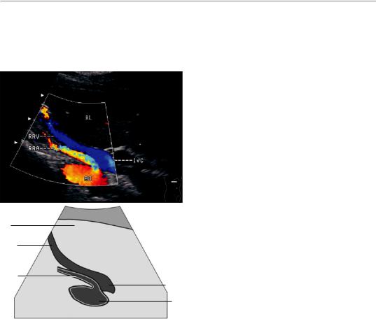
9.2\ |
Longitudinal Section |
9.2.1\ |
Scanning Method |
|
of the Right Renal Vein: |
The method is the same as that used to show the |
|
|
Transverse Section |
right renal artery using the transverse section in |
|
|
of the Right Subcostal Region |
the right subcostal region. The right renal vein |
|
|
|
lies anterior to the right renal artery. Pressing the |
|
|
|
cephalic part of the probe can make both gray- |
|
|
|
scale and color Doppler flow imaging more clear. |
|
|
|
9.2.2\ |
Section Structure |
|
|
Transverse section of the Ao, IVC, and right kid- |
|
|
|
ney; longitudinal section of the right renal vein; |
|
sometimes a longitudinal section of the right renal artery can also be shown.
9.2.3\ Measurement Method
and Normal Value
RL
RRV
RRA
The diameter is about 1 cm. There is a slight fluctuation of the spectrum due to the influence of IVC flutter and respiratory movement.
9.2.4\ Clinical Application Value
To observe if there is a partial or total embolism in the right renal vein; the details are the same as
IVC those for the left renal vein.
Ao