

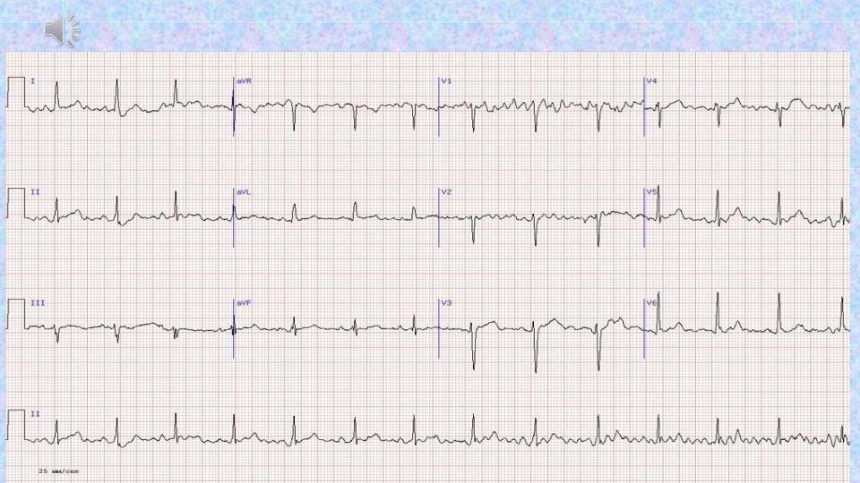
Normal sinus rhythm.
Waves P in front of each complex QRS
Normal sinus rhythm characteristics :
- HR (heart rate) equals 60-80 in min, regular rhythm (differences between minimal and maximal R-R intervals is not more than 15%); - P wave - positive in I, II, aVF, P wave – negative in aVR, PQ ≥0.12 s.

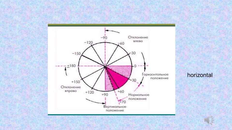
ELECTRICAL AXIS
Electric axis of the heart is a vector indicating the predominant direction of the electromotive force during ventricular depolarization.
Electric axis can coincide with the anatomical axis of the heart, and depends on the constitution and ventricular hypertrophy.

1)normal electrical axis position:α angle = +30°… +69°;
2)horizontal electrical axis position: α angle = 0°… +29°;
3)vertical electrical axis position: α angle = +70° +90°;
4)left electrical axis deviation: α angle = − 1°…−90°;
5)right electrical axis deviation: α angle = +91°…+180°.

normal electrical axis
left electrical axis
right electrical axis
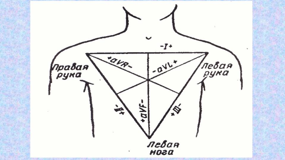
The 6 frontal plane ECG leads form a hexaxial reference system
Deviation to the left
deviation
to the right normal position of the electrical axis of
the heart, vertical,
vertical

normal electrical axis position: = +30 +69 ,
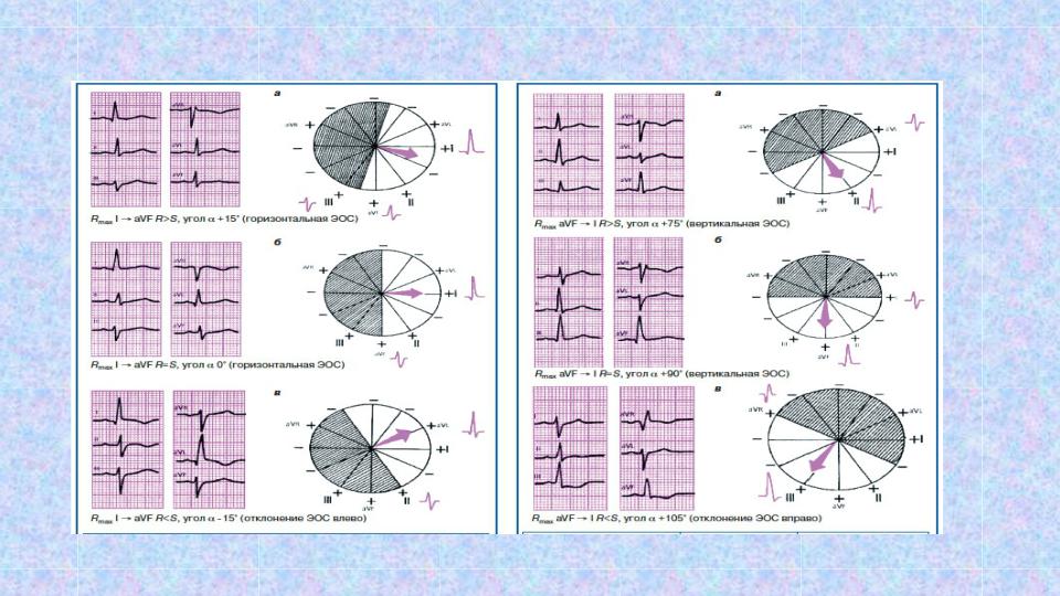
horizontal electrical axis position: = 0 +29 |
vertical electrical axis position: = +70 +90 |
(а,б). |
(а,б). |
left electrical axis deviation: = -1 -90 (в) |
right electrical axis deviation: = +91 +180 (в) |