

Cardiology / Пропедевтика внутренних болезней (кардиология, часть 1) Ослопова 2006 года
.pdfГОСУДАРСТВЕННОЕ ОБРАЗОВАТЕЛЬНОЕ УЧРЕЖДЕНИЕ ВЫСШЕГО ПРОФЕССИОНАЛЬНОГО ОБРАЗОВАНИЯ «КАЗАНСКИЙ ГОСУДАРСТВЕННЫЙ МЕДИЦИНСКИЙ УНИВЕРСИТЕТ
ФЕДЕРАЛЬНОГО АГЕНСТВА ПО ЗДРАВООХРАНЕНИЮ И СОЦИАЛЬНОМУ РАЗВИТИЮ»
КАФЕДРА ПРОПЕДЕВТИКИ ВНУТРЕННИХ БОЛЕЗНЕЙ
Пропедевтика внутренних болезней
Учебно-методическое пособие Часть VI
Introduction to Internal Diseases
Manual
Part VI
Казань – 2006
УДК 616 – 07:616.1/.9 ББК 54.1
Печатается по решению учебно-методического совета по преподаванию на английском языке Казанского государственного медицинского университета
Авторы-составители:
заведующий кафедры пропедевтики внутренних болезней, профессор В.Н.Ослопов, доц. А.Р.Садыкова, ст. преп. кафедры иностранных языков И.В.Карамышева.
Рецензенты:
декан отделения по работе с иностранными студентами, к.м.н., доцент кафедры эпидемиологии
Н.М.Хакимов
методист УМУ КГМУ, к.м.н., доцент кафедры пропедевтики внутренних болезней
О.В.Богоявленская
Пропедевтика внутренних болезней. Учебно-методическое пособие.
Часть VI.. Introduction to internal diseases. Manual. Part VI. / Ослопов В.Н., Садыкова А.Р., Карамышева И.В. – Казань: КГМУ, 2006. -75 с.
Учебно-методическое пособие составлено в соответствии с Государственным образовательным стандартом высшего профессионального образования (2000), Государственными требованиями к минимуму содержания и уровню подготовки выпускника вуза по специальности 040100 «Лечебное дело», типовой и рабочей программами по дисциплине «Пропедевтика внутренних болезней» (2003). В учебнометодическом пособии подробно освещается содержание занятий, даны теоретические и справочные материалы, описываются практические умения в четкой последовательности действий у постели больного. Пособие предназначено для иностранных студентов медицинских вузов.
© Казанский государственный медицинский университет, 2006
CONTENTS
PART VI
DIAGNOSTICS OF CARDIOVASCULAR DISEASES DIAGNOSTICS OF ACQUIRED VALVE DISEASE
Theme24. Rheumatic fever. Diagnostics of acquired mitral valve diseases: mitral stenosis and mitral incompetence. Diagnostic significance of ECO, PhonoCG………………..……….……..….……..4 Theme 25. Rheumatic fever. Diagnostics of acquired aortic valve diseases: aortic stenosis and aortic incompetence. Diagnostic significance of ECO, PhonoCG………………..………………………35 Theme 26. Diagnostics of tricuspid incompetence (organic and functional). Heart failure (acute and chronic). Emergency in acute leftsided heart failure…………………………………………………...….47 References……………………………….……….……………….75

4
Theme24. RHEUMATIC FEVER. DIAGNOSTICS OF ACQUIRED MITRAL VALVE DISEASES: MITRAL STENOSIS
AND MITRAL INCOMPETENCE. DIAGNOSTIC SIGNIFICANCE
OF ECO, PHONOCG
Goal: to get a notion about the main cardiovascular diseases, their symptoms and signs, diagnostic meanings of additional diagnostic methods data; instrumental diagnostics of cardiovascular diseases; to master skills.
Knowledge objectives:
- to know symptoms and signs of main cardiovascular diseases, appropriate changes of physical examination data, diagnostic meaning of additional diagnostic methods and their findings in these diseases.
Skill objectives:
- to collect interviewing data, to perform physical examination of patients with cardiovascular diseases (inspection, palpation, percussion and auscultation), to interpret data of additional diagnostic methods, to establish diagnosis of main cardiovascular diseases.
Subject-matter:
1.complaints of patients with cardiovascular diseases
2.basic signs of rheumatic fever
3.diagnostic criteria of rheumatic fever
4.hemodynamics changes in mitral valve disease
5.physical examination data in patients with mitral stenosis
6.physical examination data in patients with mitral incompetence
7.laboratory diagnostics of rheumatic fever activity
8.instrumental diagnostics of mitral stenosis
9.instrumental diagnostics of mitral incompetence
Equipment required: stethoscope.
5
Noninvasive Diagnostic Cardiovascular Procedures
ECHOCARDIOGRAPHY
Echocardiography (ECO) is an ultrasound technique for diagnosing cardiovascular disorders (see Table 1). It is subdivided into M-mode, two-dimensional (2-D), spectral Doppler, color Doppler, contrast, and stress echocardiography.
Table 1. Clinical uses of ECO
6 |
7 |
Echocardiography is usually performed by placing a transducer over the thorax, along the left or right sternal border, at the cardiac apex, at the suprasternal notch, or over the subcostal region. In transesophageal echocardiography, however, the transducer is placed at the tip of an endoscope, and the heart is visualized via the esophagus. Even smaller transducers can be placed on intravascular catheters, permitting intravascular recordings of vessel anatomy and blood flow.
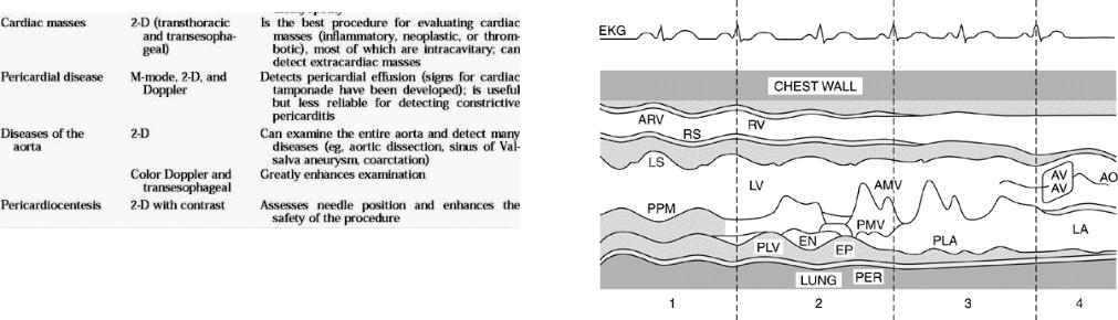
M-mode echocardiography is performed by directing a stationary pulsed ultrasound beam at some portion of the heart. Fig. 1 shows an M- mode echocardiogram as the ultrasound beam is gradually moved from the cardiac apex (position 1) toward the base of the heart (position 4). As the beam passes through the heart, structures that border the right and left ventricles, the mitral and aortic valves, and the aorta and left atrium can be seen. Changing the direction of the ultrasound beam allows echoes from the tricuspid and pulmonic valves to be recorded.
Fig. 1. Diagram of M-mode echocardiography of the heart from the apex (1) to the base (4) of the heart. ARV = anterior right ventricular wall; RV = right ventricular cavity; RS = right side of the interventricular septum; LS = left side of the interventricular septum; LV = left ventricular cavity; PPM = posterior papillary muscle; PLV = posterior left ventricular wall; EN = posterior left ventricular endocardium; EP = posterior left ventricular epicardium; PER = pericardium; AMV = anterior mitral valve leaflet; PMV = posterior mitral valve leaflet; PLA = posterior left atrial wall; AV = aortic valve; AO = aorta; LA = cavity of the left atrium. (From Feigenbaum H: "Clinical applications of echocardiography." Progress in Cardiovascular Diseases 14:531-558, 1972; used by permission of WB Saunders Company.)
2-D (or cross-sectional) echocardiography has become the dominant echocardiographic technique. It uses pulsed, reflected ultrasound to provide spatially correct real time images of the heart, which are recorded on videotape and resemble cineangiograms. Four commonly used 2-D echocardiographic views are shown in Fig. 2.
2-D echocardiography can provide multiple tomographic views of the heart and great vessels.

8 |
9 |
Fig. 2. Four commonly used views for two-dimensional echocardiography. LX = long axis; SX = short axis; 4C = four chamber; 2C = two chamber; LV = left ventricle; AO = aorta; LA = left atrium; RV = right ventricle; RA = right atrium. (From Feigenbaum H: Echocardiography, ed. 5, Lea & Febiger, Malvern, PA, 1994; used with permission.)
Spectral Doppler echocardiography uses ultrasound to record the velocity, direction, and type of blood flow in the cardiovascular system. The spectral Doppler signal is displayed on a strip chart recorder or videotape. Fig. 3 demonstrates spectral Doppler echocardiography and 2- D echocardiography recording of flow through the mitral valve.
Fig. 3. Spectral Doppler echocardiographic examination of blood flowing through the mitral orifice. RV = right ventricle; RA = right atrium; LV = left ventricle; LA = left atrium; E = early diastolic flow; A = diastolic flow secondary to atrial contraction. (From Feigenbaum H: Echocardiography, ed. 5, Lea & Febiger, Malvern, PA, 1994; used with permission.)
Color Doppler echocardiography is essentially 2-D Doppler echocardiography with flow encoded in color to show its direction (red is toward and blue is away from the transducer).
Contrast echocardiography is an M-mode or 2-D echocardiographic examination during which contrast medium is injected into the cardiovascular circulation. Almost any liquid contrast medium that is rapidly injected into the cardiovascular space acquires microbubbles in suspension, which produce a cloud of echoes within the cardiac chambers. These microbubbles usually do not traverse the capillary bed; however, an agent that does it now commercially available (Albunex), and more are being developed.

10
Stress echocardiography is performed during or after physical or pharmacologic stress
PHONOCARDIOGRAPHY
Phonocardiography (PhonoCG) is a method of graphic registering of heart sounds and murmurs, and registered curve is called phonocardiogramm.
Its preferences as additional method of examination are beyond controversy: PhonoCG gives objective heart sounds and murmurs data, permits to count timing and some indices, gives an opportunity to observe the dynamics of heart sounds changes due to pathologic processes course, their treatment and heart surgery.
Technique of PhonoCG recording. PhonoCG is recorded in special room situated far from strange noise and devices having electromagnetic and sound effects (engines, physiotherapy and roentgen devices etc.). A microphone should set up on the chest hermetically.
Commonly sounds registration is performed in 5 classic heart auscultation points (Fig.4).
11
The human ear can perceive oscillations from 20 to 20000 Hz, but the sounds within 20 to50 Hz are hardly perceived. Phonocardiograph has few sound recording channels permitting with using electric filters to select only sounds of necessary frequency. Usually phonocardiograph has 5 sound channels:
1st — auscultatory (А), permissive to record the heart sounds close like they are perceived by human ear;
2nd — low-frequency (Н), pervious sounds with frequency about 35
Hz ;
3d — the first middle-frequency (С1) — within 35 - 70 Hz ;
4th —the second middle-frequency (С2) — within 70 - 140 Hz ;
5th — high-frequency, mainly recording sounds with frequency above 140 Hz (maximum about 250 Hz ).
Some general rules of phonocardiogram analysis Conditionally PhonoCG is registered simultaneously with ECG for better orientation. PhonoCG analysis is performed in following sequence:
1)recorded heart sounds are characterized, their amplitude (by maximal oscillations in mm) and duration (in hundredths of second) is measured, notion about their frequency is made;
2)available on PhonoCG murmurs are analyzed in the same order, also their shape and attitude to the heart cycle phases are defined;
3)several intervals are measured: duration of auscultatory systole, diastole etc.
Normal PhonoCG (Fig.5) consists of 2 registering without fail S1 and S2, which are heard on auscultation, and casual registering S3 and S4.
Fig.4. Standard points of PhonoCG recording: 1 — apex,- 2—aorta; 3— pulmonary artery; 4—tricuspid valve; 5 — Botkin-Erb's point.
Sounds are recorded frequently selective due to their frequency characteristic. The heart sounds acoustical spectrum is within diapason from 10 to 1000 Hz (1 Hz = 1 oscillation per second).
Fig.5. Normal PhonoCG (sketch)
12
S1 is situated in the systole beginning. It is easily found on PhonoCG using 2 main reference points: firstly, it is registered after long pause and secondly, it begins a little bit later R-wave on ECG (approximately on the level of S-wave). The first heart sound is formed due to fusion of 3 sound phenomena. Its beginning, recording as few low-amplitude waves, is caused by ventricular walls vibrations as a result of myocardium switch to the strained condition – the muscular component of S1. The second component of S1 (central part) in general is caused by vibrations of mitral and tricuspid valves in the moment of their shutting – valvular component of S1. However, the muscular component is lasting in the same time, merging with the valvular one. The end of S1, presented with few low-amplitude waves is the vascular component of the first heart sound, caused by aorta and pulmonary artery walls vibrations during the blood ejection period. Because S1 is basically linked with mitral valve and myocardium of left ventricle (owing to its high power capacity), it is better recorded at the apex. Its normal amplitude varies within 10 - 25 mm and duration – 0,10-0,15".
More important meaning has nor the absolute value of S1 but its ratio to S2 amplitude: normally at the apex S1 should be one and half or twice as much intensive as S2. After S1 the rest part of systole, accompanying by blood ejection into aorta and pulmonary artery, is silently performed, and phonocardiograph registers zero line (isoline).
S2 is recorded in the beginning of diastole and normally corresponds to the end of T-wave. It is generally connected with aortic and pulmonary valves vibrations, appeared on their shutting. As aortic valve in physiologic conditions is shut up 0,04—0,06‖ earlier than pulmonary, so normal S2 is somewhat splitted. Physiologic splitting of the second heart sound is more often met on pulmonary artery on the height of inspiration, as right ventricle blood inflow and systole duration are increased on inspiration. S2 duration fluctuates in wide range within 0,04 - 0,11‖and its amplitude is 6—15 mm on aorta and pulmonary artery. S2 intensity normally exceeds approximately twice as much S1 in these points.
S3 is recorded at the beginning of diastole and is away 0,12—0,19‖ from S2. This heart sound is unsteady and is generally met in child's and junior age. On PhonoCG it looks as few low-frequency oscillations, revealed on low-frequency channel. Its origin is connected with
13
ventricular walls vibrations on their rapid filling at the beginning of diastole. Its frequent detection in young people is apparently connected with greater elasticity and oscillating ability of myocardium. In some cases the third heart sound is heard on auscultation, when it acquires more high frequency, and heart rhythm becomes trinomial. This phenomenon is called ―innocent gallop‖ as long as it is listened in healthy people.
S3 appearance in adults, particularly in old people with heart disease is considered as pathology and is called protodiastolic gallop. Its origin is connected with worsening of left ventricle relaxation what leads to increase of ventricle rigidity. In this connection blood blow in that ventricle wall on early diastole causes vibrations more clear than normally, which are considered as gallop sound.
S4, registered in healthy people very rare, is situated before S1 at the end of P-wave ECG. It is recorded only on low-frequency channel as few small oscillations. Its origin is connected with atrial contraction and myocardium vibrations due to blood inflow. In some pathologic conditions, such as atrial hypertrophy, delay of atrio-ventricular conduction this sound acquires greater intensity and frequency, becomes high-pitched and may be listened even on auscultation. In these cases heart rhythm becomes trinomial – presystolic gallop. Its origin may be connected with the same diastole fault (insufficient ventricular relaxation), which was described on protodiastolic gallop explanation.Gallop rhythms are more frequently listened in tachycardia.
At systole on aorta or pulmonary artery auscultation analogous auscultatory phenomenon may be detected, causing by blood blow in the wall of consolidated and extended pulmonary artery or aorta in pulmonary hypertension and aortic valve disease, severe arterial hypertension. It is usually registered 0,04—0,08‖ after S1 and is called systolic ejection sound (EjS).
The main PhonoCG intervals.
1.Auscultatory ventricles systole phase is measured between the
beginning of S1 to the beginning of S2. After S1 on the ejection period the heart contracts silently.
2.Auscultatory diastole phase is measured between the beginning of S2 to the beginning of following heart cycle S1. This phase is divided

14
on 3 periods: protodiastole – from S2 to S3, mesodiastole – fromS3 to S4, presystole – from S4 to S1.
3. “Q – S1” interval (transformation period) — time from the beginning of electrical ventricles excitation to atrio-ventricular valves closure — is measured from the beginning of Q-wave to the beginning of S1 (the beginning of S1 main oscillations). This period depends on the rate of myocardium passage to the strain condition and atrio-ventricular valves shutting, i.e. transformation of electrical events in mechanical. Normally it lasts 0,04—0,06‖ (Fig.6).
15
Etiology. Group A streptococci did not recover from tissue lesions.
•Does not occur in the absence of a streptococcal antibody response.
•Immunologic cross-reactivity between certain streptococcal antigens and human tissue antigens, suggests an autoimmune etiology.
•Both initial attacks and recurrences can be completely prevented by prompt treatment of streptococcal pharyngitis with penicillin.
The cardinal skin signs are erythema marginatum (Fig.7) and subcutaneous nodules, which are firm, painless and discrete, about 0.5—1 cm in diameter, and are found mainly over bony prominences and tendons. They resolve after a few weeks.
Fig.6. Normal phonocardiogram from the apex.
RHEUMATIC FEVER
Rheumatic fever is an acute inflammatory disease of connective tissue that is a sequel to infection with Group A streptococci may involve the heart, skin, CNS and joints. It is now a rare disease in the developed world but is still endemic elsewhere; even in the West there is still a large residue of patients with rheumatic valve disease that has resulted from childhood infection.
Fig.7. Erythema marginatum is a characteristic skin rash which may follow any streptococcal infection, especially tonsillitis, It is one of the common signs of acute rheumatic fever, and its presence should raise the possibility of cardiacinvolvement.
The arthritis varies from arthralgia to a flitting polyarthritis, mainly affecting the larger joints such as the knees, ankles, wrists and elbows. These joints may become acutely swollen, hot and tender, and the synovial fluid is full of polymorphs.

16
Carditis is the most important aspect of this disease as it has major long-term implications. Endocarditis, myocarditis and pericarditis are all often present. The diagnosis of carditis requires the finding of
•new cardiac murmurs
•cardiomegaly
•pericarditis
•congestive cardiac failure.
The murmurs may include an apical systolic murmur (caused by mitral regurgitation), a transient apical mid-diastolic (Carey—Coombs) murmur (caused by turbulent flow across the inflamed mitral valve), and a basal diastolic murmur (caused by aortic regurgitation). Other cardiac signs may include tachycardia, pericardial friction rub, muffled heart sounds resulting from pericardial effusion and evidence of heart failure.
Neurological involvement (Sydenham’s chorea) is uncommon and develops after a latent period of several weeks. The patient develops rapid purposeless involuntary movements mostly in the limbs and face.
Investigations should include throat-swab culture and the measurement of antibody response to Streptococcus (anti-streptolysin ‗0‘ titer). There is usually a leucocytosis and elevation of the ESR and C- reactive protein levels. X-ray of the chest may show a pericardial effusion and rarely pneumonia or lobar collapse. ECG often shows first degree heart block (Fig.8 & 9).
Fig.8 & 9. First degree atrioventricular block. The PR interval exceeds 0.22 seconds, so there is a delay in atrioventricular conduction, but all impulses pass on and result in a QRS complex and ventricular contraction. In Fig.8 the PR interval is easily measured, but in Fig.9 the P wave is hidden in the T wave of the preceding beat and could easily be missed.
A range of diagnostic criteria (the Jones criteria) may be used (Fig.10) as the aid to the clinical diagnosis and more accurate classification of rheumatic fever.
17
Fig.10. Criteria for the diagnosis of rheumatic fever (Jones criteria). The definitive diagnosis requires two major findings or one major and two minor findings plus evidence of recent infection.
Application. ―Jones Criteria (revised) for Guidance in the Diagnosis of Rheumatic Fever‖ published by the American Heart Association:
1.Two major criteria
2.One major and two minor criteria if supported by evidence of an antecedent group A streptococcal infection
a. Increased titer of antistreptococcal antibodies such as ASO b. Positive throat culture
c. Recent scarlet fever
• Minor criteria
1.Previous rheumatic fever or rheumatic heart disease
2.Fever
3.Arthralgia
4.Increased acute-phase reactants
a.ESR
b.C-reactive protein
c.Leukocytosis
5. Prolonged P-R interval
Treatment should be directed towards the elimination of any residual streptococcal infection with penicillin. Aspirin is an effective anti-inflammatory and antipyretic agent for the other features. Prevention of recurrence may be necessary with longterm oral penicillin.