

Cardiology / Пропедевтика внутренних болезней (кардиология, часть 1) Ослопова 2006 года
.pdf38
Fig. 30. Chest x-ray shows left ventricle enlargement and aortic heart configuration in aortic stenosis.
On ECG there are features of left ventricular hypertrophy (Fig.24) and not rarely – signs of coronary insufficiency.
PhonoCG reflects typical for this disease changes of the heart sounds: a decrease of S1 amplitude, registered at the apex, and a decrease of S2 amplitude, registered at aorta. Recording of very intensive systolic murmur with the maximum at aorta is the basic auscultative phenomenon in aortic stenosis (Fig.31). This murmur is connected with blood pass from left ventricle into aorta across narrow aortic ring. As classical ejection murmur it has typical diamond-shape on PhonoCG and is registered on all channels. Murmur intensity to a great extent correlates with stenosis degree.
On carotid sphygmogram slowly rising, low-amplitude, latepeaking arterial pulse wave with characteristic crenation of the peak (sphygmogram in the ― ‖ form, reflecting vibrations, connected with
39
murmur transmission on neck arteries) is marked due to severe aortic outflow tract obstruction.
Fig.31. Phonocardiogramm in aortic stenosis.
Echocardiography confirms the diagnosis by showing thickened and calcified valve cusps (Fig.32).
Fig.32. Aortic stenosis with calcification. This M-mode parasternal long-axis view shows the characteristic box shape of valve opening during systole (1). Calcification of the valve and annulus is suggested by the density of whiteness of the tracing (2).
40
Doppler echocardiography (Fig.33) or cardiac catheterization (Fig.34), or both, establish the severity of the stenosis, and Doppler echocardiography is extremely valuable in following the course of the disease in individual patients. Ultrasound has removed the need for repeated cardiac catheterization. The peak aortic pressure gradient can be calculated, and it correlates well with the degree of severity of stenosis. Other formulae may be used to measure the aortic valve orifice area.
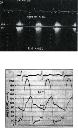
Fig.33. Doppler flow study of the aortic valve in aortic stenosis. There is a mosaic pattern at the aortic valve suggesting the valve is narrowed. The velocity of blood flow across the valve is 5 m/s which gives an estimated valve gradient of 100 mmHg by the Bernouilli equation, confirming aortic stenosis.
Fig.34. Pressure gradient across the aortic valve in aortic stenosis, as measured at cardiac catheterization. Note the low aortic (Ao) pressure compared with the left ventricular pressure (LV) and the delayed peak in aortic pressure — both characteristic of severe aortic stenosis. Such pressure studies are less commonly performed than in the past, because echocardiography and Doppler flow studies can provide much of the relevant information noninvasively.
41
Aortic stenosis is in compensated stage of disease for a long time. Heart failure develops in decrease of left ventricular contractile capacity and manifests as in aortic incompetence.
The valve is usually replaced with a prosthetic valve. Alternatively, the narrowed valve may be stretched by balloon valvuloplasty, although the long-term benefits of this procedure are unclear.
AORTIC INCOMPETENCE
Aortic incompetence (insufficientia valvulae aortae) is valve disease in which the semilunar cusps close incompletely the aortic orifice and during diastole blood regurgitates from aorta into left ventricle. Aortic regurgitation occurs if the aortic valve ring dilates, as a result of dissecting aneurysm, ankylosing spondylitis or syphilis for example, or if the valve cusps degenerate, such as after rheumatic fever, atherosclerotic lesion or endocarditis.
Anatomical changes depend on etiology of aortic lesion. After rheumatic fever inflammatory-sclerotic process at the base of valvular cusps leads to their wrinkling and shortening. In syphilis and atherosclerosis pathologic process may affect only aorta itself, causing its dilatation and taking-up of valvular cusps without their affecting, or connective tissue expands on valvular cusps and deforms them. In sepsis ulcerous endocarditis leads to destruction of valve parts, defects forming in the cusps and their subsequent scarring and shortening.
Hemodynamics abnormalities
In aortic incompetence during diastole blood comes in the left ventricle not only from left atrium but regurgitates from aorta too. It causes left ventricle overfilling and stretching on diastole. During systole left ventricle contracts with grater force to eject into aorta increased stroke volume. Left ventricle overwork leads to its hypertrophy, and increase of systolic blood volume in aorta causes its dilatation (Fig.35). Sharp fluctuation of blood pressure within aorta during systole and diastole is characteristic for aortic incompetence. Increased as compared with normal blood volume within aorta during systole causes increase of systolic blood pressure, and as a part of blood volume regurgitates into ventricle during diastole, diastolic pressure rapidly decline.

42
Fig.35. Intracardiac hemodynamics changes in aortic incompetence (sketch). Hypertrophied chambers are shaded.
Clinical manifestations. Clinical symptoms often occur later, because this valve disease is compensated by overwork of powerful left ventricle. In time symptoms of heart failure: dyspnea, palpitation, weakness etc. gradually appear. Angina-like pains in the precordium are typical. They are caused by coronary insufficiency due to severe myocardial hypertrophy and worsening of coronary arteries blood supply upon low diastolic pressure within aorta. Dizziness is observed owing to worsened cerebral blood supply, also connected with low diastolic pressure.
On general examination skin pallor, caused by small blood filling of arterial system during diastole, is marked. The peripheral arteries pulsation: carotids (―carotid's dance‖), clavicular, brachial, temporal arteries, caused by sharp pressure fluctuation in arterial system, draws attention. Such signs as head bobbing (Musset's sign) – rhythmical, synchronous with pulse head jiggles, rhythmical changes of nail bed colouring in light press at the nail edge, so-called capillary pulsation (Quinckes' sign), rhythmical increase and decrease of hyperemia zone (got after rubbing) etc.
On precordium inspection increased and displaced downward and to the left apical impulse is almost always obvious. Sometimes along with high apical beat a light retraction in the area of adjacent intercostal spaces is noted.
43
On palpation apical impulse is detected in the VI, and sometimes in the VII intercostals space, laterally from the left midclavicular line: it is diffuse, lifted, increased, dome-like, which witness a big enlargement of left ventricle (left ventricular heave).
On percussion the displacement to the left of cardiac dullness is detected; the heart acquires aortic configuration (with accentuated cardiac waist).
On auscultation diminished S1 at the apex is revealed, because during systole there is no period of closed valves. S2 at the aorta is also diminished, and in significant cusps destruction it may not to be heard. In syphilitic and atherosclerotic lesion of aorta S2 may be clear enough.
The typical auscultative sign of aortic incompetence is diastolic murmur, listened at the aorta and at the Botkin-Erb's point, which is best heard by sitting the patient upright, leaning forward in full expiration. It usually is soft, blowing protodiastolic murmur, weakening to the end of diastole as blood pressure decreasing in aorta and blood flow slowing. In aortic incompetence at the apex murmurs of functional origin may be also heard. So, in large left ventricular dilatation relative mitral incompetence occurs and systolic murmur at the apex appears. Rarely diastolic (presystolic) murmur - Flint's murmur appears due to mitral valve cusps raising by a strong stream of blood regurgitating during diastole from aorta to left ventricle. It results in difficult blood flow from LA to LV during active atrium systole. Sometimes in this valve disease two sounds (Traube's doubling sound) and the Vinogradov-Duroziez‘s doubling murmur heard over the femoral artery are revealed. They are explained by vibration of arterial wall on systole and diastole during pulse wave passing.
In aortic incompetence pulse becomes fast, high, big (pulsus celer, altus, magnus) as big pulse pressure, peculiar to this valve disease, and increased stroke volume, input into aorta during systole, influence on its character.
Blood pressure is always changed: systolic pressure is increased, diastolic – decreased, and so pulse pressure is high.
Chest X-ray (Fig.36) shows left ventricular enlargement with accentuated cardiac waist, aorta dilatation and its increased pulsation.

44
ECG also shows signs of left ventricular hypertrophy (Fig.37), axial left shift, deep S-waves in the right chest leads and high-amplitude R- waves in the left chest leads, not rarely combined with left ventricular overstrain and relative coronary insufficiency signs (ST interval depression and T-wave inversion).
Fig.36. Chest x-ray in aortic incompetence. Aortic heart configuration. Left ventricular hypertrophy and ascending aorta dilatation are noted.
45
On PhonoCG (Fig.38) diastolic murmur is registered on highfrequency and auscultatory channels. It appears just after S2 i.e. in early protodiastole, and has decrescent character. Amplitude of its oscillations is small, so it is better heard than registered. Its intensity is a reflection of the size of the leak.
Fig.38. PhonoCG in aortic incompetence, registered at the aorta: decrease of the sounds amplitude and decrescent diastolic murmur.
Echocardiogram show left ventricular enlargement. Aortography (Fig.40) or colour—flow Doppler (Fig.39) shows the regurgitant jet.
Fig.37. ECG in aortic incompetence.
Fig.39. Colour-flow Doppler mapping in a patient with mild aortic regurgitation (parasternal long-axis view). The aortic regurgitant jet (in blue) is directed posteriorly at an acute angle from the aortic valve (to the right in the picture), back into the left ventricle (to the left in the picture), impinging directly on the anterior mitral valve leaflet (in the centre of the picture) immediately below the blue jet.

46
Fig.40. Aortogram in severe aortic regurgitation. Contrast medium has only been injected into the aorta, but even in this systolic view it is clear that it has regurgitated into the left ventricle. The aortic valve is arrowed. The ‗ring‘ over the aortic root is due to an ECG skin electrode.
Aortic regurgitation leads to hypertrophy of the left ventricle and ultimately left ventricular failure. Clinical symptoms often occur later and the patient may show significant heart failure or angina. There may have been a preceding history of palpitations, syncope or headaches because of the high systolic blood pressure, especially during exercise.
In left ventricular contractile capacity weakening congestion in pulmonary circulation circle develops. Sometimes acute left ventricular weakness may appear that clinically represented as cardiac asthma attack.
In left ventricular contractile capacity weakening and its dilatation relative mitral incompetence may occur, which increases pulmonary congestion and creates additional load to the right ventricle. This is socalled mitralisation of aortic incompetence, it may be the cause of venous congestion in systemic circulation.
Medical treatment is directed at managing the angina, correcting the failure and preventing endocarditis. Definitive treatment consists of replacing the valve with a prosthetic one.
CONTROL QUESTIONS
1.Changes of intracardiac hemodynamics in aortic stenosis. Mechanisms of valve disease compensation.
47
2.Symptoms and patient' appearance in aortic stenosis.
3.Findings of precordial inspection and palpation, percussion data in patients with aortic stenosis.
4.Auscultatory signs of aortic stenosis.
5.Pulse and blood pressure in aortic stenosis
6.ECG changes in aortic stenosis.
7.PhonoCG data in aortic stenosis.
8.ECO data in aortic stenosis.
9.X-ray examination findings in aortic stenosis.
10.Changes of intracardiac hemodynamics in aortic incompetence. Mechanisms of valve disease compensation.
11.Symptoms and patient' appearance in aortic incompetence.
12.The peripheral signs of aortic incompetence
13.Findings of precordial inspection and palpation, percussion data in patients with aortic incompetence.
14.Auscultatory signs of aortic incompetence.
15.Pulse and blood pressure in aortic incompetence
16.ECG changes in aortic incompetence.
17.PhonoCG data in aortic incompetence.
18.ECO data in aortic incompetence.
19.X-ray examination findings in aortic incompetence
Theme 26. DIAGNOSTICS OF TRICUSPID INCOMPETENCE (ORGANIC AND FUNCTIONAL). HEART FAILURE (ACUTE AND
CHRONIC). EMERGENCY IN ACUTE LEFT-SIDED HEART
FAILURE
Goal: to get a notion about the main cardiovascular diseases, their symptoms and signs, diagnostic meanings of additional diagnostic methods data; instrumental diagnostics of cardiovascular diseases; to master skills.

48
Knowledge objectives:
- to know symptoms and signs of main cardiovascular diseases, appropriate changes of physical examination data, diagnostic meaning of additional diagnostic methods and their findings in these diseases.
Skill objectives:
- to collect interviewing data, to perform physical examination of patients with cardiovascular diseases (inspection, palpation, percussion and auscultation), to interpret data of additional diagnostic methods, to establish diagnosis of main cardiovascular diseases.
Subject-matter:
1.complaints of patients with tricuspid valve disease
2.hemodynamics changes in tricuspid incompetence
3.physical examination data in patients with tricuspid incompetence
4.instrumental diagnostics of tricuspid incompetence
5.notion of circulatory failure, heart failure and vascular failure
6.etiology and pathophisiology of heart failure
7.classification of heart failure
8.сlassification of chronic heart failure
9.symptoms of heart failure
10.physical examination data in patients with heart failure
11.instrumental diagnostics of heart failure
Equipment required: stethoscope.
TRICUSPID VALVE INCOMPETENCE
Tricuspid valve incompetence (insufficientia valvulae tricuspidalis) as mitral incompetence may be organic and relative. Organic incompetence is rarely met. It develops due to endocarditis, frequently rheumatic, rarely infective, or appears owing to trauma with papillary muscles of tricuspid valve rupture. As separate valve disease tricuspid incompetence is met extremely rare and usually coincides with other heart valve affecting.
49
Relative tricuspid incompetence is more frequently met. It appears in right ventricular dilatation and right atrioventricular orifice distension.
The most frequently it is observed in mitral valve disease, when due to high pressure in pulmonary circulation the right ventricle will overwork, that leads to it overstrain and dilatation.
Hemodynamics abnormalities
Hemodynamics in tricuspid incompetence is stipulated that during right ventricular systole because of incomplete closure of valve leaflets part of blood regurgitates back to the right atrium, in which usual volume of blood from venas cava simultaneously passes. Atrium extends and hypertrophies. Increased blood volume passes during diastole into right ventricle from right atrium also, because the regurgitant volume is added to usual blood volume. This causes right ventricular dilatation and hypertrophy (Fig.41).
Fig.41. Intracardiac hemodynamics changes in tricuspid incompetence.
So, in this valve disease the compensation is maintained by right atrium and right ventricle overwork, their compensatory abilities are small, so venous congestion in the systemic circulation quite rapidly develops.
Clinical manifestations. Significant venous congestion in systemic circulation in tricuspid incompetence results in edema, ascites appearance, heaviness sensation and pains in the right hypochondrium, connected with liver enlargement. Skin acquires cyanotic colouring,

50
sometimes with icteric tint. Neck veins swelling and pulsation, socalled positive vein pulse, and liver pulsation attract attention (Fig. 42). These pulsations are caused by reverse blood flow from ventricle into atrium across uncovered atrioventricular orifice, because of this pressure within atrium rises and emptying of neck and hepatic veins is laboured. If you put one hand on the liver area and other – on the right ventricle area it is possible to feel characteristic wobbling movements of both hands. The apical impulse, as a rule, is not expressed, since left ventricle is shifted backwards by hypertrophied right ventricle.
Fig. 42. Elevated external jugular venous pressure (JVP). The pressure in the internal and external jugular veins is elevated in right heart failure. Abnormalities in waveform may provide evidence of tricuspid valve disease or cardiac arrhythmia, although these abnormalities are more reliably noted in the internal than the external jugular vein, which may be affected by position.
On percussion significant displacement of cardiac dullness borders to the right owing to right atrium and right ventricular hypertrophy is detected.
On auscultation at the xyphoid process base diminished S1 is found; in the same point and also in the 3d and 4th right intercostals spaces close to the sternum systolic murmur is listened. Its intensity increases in breath-hold on the height of inhalation (Riverro-Corvallo sign). S2 at the pulmonary artery is diminished, because pressure within pulmonary circulation circle decreases in tricuspid incompetence.
Pulse does not significantly changes or becomes small and frequent, because in this valve disease there not rarely exists severe heart failure.
51
Blood pressure is often decreased. Venous pressure grossly elevates: its level may reach 200—300 mm of water column.
X-ray shows signs of the heart right chambers hypertrophy (Fig.43)
ECG shows signs of right atrium hypertrophy (―P-pulmonale‖)
(Fig.44) and right ventricular hypertrophy (Fig.15 & 45).
PhonoCG registers decrescent character systolic murmur at the base of xyphoid process and at the 3d and 4th right intercostals spaces close to the sternum (Fig.46); during PhonoCG registering in breath-hold on the height of inhalation the amplitude of oscillations corresponding to the murmur increases.
Fig.43. Chest X-ray in a patient with tricuspid regurgitation. The enlargement of the right heart shadow is caused by a grossly enlarged right atrium. Note also the calcified aortic arch (and the incidental bilateral hilar calcification, resulting from old tuberculosis). This woman‘s tricuspid regurgitation resulted from ischemic heart disease.

52
Fig.44. Right atrium hypertrophy: PII,III,aVF is tall, pointed.
53
Fig.45.Right ventricular hypertrophy (R-type): QRS complex in V1is qR-type, tall RIII, aVF; deep S1,aVL,V5-6; prominent RaVR; α=+1ОО°.
Fig.46.Tricuspid incompetence: PhonoCG shows systolic murmur and diminished S1, registered at the base of xyphoid process. Phlebogramm is typical for positive vein pulse.
Phlebogramm, registered at the jugular vein shows high positive a- wave, corresponding to hypertrophied right atrium overwork or acquires appearance, typical for positive vein pulse (Fig.46).
Echocardiography is the most effective method of diagnosing tricuspid valve disease (Fig.28).
Tricuspid incompetence, as a rule, is accompanied by severe circulatory insufficiency. Long-term venous congestion in systemic circulation leads to functional abnormalities of many organs: liver, kidneys, gastrointestinal tract. Liver particularly suffers: long-term venous congestion in it is accompanied by connective tissue development and results in so-called cardiac cirrhosis. This in its turn even greater embarrasses functions of organs and causes severe metabolic disturbance.

54
CIRCULATORY FAILURE
Circulatory failure is an extremely common problem with an incidence of 2% at age 50 years, rising to 10% at age 80 years. There is still a high mortality: 10—30% per year.
Circulatory failure occurs when an adequate blood flow to the tissues cannot be maintained. This maybe caused by inadequate cardiac output (heart failure) or by a markedly reduced intravascular volume, for example after major haemorrhage, acute dehydration or in septicaemic shock (vascular failure).
Heart failure (congestive heart failure): Symptomatic myocardial dysfunction resulting in a characteristic pattern of hemodynamic, renal, and neurohormonal responses.
No definition of heart failure (HF) is entirely satisfactory. Congestive heart failure (CHF) develops when plasma volume increases and fluid accumulates in the lungs, abdominal organs (especially the liver), and peripheral tissues.
Physiology At rest and during exercise, cardiac output (CO), venous return, and distribution of blood flow with O2 delivery to the tissues are balanced by neurohumoral and intrinsic cardiac factors. Preload, the contractile state, afterload, the rate of contraction, substrate availability, and the extent of myocardial damage determine left ventricular (LV) performance and myocardial O2 requirements. The Frank-Starling principle, cardiac reserve, and the oxyhemoglobin dissociation curve play a role.
Preload (the degree of end-diastolic fiber stretch) reflects the enddiastolic volume, which is influenced by diastolic pressure and the composition of the myocardial wall. For clinical purposes, the enddiastolic pressure, especially if above normal, is a reasonable measure of preload in many conditions. LV dilatation, hypertrophy, and changes in myocardial distensibility or compliance modify preload.
The contractile state in isolated cardiac muscle is characterized by the force and velocity of contraction, which are difficult to measure in the intact heart. Clinically, the contractile state is often expressed as the ejection fraction (LV stroke volume/end-diastolic volume).
55
Afterload (the force resisting myocardial fiber shortening after stimulation from the relaxed state) is determined by the chamber pressure, volume, and wall thickness at the time of aortic valve opening. Clinically, afterload approximates systemic BP at or shortly after aortic valve opening and represents peak systolic wall stress. The heart rate and rhythm also influence cardiac performance.
Reduced substrate availability (eg, of fatty acid or glucose), particularly if O2 availability is reduced, can impair the vigour of cardiac contraction and myocardial performance.
Tissue damage (acute with myocardial infarction or chronic with fibrosis due to various diseases) impairs local myocardial performance and imposes an additional load on viable myocardium.
The Frank-Starling principle states that the degree of end-diastolic fiber stretch (preload) within a physiologic range is proportional to the systolic performance of the subsequent ventricular contraction (Fig. 47).
Fig.47. Frank-Starling relationship. The ordinate (systolic pressure, stroke volume, cardiac output, stroke work, cardiac work) represents the ability of the ventricle to function as a pump. The abscissa (diastolic muscle length, end-diastolic pressure, end-diastolic volume) depicts the direct or indirect measurements of length or stretch of myocardial fiber. Dotted lines depict resting normal values; the normal curve includes point of intersection. Under normal conditions, left ventricular systolic

56
pressure, stroke volume, and work increase rapidly as the myocardial fiber is lengthened at end-diastole. There is a family of ventricular function curves depicting cardiac performance under normal and abnormal conditions. During heart failure (HF) consequent to reduced contractility, ventricular performance falls sharply (point A). Reduced stroke volume causes increased end-diastolic volume with consequent stretching of diastolic muscle length. Ventricular function moves to the right on a relatively flat ventricular function curve to achieve relatively normal resting cardiac performance (point B). Thus, adequate resting cardiac performance results from increased ventricular diastolic volume and pressure. Treatment of the failing ventricle with an inotropic drug improves the ventricular function curve (point C), which, however, remains abnormal. Afterload reduction may have similar effects. This diagram assumes a direct relationship between diastolic muscle length, end-diastolic pressure, and end-diastolic volume, a relationship that is generally true when ventricular contractility is reduced. This relationship does not apply to HF due to increased myocardial diastolic stiffness; cardiac output is usually normal, end-diastolic pressure is high, yet diastolic muscle length may be normal. The problem in disease causing increased diastolic stiffness is a markedly reduced myocardial compliance, hence abnormally high ventricular filling pressure and congestion with adequate ventricular emptying.
This mechanism operates in HF, but, because ventricular function is abnormal, the response is inadequate. If the Frank-Starling curve is depressed, fluid retention, vasoconstriction, and a cascade of neurohumoral responses lead to the syndrome of CHF. Over time, LV remodeling (change from the normal ovoid shape) with dilatation and hypertrophy further compromises cardiac performance, especially during physical stress. Dilatation and hypertrophy may be accompanied by increased diastolic stiffness.
Cardiac reserve (unused ability of the resting heart to deliver O2 to the tissues) is an important component of cardiac function during emotional or physical stress. Its mechanisms include increases in heart rate, systolic and diastolic volume, stroke volume, and tissue extraction of O2. For example, in well-trained young adults during maximal exercise, heart rate may increase from 55 to 70 at rest to 180 beats/min; cardiac output - CO (stroke volume × heart rate) may increase from its normal resting value of 6 to >= 25 L/min; and O2 consumption can increase from 250 to >= 1500 mL/min. In the normal young adult at rest, arterial blood contains about 18 mL O2/dL of blood, and mixed venous or pulmonary artery blood contains about 14 mL/dL. The arteriovenous O2 difference (A-VO2) is thus about 4.0 ± 0.4 mL/dL. Even maximal CO
57
during exercise is insufficient to meet tissue metabolic needs; hence, the tissues extract more O2, and mixed venous blood O2 content falls considerably. A-VO2 may increase to about 12 to 14 mL/dL. Increased A-VO2 due to lower venous O2 content is a common adaptive mechanism in HF.
The oxyhemoglobin dissociation curve (see Fig. 48) influences O2 availability to the tissues and can provide another reserve mechanism in HF.
Fig. 48. Oxyhemoglobin dissociation curve. Arterial oxyhemoglobin saturation (ordinate) is related to partial pressure of O2 (abscissa). P50 (PO2 at 50% saturation) is normally 27 mm Hg. The dissociation curve is shifted to the right by increased hydrogen ion (H+) concentration and increased RBC diphosphoglycerate (DPG). The curve is shifted to the left by decreased H+ and lower RBC diphosphoglycerate. Hb characterized by rightward shifting of the curve has a decreased affinity for O2; Hb characterized by a leftward shifting of the curve has an increased affinity for O2.
The position of this curve is frequently expressed as P50 (the partial pressure of O2 in blood at 50% oxyhemoglobin saturation). An increase in the normal P50 (27 ± 2 mm Hg) indicates a rightward shift of the oxyhemoglobin dissociation curve (decreased affinity of Hb for O2). For a given PO2, less O2 is combined with Hb, and the saturation is lower;