

Federal State Budgetary Educational Institution of Higher Education "Kuban State Medical University" of the Ministry of Health of the Russian Federation
Department of Internal Medicine Propedeutics
ECG

Electrocardiography (ECG)
•is the most important method for examining the heart and diagnosing diseases of the cardiovascular system
•является важнейшим методом исследования сердца и диагностики заболеваний сердечнососудистой системы
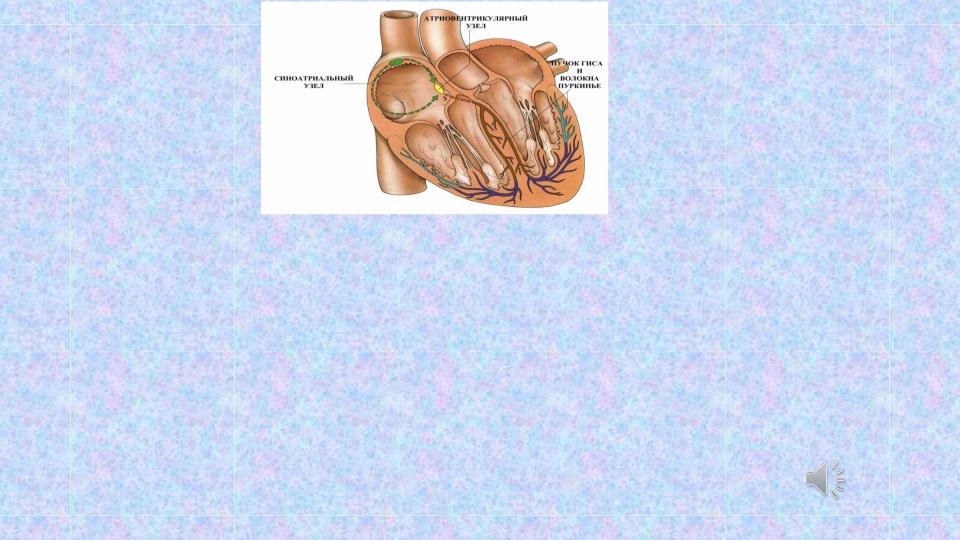
•Registration of electrical processes occurring in the heart to one degree or another reflects its main functions:
•1. Automatism.
•2. Conductivity.
•3. Excitability and refractoriness.
•4. Contractility.
•Регистрация электрических процессов происходящих в сердце в той или иной степени отражает его основные функции :
•1. Автоматизм.
•2. Проводимость.
•3. Возбудимость и рефрактерность.
•4. Сократимость.

Первые электрокардиограммы были записаны Габриелем Липпманом
( 1845 – 1921) с использованием ртутного электрометра.
The first electrocardiogram were recorded
Gabriel Lippmann (1845 - 1921) using a mercury electrometer.

Виллем Эйнтховен (Willem Einthoven; 1860 - 1927) |
||
́ |
́ |
|
нидерландский |
|
физиолог, |
основоположник электрокардиографии, |
1903 году |
|
- первый прибор для регистрации ЭКГ, в 1906 году -
использовал ЭКГ |
в диагностических целях, |
получил Нобелевскую |
премию по физиологии или |
медицине в 1924 году. |
|
Willem Einthoven (1860 - 1927) Dutch physiologist, founder of electrocardiography, 1903 - the first device for recording ECG, in 1906 - used ECG for diagnostic purposes, received the Nobel Prize in Physiology or Medicine in 1924

The ECG standard provides 12-lead recording:
6 limb leads
3 standard (I, II, III)
+ 3 augmented (aVR, aVL, aVF);
6 chest leads (V1-V6).
Стандарт ЭКГ предусматривает запись 12 отведений:
6 отведений от конечностей (3 стандартных + 3 усиленных); 6 грудных отведений.
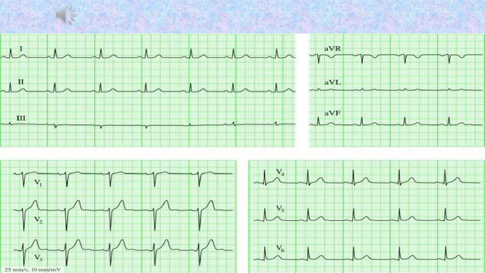
Normal ECG (12-leads)
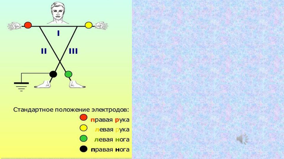
Recording of 6 limb leads.
3 standard (I, II, III)
+ 3 augmented (aVR, aVL, aVF);
Color coding of electrodes:
Right arm, left arm, left foot, right leg
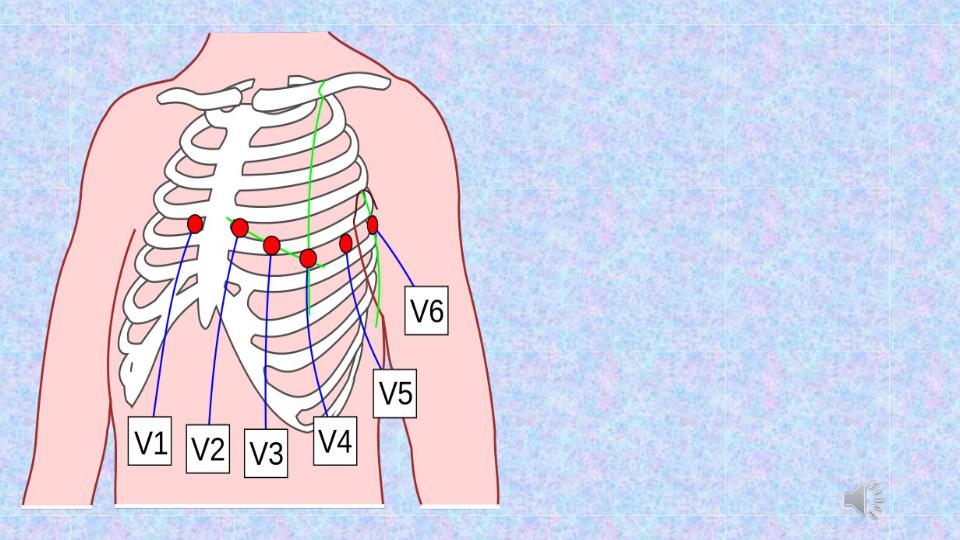
ГРУДНЫЕ отв. chest leads
V1 - 4 intercostal space D. справа
V2 – 4 intercostal space S.
V3 - 5 rib, between
V2 и V4
V4 - 5 intercostal space – mediaclavicularis linea V5 - 5 intercostal space anterior axillary line
V6 - 5 intercostal space middle axillary line
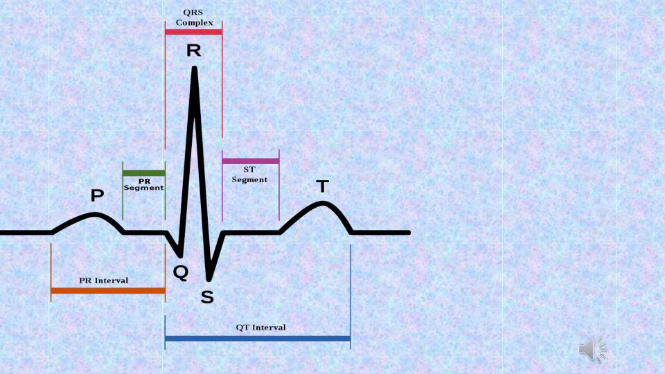
An ECG of a healthy subject has the following elements:
-positive waves P, R, and T,
-negative waves Q andS; the positive wave U is accidental;
-P-Q, S-T, T-P, and R-R intervals; -QRS and QRST complexes.
Each of these elements characterizes the time and sequence of excitation of various parts of the myocardium.