

PS-2020a / part17
.pdfDICOM PS3.17 2020a - Explanatory Information |
Page 721 |
UUU.1.3.4 Transformation Method Code Sequence
If Transformation Method Code Sequence (0022,1512) is (111791, DCM, "Spherical projection") is used then all coordinates in the Two Dimensional to Three Dimensional Map Sequence (0022,1518) are expected to lie on a sphere with a diameter that is equal to Ophthalmic Axial Length (0022,1019).
The use of this model for representing the 3D retina enables the calculation of the shortest distance between two points using great circles as per section UUU.1.3.2.
UUU.2 Relationship Between Ophthalmic Tomography Image and Ophthalmic Optical Coherence Tomography B-scan Volume Analysis IODs
This Section provides examples of the relationship between the Ophthalmic Tomography Image SOP Instance(s) and Ophthalmic Optical Coherence Tomography B-scan Volume Analysis SOP Instance(s).
Below is a typical example.
•Ophthalmic Tomography Image SOP Instance UID is "1.2.3.4.5" and contains five frames.
•Ophthalmic Optical Coherence Tomography B-scan Volume Analysis SOP Instance encodes five frames (e.g., one frame for each ophthalmic tomography frame).
•References are encoded via the Per-frame Functional Groups Sequence (5200,9230) using Attributes Derivation Image Sequence (0008,9124) and Source Image Sequence (0008,2112).
- Standard -

Page 722 |
|
|
|
|
|
DICOM PS3.17 2020a - Explanatory Information |
|||
|
... |
|
|
|
|
|
|
|
|
|
|
|
|
|
|
|
|
|
|
|
Per-Frame Functional Groups Sequence |
5200,9230) |
|
|
|||||
|
|
|
|
|
|
|
|
|
|
|
|
Item 1 |
|
Frame 1 in OCT B-Scan Volume |
|
||||
|
|
|
>Derivation Image Sequence |
(0008,9124) |
|
|
|||
|
|
|
|
|
|
|
|
|
|
|
|
|
|
Item 1 |
|
|
|
||
|
|
|
|
|
>>... |
|
|
|
|
|
|
|
|
|
>>Source Image Sequence |
(0008,2112) |
|
|
|
|
|
|
|
|
Item 1 |
|
For Frame 1 |
|
|
|
|
|
|
|
|
>>>Referenced SOP Class UID |
(0008,1150) |
= "1.2.840.10008.5.1.4.1.1.77.1.5.4" |
Opthalmic Tomography Image |
|
|
|
|
|
|
>>>Referenced SOP Instance UID |
(0008,1155) |
= "1.2.3.4.5" |
|
|
|
|
|
|
|
>>>Referenced Frame Number |
(0008,1160) |
= 1 |
|
|
|
|
|
|
|
... |
|
|
|
|
|
Item 2 |
|
Frame 2 in OCT B-Scan Volume |
|
||||
|
|
|
>Derivation Image Sequence |
(0008,9124) |
|
|
|||
|
|
|
|
|
|
|
|
||
|
|
|
|
Item 1 |
|
|
|
||
|
|
|
|
|
>>... |
|
|
|
|
|
|
|
|
|
>>Source Image Sequence |
(0008,2112) |
|
|
|
|
|
|
|
|
Item 1 |
|
For Frame 2 |
|
|
|
|
|
|
|
|
>>>Referenced SOP Class UID |
(0008,1150) |
= "1.2.840.10008.5.1.4.1.1.77.1.5.4" |
Opthalmic Tomography Image |
|
|
|
|
|
|
>>>Referenced SOP Instance UID |
(0008,1155) |
= "1.2.3.4.5" |
|
|
|
|
|
|
|
>>>Referenced Frame Number |
(0008,1160) |
= 2 |
|
|
|
|
|
|
|
... |
|
|
|
|
|
Item 3 |
|
Frame 3 in OCT B-Scan Volume |
|
||||
|
|
|
>Derivation Image Sequence |
(0008,9124) |
|
|
|||
|
|
|
|
|
|
|
|
||
|
|
|
|
Item 1 |
|
|
|
||
|
|
|
|
|
>>... |
|
|
|
|
|
|
|
|
|
>>Source Image Sequence |
(0008,2112) |
|
|
|
|
|
|
|
|
Item 1 |
|
For Frame 3 |
|
|
|
|
|
|
|
|
>>>Referenced SOP Class UID |
(0008,1150) |
= "1.2.840.10008.5.1.4.1.1.77.1.5.4" |
Opthalmic Tomography Image |
|
|
|
|
|
|
>>>Referenced SOP Instance UID |
(0008,1155) |
= "1.2.3.4.5" |
|
|
|
|
|
|
|
>>>Referenced Frame Number |
(0008,1160) |
= 3 |
|
|
|
|
|
|
|
... |
|
|
|
|
|
Item 4 |
|
Frame 4 in OCT B-Scan Volume |
|
||||
|
|
|
>Derivation Image Sequence |
(0008,9124) |
|
|
|||
|
|
|
|
|
|
|
|
||
|
|
|
|
Item 1 |
|
|
|
||
|
|
|
|
|
>>... |
|
|
|
|
|
|
|
|
|
>>Source Image Sequence |
(0008,2112) |
|
|
|
|
|
|
|
|
Item 1 |
|
For Frame 4 |
|
|
|
|
|
|
|
|
>>>Referenced SOP Class UID |
(0008,1150) |
= "1.2.840.10008.5.1.4.1.1.77.1.5.4" |
Opthalmic Tomography Image |
|
|
|
|
|
|
>>>Referenced SOP Instance UID |
(0008,1155) |
= "1.2.3.4.5" |
|
|
|
|
|
|
|
>>>Referenced Frame Number |
(0008,1160) |
= 4 |
|
|
|
|
|
|
|
... |
|
|
|
|
|
Item 5 |
|
Frame 5 in OCT B-Scan Volume |
|
||||
|
|
|
>Derivation Image Sequence |
(0008,9124) |
|
|
|||
|
|
|
|
|
|
|
|
||
|
|
|
|
Item 1 |
|
|
|
||
|
|
|
|
|
>>... |
|
|
|
|
|
|
|
|
|
>>Source Image Sequence |
(0008,2112) |
|
|
|
|
|
|
|
|
Item 1 |
|
For Frame 5 |
|
|
|
|
|
|
|
|
>>>Referenced SOP Class UID |
(0008,1150) |
= "1.2.840.10008.5.1.4.1.1.77.1.5.4" |
Opthalmic Tomography Image |
|
|
|
|
|
|
>>>Referenced SOP Instance UID |
(0008,1155) |
= "1.2.3.4.5" |
|
|
|
|
|
|
|
>>>Referenced Frame Number |
(0008,1160) |
= 5 |
|
|
|
|
|
|
|
... |
|
|
|
FigureUUU.2-1.OphthalmicTomographyImageandOphthalmicOpticalCoherenceTomographyB-scan
Volume Analysis IOD Relationship - Simple Example
Below is a more complex example.
•Ophthalmic TomographyImage SOP Instance UID is "2.3.4.5" and contains 3 frames.
•Ophthalmic TomographyImage SOP Instance UID is "1.6.7.8.9" and contains 2 frames.
•Ophthalmic Optical Coherence Tomography B-scan Volume Analysis SOP Instance encodes five frames (e.g., one frame for each Ophthalmic TomographyFrame from the two Ophthalmic Tomography Image SOP Instances).
- Standard -

|
|
|
|
|
DICOM PS3.17 2020a - Explanatory Information |
Page 723 |
|||
... |
|
|
|
|
|
|
|
|
|
|
|
|
|
|
|
|
|
|
|
Per-Frame Functional Groups Sequence |
5200,9230) |
|
|
|
|||||
|
|
|
|
|
|
|
|
|
|
|
Item 1 |
|
Frame 1 in OCT B-Scan Volume |
|
|
||||
|
|
>Derivation Image Sequence |
(0008,9124) |
|
|
|
|||
|
|
|
|
|
|
|
|
|
|
|
|
|
Item 1 |
|
|
|
|
||
|
|
|
|
>>... |
|
|
|
|
|
|
|
|
|
>>Source Image Sequence |
(0008,2112) |
|
|
|
|
|
|
|
|
Item 1 |
|
For Frame 1 |
|
|
|
|
|
|
|
|
>>>Referenced SOP Class UID |
(0008,1150) |
= "1.2.840.10008.5.1.4.1.1.77.1.5.4" |
Opthalmic Tomography Image |
|
|
|
|
|
|
>>>Referenced SOP Instance UID |
(0008,1155) |
= "2.3.4.5" |
|
|
|
|
|
|
|
>>>Referenced Frame Number |
(0008,1160) |
= 1 |
|
|
|
|
|
|
|
... |
|
|
|
|
|
Item 2 |
|
Frame 2 in OCT B-Scan Volume |
|
|
||||
|
|
>Derivation Image Sequence |
(0008,9124) |
|
|
|
|||
|
|
|
|
|
|
|
|
||
|
|
|
Item 1 |
|
|
|
|
||
|
|
|
|
>>... |
|
|
|
|
|
|
|
|
|
>>Source Image Sequence |
(0008,2112) |
|
|
|
|
|
|
|
|
Item 1 |
|
For Frame 2 |
|
|
|
|
|
|
|
|
>>>Referenced SOP Class UID |
(0008,1150) |
= "1.2.840.10008.5.1.4.1.1.77.1.5.4" |
Opthalmic Tomography Image |
|
|
|
|
|
|
>>>Referenced SOP Instance UID |
(0008,1155) |
= "2.3.4.5" |
|
|
|
|
|
|
|
>>>Referenced Frame Number |
(0008,1160) |
= 2 |
|
|
|
|
|
|
|
... |
|
|
|
|
|
Item 3 |
|
Frame 3 in OCT B-Scan Volume |
|
|
||||
|
|
>Derivation Image Sequence |
(0008,9124) |
|
|
|
|||
|
|
|
|
|
|
|
|
||
|
|
|
Item 1 |
|
|
|
|
||
|
|
|
|
>>... |
|
|
|
|
|
|
|
|
|
>>Source Image Sequence |
(0008,2112) |
|
|
|
|
|
|
|
|
Item 1 |
|
For Frame 3 |
|
|
|
|
|
|
|
|
>>>Referenced SOP Class UID |
(0008,1150) |
= "1.2.840.10008.5.1.4.1.1.77.1.5.4" |
Opthalmic Tomography Image |
|
|
|
|
|
|
>>>Referenced SOP Instance UID |
(0008,1155) |
= "2.3.4.5" |
|
|
|
|
|
|
|
>>>Referenced Frame Number |
(0008,1160) |
= 3 |
|
|
|
|
|
|
|
... |
|
|
|
|
|
Item 4 |
|
Frame 4 in OCT B-Scan Volume |
|
|
||||
|
|
>Derivation Image Sequence |
(0008,9124) |
|
|
|
|||
|
|
|
|
|
|
|
|
||
|
|
|
Item 1 |
|
|
|
|
||
|
|
|
|
>>... |
|
|
|
|
|
|
|
|
|
>>Source Image Sequence |
(0008,2112) |
|
|
|
|
|
|
|
|
Item 1 |
|
For Frame 4 |
|
|
|
|
|
|
|
|
>>>Referenced SOP Class UID |
(0008,1150) |
= "1.2.840.10008.5.1.4.1.1.77.1.5.4" |
Opthalmic Tomography Image |
|
|
|
|
|
|
>>>Referenced SOP Instance UID |
(0008,1155) |
= "1.6.7.8.9" |
|
|
|
|
|
|
|
>>>Referenced Frame Number |
(0008,1160) |
= 1 |
|
|
|
|
|
|
|
... |
|
|
|
|
|
Item 5 |
|
Frame 5 in OCT B-Scan Volume |
|
|
||||
|
|
>Derivation Image Sequence |
(0008,9124) |
|
|
|
|||
|
|
|
|
|
|
|
|
||
|
|
|
Item 1 |
|
|
|
|
||
|
|
|
|
>>... |
|
|
|
|
|
|
|
|
|
>>Source Image Sequence |
(0008,2112) |
|
|
|
|
|
|
|
|
Item 1 |
|
For Frame 5 |
|
|
|
|
|
|
|
|
>>>Referenced SOP Class UID |
(0008,1150) |
= "1.2.840.10008.5.1.4.1.1.77.1.5.4" |
Opthalmic Tomography Image |
|
|
|
|
|
|
>>>Referenced SOP Instance UID |
(0008,1155) |
= "1.6.7.8.9" |
|
|
|
|
|
|
|
>>>Referenced Frame Number |
(0008,1160) |
= 2 |
|
|
|
|
|
|
|
... |
|
|
|
|
FigureUUU.2-2.OphthalmicTomographyImageandOphthalmicOpticalCoherenceTomographyB-scan
Volume Analysis IOD Relationship - Complex Example
UUU.3 Ophthalmic Tomography Angiography Examples
OCT en face images are derived from images obtained using OCT technology (i.e., structural OCT volume images plus angiographic flow volume information). With special image acquisition sequences and post hoc image processing algorithms, OCT-A detects the motion of the blood cells in the vessels to produce images of retinal and choroidal blood flow with capillary level resolution. En face images derived from these motion contrast volumes are similar to images obtained in retinal fluorescein angiography with contrast dye administered intravenously, though differences are observed when comparing these two modalities. This technology enables a high resolution visualization of the retinal and choroidal capillary network to detect the growth of abnormal blood vessels to provide additional insights in diagnosing and managing a variety of retinal diseases including diabetic retinopathy, neovascular age-related macular degeneration, retinal vein occlusion and others.
The following are examples of how the ophthalmic tomography angiography DICOM objects may be used.
- Standard -
Page 724 |
DICOM PS3.17 2020a - Explanatory Information |
UUU.3.1 Clinical Examples
UUU.3.1.1 Diabetic Macular Ischemia
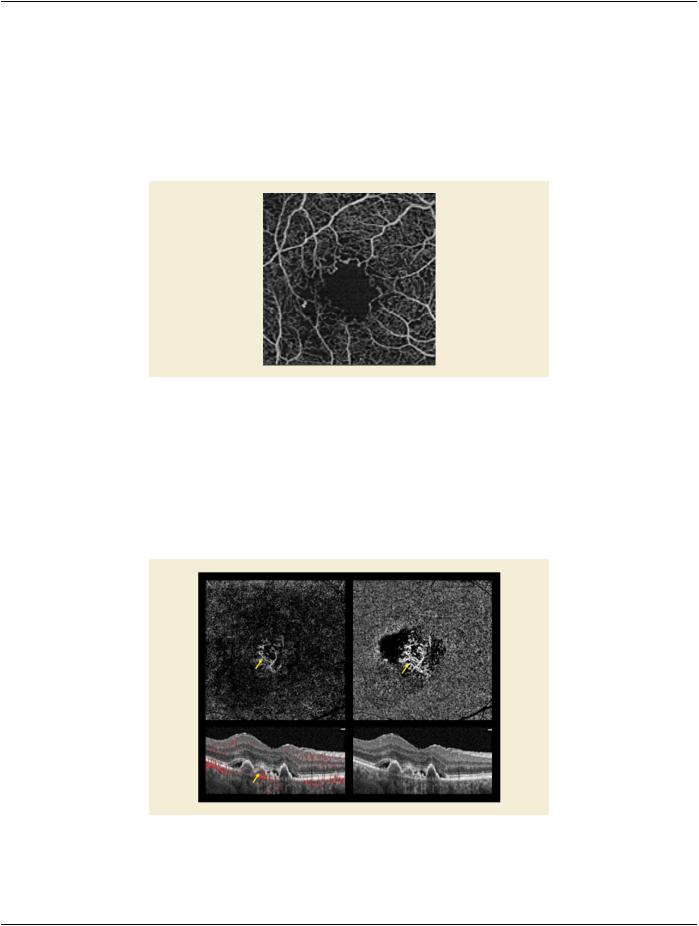
A 54 year old female patient with an 18 year history of DM2 presents with unexplained painless decreased visual acuity in both eyes. The patient was on hemodialysis (HD) for diabetes related renal failure. She had a failed HD shunt in the right arm and a functioning shunt in the left. SD-OCT testing showed no thickening of the macula. Because of her renal failure and HD history IVFA was deferred andOCTangiographyofthemaculaewasperformed.Thisshowedsignificantwideningofthefovealavascularzone(FAZ)explaining her poor visual acuity and excluding treatment opportunities.
Figure UUU.3.1-1. Diabetic Macular Ischemia example
UUU.3.1.2 Age Related Macular Degeneration
A 71 Year Old Male Patient Presents With A 3 Month History of Decreased Visual Acuity and Distorted Vision in The Right Eye. He Demonstrates A Well-defined Elevation of The Deep Retina Adjacent to The Fovea Od by Biomicroscopy That Correlates to A Small Pigment Epithelial Detachment (ped) Shown by Sd-oct. OCT Angiography Demonstrated A Subretinal Neovascular Network in The Same Area. This Was Treated With Intravitreal Anti-vegf Injection Monthly For Three Months With Resolution of The Ped and Incre- mentalRegressionofTheSubretinalNeovascularMembranebyPointtoPointRegistrationOCTAngiographyandFinallyNon-perfusion of The Previous Srn.
Figure UUU.3.1-2. Age related Macular Degeneration example
- Standard -

DICOM PS3.17 2020a - Explanatory Information |
Page 725 |
UUU.3.1.3 Branch Retinal Vein Occlusion
A 59 y/o male patient with hypertension and long smoking history presents with a six week history of painless decrease in vision in therighteye.Ophthalmoscopyshoweddilatedandtortuousveinsinferiortemporallyintherighteyewithasuperiortemporaldistribution of deep retinal hemorrhages that extended to the mid-periphery, but did not include the macula. SD-OCT showed thickening of the macula and OCT angiography showed rarefaction of the retinal capillaries consistent with ischemic branch retinal vein occlusion and macular edema.
Figure UUU.3.1-3. Branch Retinal Vein Occlusion example
UUU.3.2 Research Examples
UUU.3.2.1 Proliferative Diabetic Retinopathy
A38-year-oldmalepatientwith26yearhistoryoftype1diabetesexaminedforevaluationof10-dayhistoryofscantvitreoushemorrhage due to neovascularization of the optic disc.
Figure UUU.3.2-1. Proliferative Diabetic Retinopathy example
- Standard -

Page 726 |
DICOM PS3.17 2020a - Explanatory Information |
- Standard -

DICOM PS3.17 2020a - Explanatory Information |
Page 727 |
VVV Segmentation of Images of Groups of Animals (Informative)
Imaging of small animals used for preclinical research may involve acquiring images of multiple animals simultaneously (i.e., more than one animal is present in the same image).
This Annex describes methods of cross-referencing image and other composite SOP instances produced in the acquisition and seg- mentation process and how the provenance of each may be recorded.
Onlybackwardreferencesaredescribed,allowingforasequentialworkflowwithprocessingperformedbysuccessivedevices,without modification of earlier instances.
VVV.1 Use Case
The relevant Attributes are described in Section C.7.6.1.1.3 “Derivation Description” in PS3.3 and Section C.7.6.1.1.4 “Source Image Sequence” in PS3.3 of the General Image Module. The same principles apply if the General Image Module is not used (e.g., for En- hanced Multi-frame Images, in which the same Attributes are present, but nested in the appropriate Functional Group Macros).
For the purpose of illustration, three successive steps are assumed:
1.acquisition of an image of several animals
2.processing of that image to detect (manually or automatically) the regions containing each animal, and storing the region as an appropriate composite instance
3.creation of derived images for each animal using as input the acquired image and the stored regions for each animal
Various DICOM composite objects could be used to encode the segmented region. If the form of the segmented region is a
•rasterized (bitmap), then the Segmentation Storage SOP Class is appropriate
Note
A bitmap overlay in a Grayscale or Color Softcopy Presentation State Storage SOP Class could also be used, though there are no defined semantics for this unintended use of bitmap overlays.
•surface (mesh), then the Surface Storage SOP Class is appropriate
•set of isocontours, then:
•for 3D patient-relative coordinates, the RT Structure Set Storage SOP Class is appropriate
•for 2D or 3D coordinates (and geometric shapes), a Structured Report Storage SOP Class may be appropriate, if a template with the appropriate semantics (what the contours "mean") is defined
•for2Dcoordinates(andgeometricshapes),aGrayscaleorColorSoftcopyPresentationStateStorageSOPClassmaybeappro- priate, though there are no defined semantics for recognizing what to do with which graphic objects
Forillustrativepurposes,theuseoftheSegmentationStorageSOPClassisassumed,andaconsistentFrameofReferenceisassumed.
Note
If images from different modalities are acquired, on separate devices, but with the same physical arrangement of animals, a more complex workflow might involve the use of one segmentation derived from one modality applied to images from a different modality with a different Frame of Reference, in which case use of the Spatial Registration Storage SOP Class or DeformableSpatialRegistrationStorageaspersistentobjectmightbeappropriate,andappropriatereferencestoitincluded. The same might apply if registration were necessary between images acquired on the same device, but given that research small animals are normally anesthetized, this is usually not required.
- Standard -

Page 728 |
DICOM PS3.17 2020a - Explanatory Information |
VVV.1.1 Reference Attributes
VVV.1.1.1 Acquired Images of Multiple Animals
No references are present, since forward references are not used.
The Frame of Reference UID is present for cross-sectional modalities.
Iftheanimalsarenotallalignedinthesamedirection,PatientPosition(0018,5100)foreachanimalispresentwithinGroupofPatients Identification Sequence (0010,0027) and a nominal Patient Position (0018,5100) is present in the General Series Module, and the coordinate system dependent position and orientation Attributes of the Image Plane Module Attributes (or corresponding Functional Groups) are relative to the nominal Patient Position (0018,5100) present in the General Series Module.
VVV.1.1.2 Segmentation Instances
Segmentations are Enhanced Multi-frame Images, so the Derivation Image Functional Group (Section C.7.6.16.2.6 in PS3.3) is used.
As required by the Segmentation IOD (Section A.51.5.1 in PS3.3):
•the value of Purpose of Reference Sequence (0040,A170) within the Source Image Sequence (0008,2112) within Derivation Image Sequence (0008,9124) is (121322, DCM, "Source Image for Image Processing Operation")
•the value of Derivation Code Sequence (0008,9215) within Derivation Image Sequence (0008,9124) is (113076, DCM, "Segment- ation")
•though not required, the value of Derivation Description (0008, 2111) may contain additional detail describing the image processing operation
The Frame of Reference UID is the same as that for the images from which the segmentation was derived.
There is no requirement that application of the Segmentation be restricted to the image referenced in the Derivation Image Functional GroupMacro,whichdescribestheimagesthatthesegmentationwasderivedfrom,nottheimagestowhichitisapplicable(potentially all of the images in the same Frame of Reference).
The Common Instance Reference Module is required to be present, which provides Study and Series Instance UIDs for all referenced instances.
A segmentation instance may contain multiple segments, thus multiple animals could be described in a single segmentation instance, or each animal could be described in one of multiple segments within a single segmentation instance. The manner in which each segment is numbered, labeled and categorized is thus important. Each segment may be described as follows:
•Segment Number (0062,0004) from 1 to the number of animals (since the Attribute definition requires starting at 1, incrementing by 1)
•SegmentLabel(0062,0005)usingahuman-readablelabelthatappropriatelyidentifieseachanimalinthecontextoftheexperiment, e.g., it may have the same value as the Patient ID (0010,0020) used for each separate animal.
•Segmented Property Category Code Sequence (0062,0003) value of (309825002, SCT, "Spatial and Relational Concept")
•Segmented Property Type Code Sequence (0062,000F) value of (113132, DCM, "Single subject selected from group")
Note
The properties of (309825002, SCT, "Spatial and Relational Concept") and (113132, DCM, "Single subject selected from group") are suggested instead of a more generic description, such as (123037004, SCT, "Anatomical Structure") and (38266002, SCT, "Entire Body"), since though the latter would be accurate, it would not convey the additional implication of selectionofonefrommany.Further,insomecases,theentirebodymaynotactuallybeimaged(e.g.,justtheheadofmultiple subjects may be imaged simultaneously for brain studies).
- Standard -

DICOM PS3.17 2020a - Explanatory Information |
Page 729 |
VVV.1.1.3 Derived Images of Single Animals
It is recommended that the source image(s) be referenced using Source Image Sequence (0008,2112), either in the top level Data Set or within the Derivation Image Functional Group (Section C.7.6.16.2.6 in PS3.3) as appropriate for the IOD, with:
•the value of Purpose of Reference Sequence (0040,A170) within the Source Image Sequence (0008,2112) being (113130, DCM, "Predecessor containing group of imaging subjects")
•the value of Derivation Code Sequence (0008,9215) being (113131, DCM, "Extraction of individual subject from group")
•the value of Derivation Description (0008,2111) containing additional detail describing the image processing operation
It is recommended that the segmentation used be referenced using Referenced Image Sequence (0008,1140), either in the top level Data Set or within the Referenced Image Functional Group (Section C.7.6.16.2.5 in PS3.3) as appropriate for the IOD, with:
•the value of Purpose of Reference Sequence (0040,A170) within Referenced Image Sequence (0008,1140) being (121321, DCM, "Mask image for image processing operation")
Note
If instead of a segmentation (which is a form of image), a non-image object were used to encode the segmented regions, then use of Referenced Instance Sequence (0008,114A) instead of Referenced Image Sequence (0008,1140) would be appropriate.
The Frame of Reference UID is the same as the source images and the segmentation.
Ifalltheanimalsarenotalignedinthesamedirection(i.e.,donothavethesamevalueforPatientPosition(0018,5100)),thecoordinate system dependent position and orientation Attributes of the Image Plane Module Attributes (or corresponding Functional Groups) may have been recomputed. If the animals are aligned in different directions, and Patient Position (0018,5100) from within Group of Patients Identification Sequence (0010,0027) in the source images is compared against Patient Position (0018,5100) from the Gen- eral Series Module in the source images, the difference may be used to recompute (rotate, flip and translate) new patient-relative vectors and offsets within the same Frame of Reference. The value in the Patient Position (0018,5100) from the General Series Module in the derived images are appropriate for the selected animal.
It is recommended that the Common Instance Reference Module be present even if it is not required by the IOD, to provide Study and Series Instance UIDs for all referenced instances.
VVV.1.2 Propagation of Composite Context
Propagation and replacement of the appropriate patient-level and study-level identifying and descriptive Attributes is also required.
The issues related to the identification of the "patient" in such cases are addressed in Section C.7.1.4.1.1 “Groups of Subjects” in PS3.3.
New studies are required if the patient identifiers have changed.
New series are required for each of the derived (types) of objects, since they are created by different equipment and have different values for Modality.
VVV.1.3 Propagation of History
The history of operations applied to a composite instance and its predecessors may be recorded in multiple items of Derivation Code Sequence (0008,9215). It is preferable, when creating a new derived object, to add to the end of the existing sequence of items, rather than to completely replace them. It is also common to add to the plain text that is contained in Derivation Description (0008, 2111), rather than replacing it (maximum length permitting).
The history of which devices (and human operators) have operated on a composite instance and its predecessors may be recorded in Contributing Equipment Sequence (0018,A001). Again, it is preferable that the existing sequence of items be extended rather than replaced, if possible.
For both Derivation Code Sequence (0008,9215) and Contributing Equipment Sequence (0018,A001), if multiple predecessors are applicable (e.g., the source image and a segmentation mask), then the sequence of items of both predecessors may be merged.
- Standard -

Page 730 |
DICOM PS3.17 2020a - Explanatory Information |
- Standard -