Учебники / Middle Ear Mechanics in Research and Otology Huber 2006
.pdf
PreventionandTreatmentofTympanicMembraneBlunting |
177 |
Thomas L. Eby |
|
TheIntactOssicularChaininCholesteatomaSurgery |
183 |
John Hamilton |
|
TheE cacyofOne-StageTympanoplastywithMastoidObliteration |
189 |
andTympanoplastybyTranscanalApproach |
|
Ken Hayashi, Atsushi Shinkawa |
|
“ConceptualDesign”oftheHumanMiddleEar |
197 |
Herbert Hudde |
|
Acoustic-StructuralCoupledFiniteElementAnalysisforSound |
205 |
TransmissioninHumanEar–MiddleEarTransferFunction |
|
Rong Z. Gan, Tao Cheng, Mark W. Wood |
|
E ectsofMiddleEarSuspensoryLigamentsonAcoustic-Mechanical |
212 |
TransmissioninHumanEar |
|
Rong Z. Gan, Tao Cheng, Don Nakmali, Mark W. Wood |
|
EvaluationofLaserVibrometryasDiagnosticUtilitybyMeans |
222 |
ofaSimulationModeloftheMiddleEar |
|
Matthias Bornitz, Nikoloz Lasurashvili, Hans-Jürgen Hardtke, |
|
Thomas Zahnert |
|
BasilarMembraneDisplacementwithOpenedandOccludedOval |
230 |
WindowandBoneConduction |
|
F. Böhnke, A. Arnold, T. Fawzy |
|
x |
|
DevelopmentofaNewClip-PistonProsthesisfortheStapes |
237 |
G. Schimanski, U. Steinhardt, A. Eiber |
|
OntheOptimalCouplingofanImplantableHearingAid– |
246 |
MeasurementsandSimulations |
|
Albrecht Eiber, Christian Breuninger, Jesus Rodriguez Jorge, |
|
Hans P. Zenner, Marcus M. Maassen |
|
TheE ectofCochlearImplantElectrodeInsertiononMiddleEar |
253 |
FunctionasMeasuredbyInVivoLaserDopplerVibrometry |
|
N. Donnelly, A. Bibas, D. Jiang, C. Santulli, A. Fitzgerald O’Connor |
|

MiddleEarMorphometryfromCadavericTemporalBone |
259 |
Micro-CTImaging |
|
S. Puria, J.H. Sim, M. Shin, J. Tuck-Lee, C.R. Steele |
|
InvestigationofBoneConductionThresholdsinOtosclerosis |
269 |
Andreas Arnold, Tamer Fawzy, Frank Böhnke |
|
TranscranialTransmissionofBoneConductedSoundMeasured |
276 |
AcousticallyandPsychoacoustically |
|
Sabine Reinfeldt, Stefan Stenfelt, Bo Håkansson |
|
High-Resolution3-DImagingofMiddleEarOssiclesandtheirSoft |
282 |
TissueStructuresinIntactGerbilTemporalBones,Using |
|
OrthogonalFluorescenceOptical-Sectioning |
|
J.A.N. Buytaert, J.J.J. Dirckx |
|
HyperelasticWarpingAppliedtoX-RayMicroCTImagesforthe |
289 |
Studyof HumanMiddleEarChainDeformationsUnderStatic |
|
PressureLoad |
|
S.L. Gea, S.A Maas, W.F. Decraemer, H. Maier, J.J.J. Dirckx |
|
Real-TimeOpto-ElectronicHolographicMeasurementsof |
295 |
Sound-InducedTympanicMembraneDisplacements |
|
J.J. Rosowski, C. Furlong, M.E. Ravicz, M.T. Rodgers |
|
AnAnimalModelofSuperiorCanalDehiscence |
306 |
J.E. Songer, M.L. Wood, J.J. Rosowski |
|
EpidemiologyofPressureRegulation.IncidenceofVentilationTube |
314 |
TreatmentsanditsPreliminaryCorrelationtoSubsequentEarSurgery |
xi |
Michael Gaihede, Kirsten Hald, Mette Nørgaard, Pia Wogelius, |
|
Daniel Buck, Kjell Tveterås |
|
Morpho-FunctionalPartitionoftheMiddleEarCleft |
322 |
B.M.P.J. Ars, J.J.J. Dirckx, N.M. Ars-Piret |
|
LaserDopplerVibrometryDataoftheCliPPistonMVP |
330 |
A. Arnold, Ch. Stieger, R. Häusler |
|

This page intentionally left blank

OVERVIEW AND RECENT ADVANCES IN BONE CONDUCTION PHYSIOLOGY
Stefan Stenfelt, Department of Neuroscience and Locomotion,
Division of Technical Audiology, Linköping University, Linköping, Sweden Email: stefan.stenfelt@inr.liu.se
During the mid 18th century it was found that sound could be transmitted through |
|
solidsandinthe19thcenturyitwasgenerallyacceptedthatapersoncanperceivesound |
|
by bone conduction (BC). Since then, the research community has tried to understand |
|
its fundamental mechanisms. This report provides an overview of the present state |
|
in BC physiology. Five factors contributing to BC hearing are identified: 1) sound |
|
radiatedintotheearcanal,2)middleearossicleinertia,3)inertiaofthecochlearfluids, |
|
4)alterationofthecochlearspace,and5)pressuretransmissionfromthecerebrospinal |
|
fluid.Ofthese,inertiaofthecochlearfluidseemsmostimportant.Thevibrationmodes |
|
in the skull can be divided into three frequency ranges. At the lowest frequencies, |
|
approximately below 400 Hz, the skull moves as a whole with rigid body motion. At |
|
higher frequencies, up to 1 kHz, the skull motion can be modelled as a mass-spring |
|
system whereas at frequencies above 1 kHz, wave propagation dominates the skull |
|
vibration response. The wave propagation di ers between the thin-boned cranial vault |
|
and the thick and dense bone of the skull base. Both vibration measurement of the |
|
cochlea and ear canal sound pressure can estimate BC perception changes caused by |
|
stimulation alterations for frequencies above 0.8 kHz; below 0.8 kHz measured hearing |
|
thresholds di er from skull vibration and ear canal sound pressure data. |
1 |
1. Introduction
Bone conduction (BC) as a means for hearing stimulation has been used for nearly two centuries to distinguish between a sensorineural and a conductivehearingimpairment.Althoughextensiveresearchisreportedinthe literature, the interpretations are largely dependent on the knowledge of thenormalhearingfunction.Andastheknowledgeincreasesinthefieldof hearing function and in areas as general acoustics and sound transmission insolids,theknowledgeinthefieldofBCphysiologyincreasesaswell.This report gives an overview of the present state in BC physiology.

Through history, di erent species of research animals (cats, guinea pigs, etc) as well as human specimens and live subjects have been used for theBCresearch.ThereisapotentialproblemcomparinganimalBCresults withhumanBCresults;oneshouldbecarefuldrawinggeneralconclusions fromanimaldatatothehuman.Forexample,animalskullsdi eringeometryand compositionfromthehuman skull.Thisresults indi erent vibrationmodesoftheskullthatcanstimulatetheinnereardi erently.Further, in the human, the cochleae are positioned in the skull base surrounded by hard dense bone structure, whereas some animals have cochleae protruding out into the air-filled bulla.
One of the fundamental questions of BC sound was whether it was a cochlear stimulation or a stimulation of some other end organ. Another question was whether the BC transmission was linear or not. Several researchers have addressed these questions by cancelling one air conducted (AC) tone with one BC tone [1–3]. These studies are somewhat limited since they only use one single frequency and consequently only produce local basilar membrane stimulation. The concept of canceling a single AC tone with a BC tone was extended to include two tones; one of 0.7 and one of 1.1 kHz that was subjectively cancelled one at the time, simultaneously, and while a disturbing tone was present. These tests were conducted at three stimulation levels: 40, 50, and 60 dB HL. By comparing amplitude and phase settings for three test subjects at cancellation it was found that the deviations from a true linear response was less than 0.7 dB and 8 degrees at any of the test situations or levels used. The conclusion from these tests, together with previous reported cancellation tests, is that BC sound transmission is linear and a cochlear stimulation at frequencies and levels used for normal hearing.
22. Five contributing factors
Some researchers have identified one single main contributor for perceived BC sound [4,5] while others have reported it to be composed of several differentcontributors[6].Here,fivecomponentsarepresentedascontributing to BC hearing.
2.1 Part I. Outer ear
WhentheskullisvibratedbyBCstimulation,soundisradiatedintotheear canal that is subsequently transmitted to the cochlea by the ear drum and middle ear ossicles. With the BC stimulation applied at the mastoid and with the ear canal open, the outer ear part of the BC sound is 5 to 20 dB below other contributing parts and is not considered as a major contributing

part to BC sound [7]. This may change when the stimulation is at another position,e.g.whenthestimulationisattheforehead,theouterearseemsto contribute more at frequencies below 1 kHz.
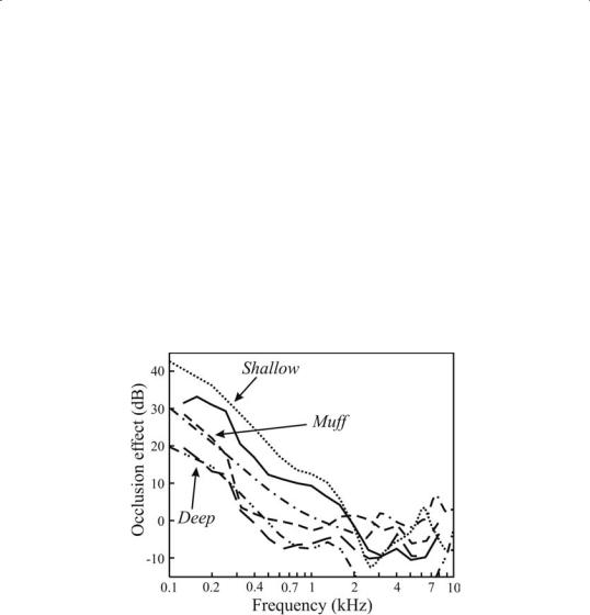
When the ear canal is closed (e.g. by a finger or an ear plug) the BC sound in the ear canal is enhanced at the low frequencies; this is termed the occlusion e ect. The exact frequency range and amount of elevation caused by the occlusion depends on the type and location of the occlusion device. However, the occlusion e ect is determined by the acoustics of the ear canal and the resulting ear canal sound pressure alteration caused by the occlusion device can be estimated with good accuracy if the position and type of device is known. An estimate of the occlusion e ect with ear plugs at two di erent depths and with one ear mu is shown in Fig 1.
Fig. 1 The graph presents the measured ear canal sound pressure occlusion e ect together with model prediction for ear plugs shallow and deeply inserted and with
an ear mu of about 30 cm3. Solid line: measured shallow inserted ear plug, dotted 3 line: model estimated ditto, long-dashed line: measured deeply inserted earplug, dash-double dotted line: model estimated ditto, dashed line: measured ear mu , dashdotted line: model estimated ditto.
2.2 Part II. Middle ear ossicle inertia
The three middle ear ossicles are suspended by several ligaments, the TM and two muscle tendons. This becomes a mechanical mass-spring system that, due to inertial forces on the masses, constitutes di erent vibration than the surrounding bone during BC stimulation. This di erence motion between the stapedius footplate and the cochlear promontory provides a stimulation input to the cochlea [8]. The importance of the ossicle inertia

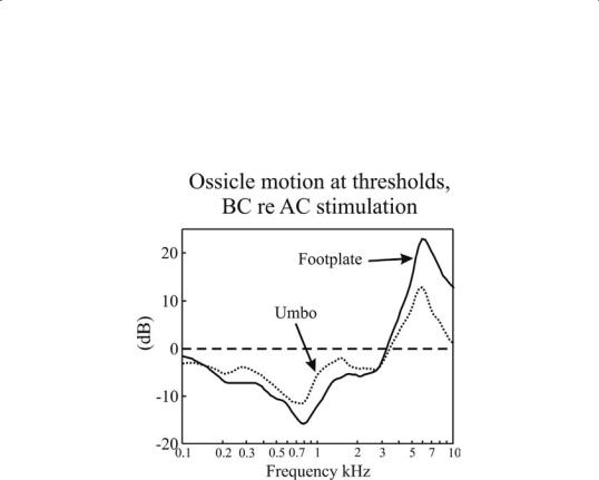
component can be estimated by comparing the stapes footplate motion at thresholdstimulationbyACandBC;thiswasdonein[9]andsomeofthose results are presented in Fig 2.
Fig. 2 Stapes footplate (solid line) and malleus umbo (dotted line) motion at BC threshold stimulation compared with the motion at AC threshold stimulation.
The vibration of the umbo and footplate at threshold vibration is 5 to 15 dB lower with BC stimulation compared with AC stimulation at frequencies between 0.2 and 1.5 kHz. This result indicates that middle ear inertia is not an important contribution to BC perception at these frequencies. At frequencies above 4 kHz, the comparison is not valid since the stimulation is probably no longer through the oval window. However, the similarity be-
4tween the ossicle threshold motion between 1.5 and 3.5 kHz indicates that the inertial e ects of the ossicles can contribute to BC sound perception within this frequency range. Also, lesions and manipulations of the ossicles provides results indicative of possible importance of ossicle inertia at the mid-frequency range [10].
2.3 Part III. Inertia of the cochlear fluids
As with the middle ear ossicles, inertial e ects cause a relative motion between the cochlear fluids and the cochlear promontory bone. This type of motion relies on several compliant fluid in and outlets in the cochlea: the oval and round windows are two such fluid pathways, and others that can be of importance are the cochlear and vestibular aqueducts, nerve fibers,

veins, and microchannels entering the cochlea. Since many BC threshold alterations with middle ear lesions corroborate the theory of fluid inertia it is believed to be the most important BC contributor for frequencies below 4 kHz [10].
2.4 Part IV. Alteration of the cochlear space
One of the first theories presented for BC perception was alteration of the cochlear space, often termed compression response. Since the cochlear fluids can be considered incompressible, any alteration of the cochlear space would force fluid to move and most probably cause a stimulation of the basilar membrane. First, alteration of the cochlear space requires wave propagation in the skull bone which begins at around 1 kHz [11]. Second, according to the theory a compression response would benefit from reduced fluid flow at the oval window (otosclerosis), and would be reduced by an opening at the vestibular side (fenestration operation or semicircular canaldehiscence).Thisisoppositetoclinicalfindingsforfrequenciesbelow 4 kHz. Compression may well be important at higher frequencies.
2.5 Part V. Pressure transmission from the CSF
A recent identified contributor to perception of BC sound is pressure set up in the cerebrospinal fluid surrounding the brain that is transmitted to the cochlea producing a traveling wave on the basilar membrane [12]. Although this mode does provide cochlear stimulation, it fails to explain several experimental findings, including BC threshold losses with certain lesionsofthemiddleearossicles,lateralizationofaBCtone,andtranscranial attenuation. It is therefore unlikely that this contribution is significant for BC perception in the hearing frequency range.
3. BC transmission and skull vibration |
5 |
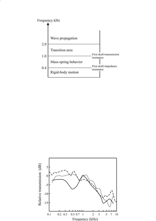
When a BC stimulation is applied to the skull, the resulting skull motion depends on the position, direction, and frequency of the stimulation. The frequency response of the skull can roughly be divided into three response modes: (1) pure rigid body motion, (2) mechanical mass-spring system, and (3) wave propagation. The pure rigid body motion is at the lowest frequencies (up to 250 to 400 Hz). At these low frequencies the skull motion is solely determined by its geometry together with three translational and three rotational components. This vibration mode can be identified by investigation of the mechanical point impedance; it is the frequency range below the first impedance resonance where the impedance shows a masslike behavior.

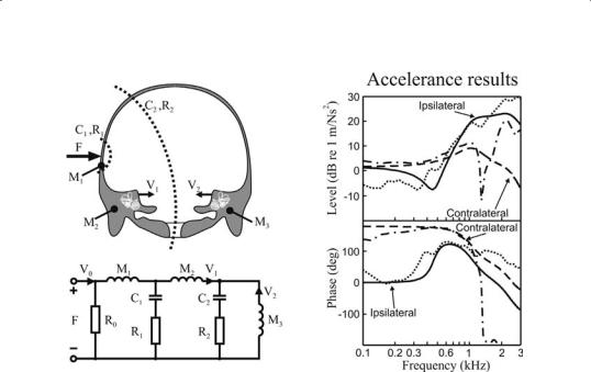
Fig. 3 The left part shows the anatomical structure and related lumped element model for estimation of the cochleae vibration during mastoid stimulation. The right part presents the level and phase of the accelerance transmission between the mastoid and cochlea. Solid line: modelled accelerance of the ipsilateral cochlea, dashed line: measured ditto, dashed line: modelled accelerance of the contralateral cochlea, dashdotted line: measured ditto.
The next response mode, where the skull behaves as a mass-spring system, is at frequencies below the first skull resonance, which is approximately at 1 kHz [11]. In this frequency range, the motion of the skull can be approximated by three masses with springs in-between (see Fig 3). At higher frequencies, above the first skull resonance, the BC sound is transmitted by wave propagation. This wave propagation depends on type and size of the bone. At the cranial vault, where the skull bone is thin, the wave trans-
6mission is a mixture of bending, longitudinal, and transverse wave propagation. These waves are dispersive and the phase velocity increases from just above 200 m/s at 2 kHz to between 300 and 350 m/s at 10 kHz. In the skullbase,wherethecochleaearepositioned,theboneisthickeranddenser and the wave transmission is primarily by longitudinal wave propagation producing a constant wave speed of approximately 400 m/s. The di erent transmission modes and frequency ranges are depicted in Fig 4.

Fig. 4 Overview of the frequency ranges of the human heads’ vibration modes.
The transcranial transmission, i.e. the sound transmitted from the mastoid to the contralateral cochlea compared with that transmitted to the ipsilateral cochlea was measured by three di erent methods; (1) mechanical vibrationofthecochleaeaobtainedincadaverheads[11],(2)earcanalsound pressure, and (3) hearing thresholds obtained in live humans. The results are presented in Fig. 5 where the three methods show similar results at frequencies above 0.8 kHz; almost monotonically decreasing transmission of –2 dB at 0.8 kHz to –15 dB at 8 kHz. At lower frequencies, the vibration and ear canal sound pressure data are similar whereas the threshold data are almost 7 dB lower at 0.5 kHz. This indicates that both vibration and ear canal sound pressure can be used to estimate changes in BC perception at frequencies at and above 1 kHz.
7
Fig. 5 The transcranial transmission with the stimulation at the mastoid estimated by three methods. Solid line: hearing thresholds, dotted line: ear canal sound pressure, dashed line: vibration of the cochlear promontory.
