

- •Overview
- •Preface
- •Translator’s Note
- •Contents
- •1. Fundamentals
- •Microscopic Anatomy of the Nervous System
- •Elements of Neurophysiology
- •Elements of Neurogenetics
- •General Genetics
- •Neurogenetics
- •Genetic Counseling
- •2. The Clinical Interview in Neurology
- •General Principles of History Taking
- •Special Aspects of History Taking
- •3. The Neurological Examination
- •Basic Principles of the Neurological Examination
- •Stance and Gait
- •Examination of the Head and Cranial Nerves
- •Head and Cervical Spine
- •Cranial Nerves
- •Examination of the Upper Limbs
- •Motor Function and Coordination
- •Muscle Tone and Strength
- •Reflexes
- •Sensation
- •Examination of the Trunk
- •Examination of the Lower Limbs
- •Coordination and Strength
- •Reflexes
- •Sensation
- •Examination of the Autonomic Nervous System
- •Neurologically Relevant Aspects of the General Physical Examination
- •Neuropsychological and Psychiatric Examination
- •Psychopathological Findings
- •Neuropsychological Examination
- •Special Considerations in the Neurological Examination of Infants and Young Children
- •Reflexes
- •4. Ancillary Tests in Neurology
- •Fundamentals
- •Imaging Studies
- •Conventional Skeletal Radiographs
- •Computed Tomography (CT)
- •Magnetic Resonance Imaging (MRI)
- •Angiography with Radiological Contrast Media
- •Myelography and Radiculography
- •Electrophysiological Studies
- •Fundamentals
- •Electroencephalography (EEG)
- •Evoked potentials
- •Electromyography
- •Electroneurography
- •Other Electrophysiological Studies
- •Ultrasonography
- •Other Ancillary Studies
- •Cerebrospinal Fluid Studies
- •Tissue Biopsies
- •Perimetry
- •5. Topical Diagnosis and Differential Diagnosis of Neurological Syndromes
- •Fundamentals
- •Muscle Weakness and Other Motor Disturbances
- •Sensory Disturbances
- •Anatomical Substrate of Sensation
- •Disturbances of Consciousness
- •Dysfunction of Specific Areas of the Brain
- •Thalamic Syndromes
- •Brainstem Syndromes
- •Cerebellar Syndromes
- •6. Diseases of the Brain and Meninges
- •Congenital and Perinatally Acquired Diseases of the Brain
- •Fundamentals
- •Special Clinical Forms
- •Traumatic Brain injury
- •Fundamentals
- •Traumatic Hematomas
- •Complications of Traumatic Brain Injury
- •Intracranial Pressure and Brain Tumors
- •Intracranial Pressure
- •Brain Tumors
- •Cerebral Ischemia
- •Nontraumatic Intracranial Hemorrhage
- •Infectious Diseases of the Brain and Meninges
- •Infections Mainly Involving the Meninges
- •Infections Mainly Involving the Brain
- •Intracranial Abscesses
- •Congenital Metabolic Disorders
- •Acquired Metabolic Disorders
- •Diseases of the Basal Ganglia
- •Fundamentals
- •Diseases Causing Hyperkinesia
- •Other Types of Involuntary Movement
- •Cerebellar Diseases
- •Dementing Diseases
- •The Dementia Syndrome
- •Vascular Dementia
- •7. Diseases of the Spinal Cord
- •Anatomical Fundamentals
- •The Main Spinal Cord Syndromes and Their Anatomical Localization
- •Spinal Cord Trauma
- •Spinal Cord Compression
- •Spinal Cord Tumors
- •Myelopathy Due to Cervical Spondylosis
- •Circulatory Disorders of the Spinal Cord
- •Blood Supply of the Spinal Cord
- •Arterial Hypoperfusion
- •Impaired Venous Drainage
- •Infectious and Inflammatory Diseases of the Spinal Cord
- •Syringomyelia and Syringobulbia
- •Diseases Mainly Affecting the Long Tracts of the Spinal Cord
- •Diseases of the Anterior Horns
- •8. Multiple Sclerosis and Other Myelinopathies
- •Fundamentals
- •Myelin
- •Multiple Sclerosis
- •Other Demyelinating Diseases of Unknown Pathogenesis
- •9. Epilepsy and Its Differential Diagnosis
- •Types of Epilepsy
- •Classification of the Epilepsies
- •Generalized Seizures
- •Partial (Focal) Seizures
- •Status Epilepticus
- •Episodic Neurological Disturbances of Nonepileptic Origin
- •Episodic Disturbances with Transient Loss of Consciousness and Falling
- •Episodic Loss of Consciousness without Falling
- •Episodic Movement Disorders without Loss of Consciousness
- •10. Polyradiculopathy and Polyneuropathy
- •Fundamentals
- •Polyradiculitis
- •Cranial Polyradiculitis
- •Polyradiculitis of the Cauda Equina
- •Polyneuropathy
- •Fundamentals
- •11. Diseases of the Cranial Nerves
- •Fundamentals
- •Disturbances of Smell (Olfactory Nerve)
- •Neurological Disturbances of Vision (Optic Nerve)
- •Visual Field Defects
- •Impairment of Visual Acuity
- •Pathological Findings of the Optic Disc
- •Disturbances of Ocular and Pupillary Motility
- •Fundamentals of Eye Movements
- •Oculomotor Disturbances
- •Supranuclear Oculomotor Disturbances
- •Lesions of the Nerves to the Eye Muscles and Their Brainstem Nuclei
- •Ptosis
- •Pupillary Disturbances
- •Lesions of the Trigeminal Nerve
- •Lesions of the Facial Nerve
- •Disturbances of Hearing and Balance; Vertigo
- •Neurological Disturbances of Hearing
- •Disequilibrium and Vertigo
- •The Lower Cranial Nerves
- •Accessory Nerve Palsy
- •Hypoglossal Nerve Palsy
- •Multiple Cranial Nerve Deficits
- •12. Diseases of the Spinal Nerve Roots and Peripheral Nerves
- •Fundamentals
- •Spinal Radicular Syndromes
- •Peripheral Nerve Lesions
- •Fundamentals
- •Diseases of the Brachial Plexus
- •Diseases of the Nerves of the Trunk
- •13. Painful Syndromes
- •Fundamentals
- •Painful Syndromes of the Head And Neck
- •IHS Classification of Headache
- •Approach to the Patient with Headache
- •Migraine
- •Cluster Headache
- •Tension-type Headache
- •Rare Varieties of Primary headache
- •Symptomatic Headache
- •Painful Syndromes of the Face
- •Dangerous Types of Headache
- •“Genuine” Neuralgias in the Face
- •Painful Shoulder−Arm Syndromes (SAS)
- •Neurogenic Arm Pain
- •Vasogenic Arm Pain
- •“Arm Pain of Overuse”
- •Other Types of Arm Pain
- •Pain in the Trunk and Back
- •Thoracic and Abdominal Wall Pain
- •Back Pain
- •Groin Pain
- •Leg Pain
- •Pseudoradicular Pain
- •14. Diseases of Muscle (Myopathies)
- •Structure and Function of Muscle
- •General Symptomatology, Evaluation, and Classification of Muscle Diseases
- •Muscular Dystrophies
- •Autosomal Muscular Dystrophies
- •Myotonic Syndromes and Periodic Paralysis Syndromes
- •Rarer Types of Muscular Dystrophy
- •Diseases Mainly Causing Myotonia
- •Metabolic Myopathies
- •Acute Rhabdomyolysis
- •Mitochondrial Encephalomyopathies
- •Myositis
- •Other Diseases Affecting Muscle
- •Myopathies Due to Systemic Disease
- •Congenital Myopathies
- •Disturbances of Neuromuscular Transmission−Myasthenic Syndromes
- •15. Diseases of the Autonomic Nervous System
- •Anatomy
- •Normal and Pathological Function of the Autonomic Nervous System
- •Sweating
- •Bladder, Bowel, and Sexual Function
- •Generalized Autonomic Dysfunction
- •Index
216 12 Diseases of the Spinal Nerve Roots and Peripheral Nerves
a
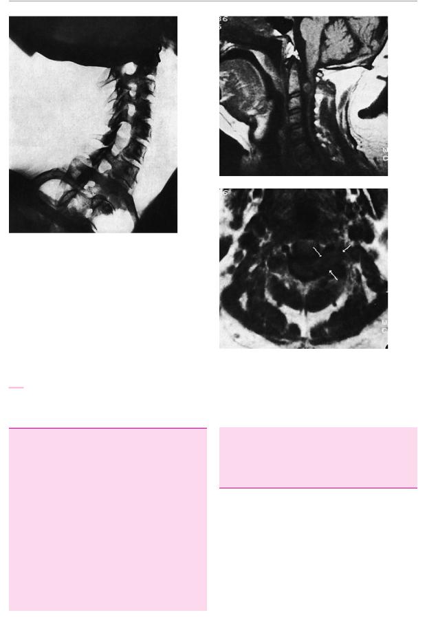
Fig. 12.13 Widened C5/6 intervertebral foramen caused by a neurinoma of the C6 root. The C5/6 foramen has opened up into the C6/7 foramen below it.
Fig. 12.14 Left C3 root neurinoma. The sagittal MR image (a) |
|
reveals a compressive lesion anterior to the spinal cord. The axial |
|
image (b) shows the deformed cervical spinal cord posterior to the |
|
dens. The arrows indicate the left C3 root, which is expanded by the |
|
tumor. |
b |
Peripheral Nerve Lesions
Fundamentals
When we speak here of the “peripheral nerves,” we are referring to the nerve plexuses formed by the junction and regrouping of fibers derived from the spinal nerve roots, as well as to the more distally lying peripheral nerve trunks and branches. The plexuses always contain mixed fiber types and the peripheral nerve trunks nearly always do, i. e., somatic motor, somatosensory, and often also autonomic (particularly sympathetic) fibers. The individual peripheral nerve trunks bear an anatomically invariant relationship to the muscles and cutaneous zones that they innervate. This pattern of innervation is fundamentally different from that of the spinal nerve roots, because, as we recall, the nerve root fibers undergo reassortment in the plexuses. This fact enables the clinical examiner to distinguish a peripheral nerve lesion from a radicular lesion based on the observed pattern of neurological deficits. The main clinical manifestations of
peripheral nerve lesions are marked paresis, extensive sensory deficits, and diminished sweating in the zone of innervation of the affected nerve or nerve branch. Pain can be produced by either a radicular lesion or a peripheral nerve lesion and is thus not a distinguishing feature of either.
Preliminary anatomical remarks. A peripheral nerve is a cablelike bundle of nerve fibers of different functional types (see p. 3). The nerve fiber is the smallest “building block” of a peripheral nerve; it consists of an axon and an encasing myelin sheath (if present), which is the membrane of a Schwann cell wrapped around the axon numerous times. Individual nerve fibers are surrounded by a delicate connective tissue called endoneurium. The nerve fibers and the endoneurium are then bundled together into larger fascicles, each of which is surrounded by a tough perineurium. Along the length of the nerve, the individual fascicles make many plexus-like intercon-
Mumenthaler / Mattle, Fundamentals of Neurology © 2006 Thieme All rights reserved. Usage subject to terms and conditions of license.

Peripheral Nerve Lesions
nections with one another; they are held together as a single peripheral nerve by an encompassing layer of epineurium. The epineurium is not a tough husk around the nerve, but rather a loose, lipid-rich layer of connective tissue, reinforced by transversely and longitudinally oriented collagen fibers. It contains not only the nerve fascicles, but also the vasa nervorum. The nerve trunks are fixed to the adjacent connective tissue at only a few points, at which they are especially vulnerable to mechanical damage. Larger nerve trunks are often found together with arteries and veins in so-called neurovascular bundles surrounded by a common connective tissue sheath. These bundles form an anatomical unit that is clearly demarcated from the surrounding structures.
Causes of peripheral nerve lesions. Most lesions of the nerve plexuses or the peripheral nerve trunks are either traumatic (caused by excessive traction, stab wounds, cuts, bony fractures, etc.), or else due to prolonged compression, which may occur through external influences, at anatomical bottlenecks, or because of space-occupying lesions in the vicinity of the nerve (especially tumors and hematomas). Less commonly, plexus and nerve lesions can be caused by infection and/or inflammation, e. g., neuralgic shoulder amyotrophy, which is probably an autoimmune disorder affecting the brachial plexus (p. 222). Nearly all lesions affecting a single peripheral nerve trunk or branch (mononeuropathy) are of mechanical origin; in contrast, most polyneuropathies (cf. Chapter 10) are of toxic, infectious/inflammatory, or paraneoplastic origin.
General clinical manifestations of peripheral nerve lesions. Depending on the particular segment of plexus or peripheral nerve trunk/branch that is affected, there may be a motor, sensory, autonomic, or (usually) mixed neurological deficit:
flaccid paresis of the muscle(s) innervated by the affected nerve;
usually marked atrophy of the affected muscle(s);
the corresponding reflex deficits;
diminished sensation and possibly also pain and paresthesia in the cutaneous area innervated by the nerve, though the pain is often felt beyond this area as well; all sensory modalities are affected to a comparable extent; in contrast to a radicular lesion, the affected area of skin is more easily demarcated by testing the sense of touch than by testing nociception;
because the sudomotor fibers travel together with the somatosensory components of the peripheral nerves, diminished sweating is often found in the hypesthetic area of skin and autonomic abnormalities of other kinds may also be present in the distribution of the affected nerve; radicular lesions affecting the limbs, in contrast, generally leave sweating intact (an important criterion for differential diagnosis);
fasciculations only in exceptional cases; these are much more common in anterior horn disease.
Grades of severity of peripheral nerve lesions. A peripheral nerve can be damaged more or less severely, with corresponding implications for treatment and prognosis. The traditional, clinically useful threefold distinction is as follows:
Neurapraxia: the nerve is dysfunctional, but its anatomical components are still in continuity (e. g., nerve dysfunction due to pressure on the nerve when the individual has slept for a prolonged period in an unusual posture); the functional deficit is completely reversible.
Axonotmesis: the axons within the nerve are interrupted, but the external structure of the nerve and its internal connective tissue sheaths remain intact; the full clinical picture of a peripheral nerve lesion results; under optimal conditions, full recovery may still be possible.
Neurotmesis: the axons and all surrounding structures are interrupted and the nerve is no longer in continuity (e. g., because of tearing or sharp transection of the nerve); a surgical procedure is needed to restore nerve integrity and the prognosis for recovery is uncertain.
Diseases of the Brachial Plexus
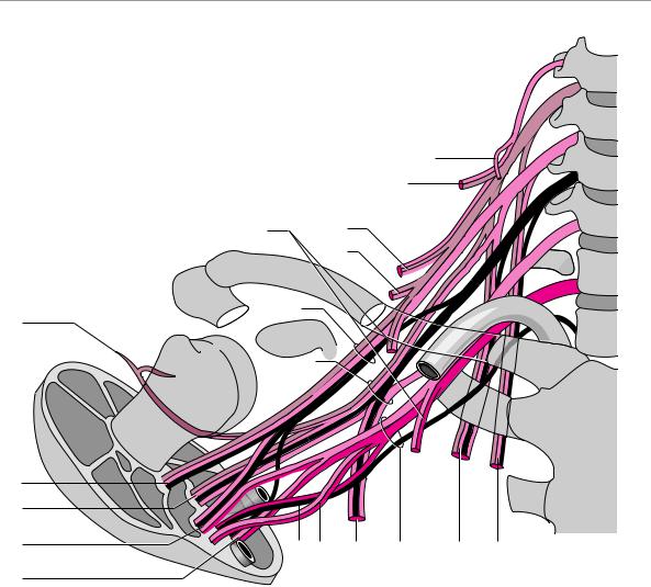
Anatomy of the brachial plexus. In the brachial plexus, the axons derived from the nerve roots of C4 to T1 (or T2) are regrouped and distributed to the various nerves that innervate the upper limb (Fig. 12.15). The brachial plexus passes through three anatomical bottlenecks on its way to the upper arm; the first two are the scalene hiatus, where it is accompanied by the subclavian a., and the costoclavicular space between the first rib and the clavicle. At this location, the caudal portion of the plexus is in close proximity to the apex of the lung. A bit further distally, the brachial plexus is covered by the pectoralis minor m., which originates from the coracoid process of the scapula, and it can be compressed at this location when the arm is elevated. These bottlenecks are sketched in Fig. 12.16.
General clinical manifestations of brachial plexus lesions. The complex structure of the brachial plexus and the redistribution of individual radicular elements within it make it very difficult to localize brachial plexus lesions exactly based on the neurological findings alone. Nonetheless, detailed functional testing of the affected muscles can reveal the root levels that are involved and this, in turn, permits localization of the lesion within the plexus with a fair degree of precision. The information provided in Fig. 12.17 will be helpful in this regard.
Classification of brachial plexus lesions. In addition to total paralysis of the entire upper limb, there are three types of partial lesion in the customary, topically oriented classification, namely, upper and lower brachial plexus lesions and C7 lesions. An alternative (or additional) classification is by etiology: traumatic, compressive, or infectious/inflammatory.
Topical Classification of Brachial Plexus Lesions
Upper brachial plexus lesion (Erb−Duchenne palsy).
This type of lesion involves the fibers originating in the C5 and C6 nerve roots. The affected muscles are the abductors and external rotators of the shoulder joint, the flexor muscles of the upper arm, the supinator m., and
ThiemeArgoOneBoldthiemeArgoOne
217
Diseases of the Spinal Nerve Roots and Peripheral Nerves
12
Mumenthaler / Mattle, Fundamentals of Neurology © 2006 Thieme All rights reserved. Usage subject to terms and conditions of license.
218 12 Diseases of the Spinal Nerve Roots and Peripheral Nerves
|
|
(C4) |
|
|
|
C5 |
IV |
|
|
C6 |
V |
|
|
18 |
|
|
|
C7 |
VI |
|
|
17 |
|
1 |
16 |
C8 |
VII |
|
|
||
|
15 |
|
|
|
|
T1 |
I |
5 |
2 |
|
|
|
(T2) |
II |
|
|
|
||
|
3 |
|
|
6
7
8 |
10 |
11 |
12 |
4 |
13 |
14 |
|
9
Fig. 12.15 The brachial plexus and its anatomical relationships to the surrounding bony structures.
1pectoral nn. (med./lat.) C5−T1 pectoralis major & minor mm.
2 lateral cord
3 dorsal (posterior) cord
4 medial cord
5axillary n. C5, 6 deltoid m. C5, 6 teres minor m. C5, 6
6 musculocutaneous n. C5−7 biceps brachii m. C5, 6 coracobrachialis m. C6, 7 brachialis m. C5, 6
7radial n. C5−T1
triceps brachii m. C7−T1 anconeus m. C7, 8 brachioradialis m. C5, 6
extensor carpi rad. long./brev. mm. C6−8 extensor digit. m. C7, 8
extensor indicis m. C7, 8 extensor digiti minimi m. C7, 8
ext. poll. long./brev. mm. C7, 8 abd. poll. long. m. C7, 8
8median n. C5−T1 pronator teres m. C6, 7
flexor carpi radialis m. C6−8 palmaris longus m. C7, 8 flexor digit. superf. m. C7−T1
flexor digit. prof. m. (radial side, II/III) C7−T1
pronator quadratus m. C7−T1 opponens pollicis m. C7, 8 abductor poll. brev. m. C7, 8
superf. head of flex. poll. brev. m. C6−8
lumbrical mm. I & II C8−T1
9ulnar n. (C7) C8−T1 flexor carpi uln. m. C8−T1
flexor digit. prof. m. (ulnar side, IV/V) C8−T1
palm./dors. interossei mm. C8−T1 lumbrical mm. III & IV C8−T1 adductor pollicis m. C8−T1
deep head of flex. poll. brev. m. C8−T1 palmaris brevis m. C8−T1
10medial brachial cutaneous n. C8−T1
11medial antebrachial cutaneous n.
C8−T1
12thoracodorsal n. C6−8 latissimus dorsi m.
13subscapular nn. C5−8 subscapular m. C5−7 teres major m. C5−6
14long thoracic n. C5−7 serratus anterior m.
15nerve to the subclavius m. C5, 6 subclavius m.
16suprascapular n. C4−6 supraspinatus m. C4−6 infraspinatus m. C4−6
17dorsal scapular n. C3−5 levator scapulae m. C4−6 rhomboid mm. C4−6
18phrenic n. C3, 4
Mumenthaler / Mattle, Fundamentals of Neurology © 2006 Thieme All rights reserved. Usage subject to terms and conditions of license.
Peripheral Nerve Lesions
Fig. 12.16 Anatomical bottlenecks in the shoulder region, at
which nerves and/or blood vessels can be compressed.
anterior scalene m.
middle scalene m.
brachial 1 plexus
subclavian a.
subclavian v.
2
clavicle
1st rib
3
1scalene hiatus costoclavicular
2passage
3subpectoralpassage
|
|
|
|
|
|
|
|
|
|
|
|
|
|
|
|
|
|
|
rhomboid muscles |
|
|
|
C6 |
|
|
|
|
|
C8 |
|
|
|
|
||
|
|
|
|
|
|
|
|
|
|
|
|
|
|
|
|
|
|
|
trapezius |
|
|
|
|
|
|
|
|
|
|
|
|
|
|
|
|
|
|
C5 |
|
|
|
C7 |
|
|
|
|
|
T1 |
|
|
|
||
|
|
|
|
|
|
|
|
|
|
|
|
|
|
||||
|
|
serratus anterior |
|
|
|
|
|
|
|
|
|
|
|
|
|
|
|
|
|
|
|
|
|
|
II III IV V |
|
|
|
|
|
|
|
|
||
|
posterior |
|
|
|
|
|
pronator teres |
flexor digitorum |
|
|
|
|
opponens |
abductor |
|
||
|
|
|
|
|
|
superficialis |
|
|
|
|
pollicis |
|
|||||
|
portion |
biceps |
|
|
|
|
|
|
|
|
|
pollicis |
|
|
|||
|
|
|
|
|
|
|
|
|
|
|
|
brevis |
|
||||
|
|
|
|
|
|
|
|
|
|
|
|
|
|
|
|||
|
|
brachii |
|
|
|
flexor carpi |
palmaris |
|
|
|
|
|
|
|
|||
|
lateral |
|
|
|
|
|
|
|
|
|
|
|
|||||
|
|
|
|
|
|
radialis |
longus |
|
|
|
|
|
|
|
|
||
|
portion |
|
|
|
|
|
|
|
|
|
|
|
|
|
|||
|
|
|
|
|
|
|
|
|
|
|
|
|
|
|
|
|
|
|
deltoid |
|
|
|
|
|
triceps brachii |
|
|
|
flexor |
flexor |
|
adductor |
|
||
|
|
|
|
|
|
|
extensor carpi |
extensor |
|
|
|
|
|
||||
|
|
|
|
|
|
|
|
|
|
pollicis |
pollicis |
|
|
||||
|
|
brachialis |
|
|
|
|
|
|
|
pollicis |
|
||||||
|
anterior |
|
|
|
radialis |
carpi ulnaris |
|
|
|
longus |
brevis |
|
|
||||
|
|
|
|
|
|
|
|
|
|
|
|
||||||
|
portion |
|
|
|
|
|
|
|
|
|
|
|
|
|
|
|
|
|
|
|
|
|
|
|
extensor |
abductor |
|
|
|
|
abductor digiti |
|
|||
|
|
brachio- |
|
|
|
digitorum |
pollicis longus |
|
|
|
flexor |
|
minimi |
|
|||
|
teres minor |
|
|
|
communis and |
|
|
|
|
|
|
||||||
|
radialis |
|
|
|
|
|
|
|
|
|
|
|
|||||
|
|
|
|
|
extensores |
extensor |
|
|
|
digitorum |
1st dorsal |
|
|||||
|
|
|
|
|
|
|
|
|
|
|
|||||||
|
|
|
|
|
|
|
digitorum |
pollicis brevis |
|
|
|
II+III |
interosseous m. |
|
|||
|
|
supinator |
|
|
|
proprii |
|
|
|
|
|
|
|
|
|
||
|
supraspinatus |
|
|
|
|
|
extensor |
|
|
|
pro- |
palmaris brevis |
|
||||
|
|
|
|
|
|
|
|
|
|
|
|
|
|||||
|
|
|
|
|
|
|
|
|
pollicis longus |
|
|
|
fundus |
|
|
|
|
|
|
|
|
|
|
|
latissimus |
|
|
|
|
IV+V |
remainder of the dorsal |
|
|||
|
infraspinatus |
teres major |
|
|
|
flexor carpi |
|
|
|
|
|
||||||
|
|
|
|
dorsi |
|
|
|
|
interosseous mm. |
|
|||||||
|
|
|
|
|
|
|
ulnaris |
|
|
|
|
|
|||||
|
|
|
|
|
|
|
|
|
|
|
|
|
|
|
|
|
|
|
|
|
|
|
|
|
M. pectoralis major |
|
|
|
|
|
|
|
|
||
|
|
|
|
|
|
|
|
|
|
|
|
|
|
|
|
|
|
|
|
|
|
|
|
|
|
|
|
|
|
|
|
|
|
|
|
Fig. 12.17 Muscles of the upper limb and the nerve roots that innervate them. With the aid of this diagram, brachial plexus lesions can be localized from the pattern of muscle weakness that they cause.
ThiemeArgoOneBoldthiemeArgoOne
219
Diseases of the Spinal Nerve Roots and Peripheral Nerves
12
Mumenthaler / Mattle, Fundamentals of Neurology © 2006 Thieme All rights reserved. Usage subject to terms and conditions of license.
220 12 Diseases of the Spinal Nerve Roots and Peripheral Nerves
a |
b |
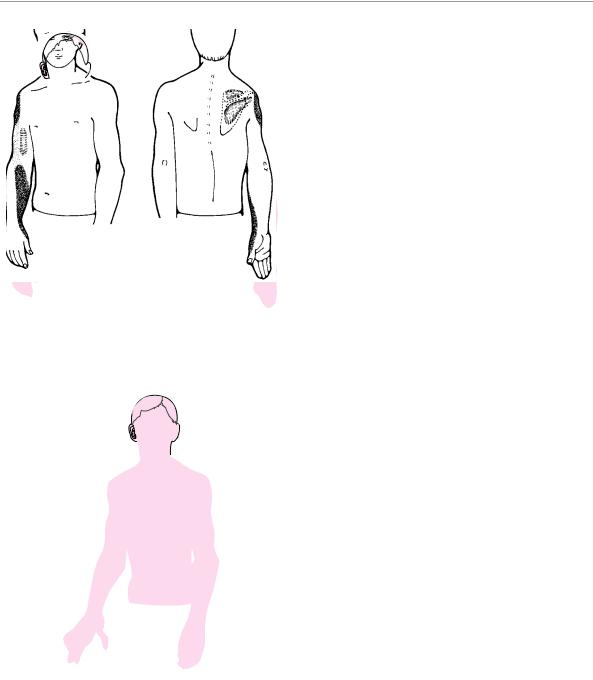
Fig. 12.18 Upper limb posture and sensory deficit in right upper brachial plexus palsy (diagram). The deltoid, biceps, and supraand infraspinatus mm. are atrophic, the arm is internally rotated, and the palm of the hand points posteriorly.
Fig. 12.19 Upper limb posture and sensory deficit in right lower brachial plexus palsy (diagram). The intrinsic muscles of the hand are atrophic, the sensory deficit corresponds to the C7 and C8 dermatomes, and there is an accompanying Horner syndrome.
sometimes the elbow extensors and the extensors of the hand. A sensory deficit is not necessarily present; if there is one, it is located in the area of the shoulder, on the outer surface of the upper arm, or on the radial edge of the forearm (Fig. 12.18).
Lower brachial plexus lesion (Dejerine−Klumpke palsy). This type of lesion involves the fibers originating in the C8 and T1 roots. Its prominent findings include weakness of the intrinsic muscles of the hand, some-
times also of the long flexors of the fingers, and rarely of the wrist flexors. The triceps brachii m. usually remains intact. The mechanism of the precipitating accident, and the anatomical relationships in this area, often lead to an accompanying dysfunction of the cervical sympathetic supply, resulting in Horner syndrome with impaired sweating. On the basis of these findings, a lesion of the T1 root is presumed to be present, proximal to the origin of its branch to the sympathetic chain. There is always a sensory deficit involving the ulnar edge of the forearm, hand, and fingers (Fig. 12.19).
C7 palsy. In the present context, we are speaking not of a lesion of the C7 root itself, but rather of the fibers derived from it that make up the C7 portion of the brachial plexus. This type of palsy involves deficits in the distribution of the radial n. (p. 226), while the function of the brachioradialis m. is preserved.
Etiologic Classification of Brachial Plexus Lesions
Traumatic Lesions
Traumatic lesions of the brachial plexus are usually due to motor vehicle accidents; rarer causes include occupational injury and direct stab or gunshot wounds. The initial clinical finding is not uncommonly a total upper limb paralysis (Fig. 12.20), which may later improve until it resembles one of the types of localized brachial plexus lesion described above. The prognosis is generally better for upper brachial plexus lesions; bloody CSF obtainable by lumbar puncture and, later, clinical evidence of myelopathy are poor prognostic signs indicating probable nerve root avulsion. In such patients, MRI may reveal empty nerve root pouches (Fig. 12.21). The treatment consists of the fitting of an abduction splint and the performance of passive exercises to prevent freezing of the shoulder joint. Brachial plexus surgery is highly complex and demanding and is occasionally resorted to in cases of upper brachial plexus injury.
Brachial plexus palsy caused by trauma during delivery is the result of obstetrical complications, such as breech delivery. When the damaged axons regenerate, they may reconnect to the “wrong” muscles and/or muscle groups, leading to pathological accessory movements and abnormal motor patterns.
Compressive Lesions of the Brachial Plexus
External compression can injure the brachial plexus in persons who carry heavy loads on their shoulders or wear heavy backpacks. Lesions of this type usually affect the upper brachial plexus and sometimes only individual branches of it. The long thoracic n. is most frequently involved (p. 224).
Compression at anatomical bottlenecks. The collective designation “thoracic outlet syndrome” (TOS) is commonly used for these conditions, usually in nonspecific fashion, and often, unfortunately, as a vague term for brachialgia of as yet undetermined origin, or for other unexplained symptoms relating to the brachial plexus. Scalene syndrome is usually due to the
Mumenthaler / Mattle, Fundamentals of Neurology © 2006 Thieme All rights reserved. Usage subject to terms and conditions of license.
Peripheral Nerve Lesions 221
Fig. 12.20 Complete right brachial plexus palsy. Atrophy of all muscles of the upper limb, internal rotation of the arm.
anomalous presence of a cervical rib, a fibrous band (which may be visible in a CT scan), or some other type of structural anomaly in the scalene hiatus. The typical manifestations of scalene syndrome include: clinical evidence of a lower brachial plexus lesion, worsening of symptoms on lowering of the arm, and fixed or motioninduced circulatory insufficiency of the subclavian a. (as revealed by a vascular bruit, and/or by disappearance of the radial pulse when certain maneuvers are performed, e. g., the Adson maneuver—turning the chin to the side of the lesion, with simultaneous backward bending of the head).
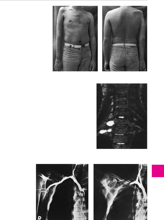
Costoclavicular syndrome. This syndrome, like the scalene syndrome, should be diagnosed only if a causative anatomical anomaly and objectifiable neurological deficits (usually, a lower brachial plexus palsy) can be found. An arteriogram is occasionally helpful in establishing the diagnosis, as it may demonstrate motiondependent compression of the subclavian artery or vein (Fig. 12.22).
Fig. 12.22 Costoclavicular syndrome in a 24-year-old patient with clinical evidence of a lower brachial plexus lesion. An arteriogram of the brachial a. reveals free passage of contrast medium (a) when the arm is dependent. When the arm is elevated, however, (b) the subclavian a. is compressed between the clavicle and the first rib.
a
ThiemeArgoOneBoldthiemeArgoOne
|
of the Spinal Nerve Roots and Peripheral Nerves |
Fig. 12.21 Right C7 and T1 nerve root avulsion after trauma to |
Diseases |
|
|
the brachial plexus. The T2-weighted coronal MR image reveals the |
|
empty nerve root sleeves containing only cerebrospinal fluid. |
|
12
b
Mumenthaler / Mattle, Fundamentals of Neurology © 2006 Thieme All rights reserved. Usage subject to terms and conditions of license.
222 12 Diseases of the Spinal Nerve Roots and Peripheral Nerves
Treatment of compression syndromes. Both the scalene syndrome and costoclavicular syndrome should be treated conservatively at first, once their diagnosis has been definitively established. Special exercises are used to strengthen the shoulder-elevating muscles. Surgical treatment is reserved for the small minority of patients with objectifiable, severe neurological deficits. A supraor transclavicular approach gives the surgeon optimal access to the anatomical structures.
Neuralgic Shoulder Amyotrophy
Pathogenesis. This disorder is presumed to be due to an inflammatory/allergic affection of the brachial plexus. It usually arises spontaneously, but is sometimes seen in the aftermath of an infectious disease, or after the administration of serum (brachial plexus neuritis).
Clinical manifestations. This disease affects men more often than women and is usually located in the right arm. It always begins with intense local pain in the shoulder, which generally lasts for a few days. Rarely, the pain will persist at a milder intensity for a longer period. Once the pain subsides, motor weakness of the muscles of the shoulder girdle and/or of the arm develops. The weakness can, in principle, affect any muscle group of the upper limb, but it tends to affect the muscles innervated by the upper portion of the brachial plexus. Paresis of the serratus anterior m. is particularly common. There may be no objectifiable sensory deficit.
Treatment. No specific treatment is required beyond analgesic medication in the initial, painful phase.
Prognosis. The prognosis is generally good, but it may take many months for the weakness to resolve completely.
Other Causes of Brachial Plexus Lesions
Radiation-induced brachial plexus lesions usually appear with a latency of one or more years after radiotherapy, usually in women who have been treated with surgery and radiotherapy for breast cancer. In 15 % of patients, pain is the main symptom; it can increase over the course of several years. The differentiation of radia- tion-induced brachial plexopathy from a recurrent malignant tumor is not always easy; a short interval between the completion of radiotherapy and the onset of pain (except when a very high radiation dose was given) and very intense pain, both tend to suggest a recurrent tumor rather than radiation injury as the cause. Imaging studies are helpful, but even these cannot always reliably distinguish scarring from new tumor tissue.
Pancoast tumors of the apex of the lung usually cause a lower brachial plexus palsy accompanied by severe pain. The sympathetic chain is usually involved as well; thus, Horner syndrome and diminished sweating in the upper body on the affected side are typical findings (Fig. 12.23).
a
b
c
Fig. 12.23 Right-sided Pancoast tumor in a 68-year-old man. There is clinical evidence of compression of the lower brachial plexus and of the sympathetic chain. a Right Horner syndrome. b Atrophy of the intrinsic muscles of the right hand, especially the thenar muscles. c Weakness of the wrist and finger extensors on the right.
Other causes of brachial plexus palsy , some of which are very rare, are usually diagnosable only by the specialist: acute palsy due to occlusion of a small artery supplying the plexus, after medical procedures, in heroin addicts, in familial brachial plexus neuritis, parainfectious and serogenic forms, etc.
Mumenthaler / Mattle, Fundamentals of Neurology © 2006 Thieme All rights reserved. Usage subject to terms and conditions of license.
Peripheral Nerve Lesions 223
Differential Diagnosis of Brachial Plexus Lesions
A precise clinical and, if necessary, electrophysiological examination of the patient should always permit a reliable differentiation between brachial plexus lesions and lesions affecting multiple nerve roots or a peripheral nerve. Experience suggests, however, that it is not always easy to distinguish a lower brachial plexus lesion (affecting fibers derived from C8 and T1) from an ulnar nerve lesion (p. 231). The same can be said for the distinction between a (traumatic) upper brachial plexus lesion and a lesion of the axillary n. or a rotator cuff injury.
Diseases of the Peripheral Nerves
of the Upper Limbs
Suprascapular N. (C4−C6)
Anatomy. This nerve innervates the supraspinatus and infraspinatus mm. It reaches them after passing through the scapular notch and then running dorsally. It receives sensory branches from the shoulder joint, but not from the skin.
Typical deficits. A lesion of the suprascapular n. produces weakness and atrophy of the two muscles on the dorsal surface of the scapula (Fig. 12.24). The first 15° of lateral elevation of the arm are weak (supraspinatus m.), as is external rotation of the arm at the shoulder joint (infraspinatus m.) (Fig. 12.25).
Causes. Overuse of the arm can lead to mechanical compromise of the nerve in the scapular notch. Other causes include trauma and a ganglion lying in the notch.
Axillary N. (C5−C6)
Anatomy. This nerve provides motor innervation to the deltoid and teres minor mm. and sensory innervation to a palm-sized patch of skin on the proximal, lateral surface of the upper arm (superior lateral brachial cutaneous n.) (Fig. 12.26).
Typical deficits. Axillary nerve palsy manifests itself as marked weakness of lateral abduction and forward elevation of the arm. The normal roundness of the shoulder is flattened. External rotation of the arm at the shoulder joint is lessened at rest, so that the dependent arm is held in mild internal rotation (Fig. 12.27).
Causes. The most common cause of axillary n. palsy is dislocation of the shoulder (forward and downward). The prognosis in such patients is favorable.
Fig. 12.25 Testing of the muscles innervated by the suprascapular n. a Weakness of the supraspinatus m. is most evident in the first 15° of lateral elevation of the arm. b Weakness of the infraspinatus m. is evident when the arm is externally rotated at the shoulder joint.
ThiemeArgoOneBoldthiemeArgoOne
Fig. 12.24 Atrophy of the supra and infraspinatus muscles due to a lesion of the left suprascapular n. in a 25-year old man. The etiology remained unclear in this case.
a
b
Diseases of the Spinal Nerve Roots and Peripheral Nerves
12
Mumenthaler / Mattle, Fundamentals of Neurology © 2006 Thieme All rights reserved. Usage subject to terms and conditions of license.
224 12 Diseases of the Spinal Nerve Roots and Peripheral Nerves
|
|
|
|
|
|
rectangular space with |
|
|
|
|
|
|
|
axillary n. and posterior |
|
articular branches |
|
|
|
humeral circumflex a. |
|||
|
|
|
|
|
|||
|
|
|
|
|
|
deltoid m. and muscular branches |
|
teres minor m. and |
|
|
|||||
|
|
|
|
|
|||
muscular branches |
|
|
|
|
|
||
|
|
|
|
|
|
superior lateral brachial |
|
|
|
|
|
|
|||
|
|
|
|
|
|
cutaneous n. |
|
|
|
|
|
|
|
autonomic zone of superior |
|
|
|
|
|
|
|
lateral brachial cutaneous n. |
|
|
|
|
|
|
|
|
|
|
teres major m. |
|
|
|
|
|
|
|
|
|
|
|
|
|
|
|
|
|
|
|
|
|
|
|
|
|
|
radial n. |
|
|
|
|
|
|
|
|
|
|
|
|
|
|
|
|
|
||
|
|
triceps brachii m., long head |
|
triceps brachii m., medial head |
|
||
|
|
|
|
|
|
|
|
|
|
|
|
|
|
|
|
Fig. 12.26 Anatomical course and distribution of the axillary n. Sensory (red) and motor branches (black). The autonomous sensory area of the superior lateral brachial cutaneous n. is shaded in red.
Fig. 12.27 Left axillary nerve lesion in a 26-year-old man. Atrophy of the left deltoid m. with “pointed” shoulder contour. The sensory deficit in the territory of the superior lateral brachial cutaneous n. is indicated.
Differential diagnosis of axillary nerve palsy includes complex brachial plexus lesions affecting the upper portion of the plexus, rotator cuff lesions, and restriction of movement by pain in humeroscapular periarthropathy.
Long Thoracic N. (C5−C7)
Anatomy. This nerve is the longest branch of the brachial plexus. It is a purely motor nerve supplying the serratus anterior m.
Typical deficits. A long thoracic nerve palsy causes winging of the scapula, which is particularly evident when the arms are held high, or when the patient extends the arms and presses with the palms of the hands against a wall (Fig. 12.28).
Causes. Lesions of the long thoracic n. are usually due to excessive mechanical strain on the shoulder (carrying heavy loads) or to neuralgic shoulder amyotrophy
(p. 222). Cryptogenic cases also occur.
Musculocutaneous N. (C5−C7)
Anatomy. This nerve innervates the biceps brachii and coracobrachialis mm. and a portion of the brachialis m.
Its sensory terminal branch, the lateral antebrachial cutaneous n., innervates the skin on the radial side of the forearm (Fig. 12.29).
Typical deficits. Lesions of the musculocutaneous n. cause marked weakness of elbow flexion. This must be tested with the forearm in the supinated position, because, if the forearm is pronated or in neutral position, the elbow can still be flexed by the powerful brachioradialis m., which is innervated by the radial n.
Mumenthaler / Mattle, Fundamentals of Neurology © 2006 Thieme All rights reserved. Usage subject to terms and conditions of license.
Peripheral Nerve Lesions
serratus anterior m.
Fig. 12.28 Lesion of the right long thoracic n. Weakness of the serratus anterior m. causes winging of the scapula, which is particularly evident when the patient extends the arms forward and pushes against a wall.
Causes. The usual cause is trauma, in which case further signs of an upper brachial plexus lesion may be present. Cryptogenic lesions of the musculocutaneous n. and palsy of this nerve in the setting of neuralgic shoulder amyotrophy, are rarer events.
Differential diagnosis. A tear of the long tendon of the biceps brachialis m. only rarely causes weakness of elbow flexion when the forearm is held in the supinated position. This condition is easy to differentiate from musculocutaneous nerve palsy for two further reasons: one is the typical appearance of the belly of the muscle on the volar surface of the forearm; the other is the absence of a sensory deficit.
Radial N. (C5−C8)
Anatomy. The anatomy of the radial n. is depicted in Fig. 12.30. It provides motor innervation to the triceps brachii, brachioradialis, and supinator mm., as well as all of the extensors of the wrist, thumb, and finger joints. Its sensory innervation is to the dorsal skin of the upper arm and forearm as well as the dorsum of the hand, with an autonomic zone located between the first and second metacarpal bones.
Typical deficits. The clinical manifestations of radial nerve palsy depend on the level of the lesion:
Lesion in the upper arm: the radial n. is particularly vulnerable to injury in the radial nerve canal of the humerus, because it lies directly on the bone at this location. The corresponding, readily apparent neuro-
lateral |
C5 |
|
ante- |
C6 |
|
brachial |
C7 |
|
|
||
cuta- |
|
|
neous n. |
|
|
|
coraco- |
|
|
brachialis m. |
|
musculocutaneous n. |
|
|
brachialis m. |
|
|
long head of biceps |
short head of |
|
brachii m. |
biceps brachii m. |
|
lateral antebrachial |
brachialis m.; |
|
muscular branch |
||
cutaneous n. |
||
of radial n. |
||
|
communicating branch to the superficial radial n.
Fig. 12.29 Anatomical course and distribution of the musculocutaneous n.
logical deficit is a wrist drop (Fig. 12.31), attributable to loss of action of the wrist and finger extensors. In addition, sensation is diminished on the radial portion of the dorsum of the hand.
“High radial nerve lesion”: if the nerve is injured more proximally in the upper arm or in the axilla, the triceps brachii m. is also weak and the elbow can no longer be actively extended against resistance.
Supinator canal syndrome: if the radial n. is compromised at the site of its passage through the supinator m., only its deeply penetrating motor terminal branch is affected. The resulting deficit is purely motor. The branch to the extensor carpi radialis m. and the brachioradialis m., which leaves the nerve proximal to its passage through the supinator m., is unaffected, but all of the other forearm muscles innervated by the radial n. are paretic. Finger extension is impaired, but wrist extension is preserved, particularly on the radial side (Fig. 12.32).
ThiemeArgoOneBoldthiemeArgoOne
225
Diseases of the Spinal Nerve Roots and Peripheral Nerves
12
Mumenthaler / Mattle, Fundamentals of Neurology © 2006 Thieme All rights reserved. Usage subject to terms and conditions of license.
226 12 Diseases of the Spinal Nerve Roots and Peripheral Nerves
C5
C6
C7
|
|
|
lateral brachial |
C8 |
|
||
|
cutaneous n. |
||
|
|
|
|
|
T1 |
|
|
|
|
|
posterior brachial |
|
|
|
cutaneous n. |
radial n. lat. brach. cut. n. deltoid m.
medial head of triceps brachii m.
lateral head of triceps brachii m.
muscular branch of musculocutaneous n.
posterior antebrachial cutaneous n.
brachialis m. extensor carpi radialis longus m.
brachioradialis m. extensor carpi radialis brevis m.
superficial branch of radial n.
posterior antebrachial cutaneous branch
tendon of extensor carpi radialis longus m.
tendon of extensor carpi radialis brevis m.
communicating branch to ulnar n.
axillary n.
posterior brachial |
posterior antebrachial |
|
|
cutaneous n. |
|
cutaneous n. |
|
|
|
|
|
long head of |
|
|
triceps brachii m. |
|
|
superficial branch
lateral intermuscular septum
deep branch
supinator m.
extensor digitorum comm. m.
extensor carpi ulnaris m. extensor digiti V m.
posterior antebrachial interosseous n.
extensor indicis m.
tendon of extensor carpi ulnaris m.
dorsal digital nn.
c
superficial branch
abductor pollicis longus m. extensor pollicis longus m. extensor pollicis brevis m.
|
a |
|
|
b |
|
|
|
|
taneous innervation of the branches of the radial n. and sensory au- |
||
Fig. 12.30 Anatomical course and distribution of the radial n. a |
|||||
Proximal muscle branches (black) and course of the sensory superfi- |
tonomous area of the superficial branch. |
||||
cial branch (red). b Course of the motor deep branch. c Zones of cu- |
|
|
|
||
Mumenthaler / Mattle, Fundamentals of Neurology © 2006 Thieme All rights reserved. Usage subject to terms and conditions of license.
Peripheral Nerve Lesions
|
Median N. (C5−T1) |
|
|
Anatomy. The anatomy of the median n. is shown in |
|
|
Fig. 12.33. All of the muscles innervated by this nerve |
|
|
are distal to the elbow. In the forearm, these include |
|
|
most of the long flexors of the fingers (with the exception |
|
|
of the deep flexors of the fourth and fifth fingers, which |
|
|
are innervated by the ulnar n.), as well as the flexor carpi |
|
|
radialis, pronator teres, and pronator quadratus mm. |
|
|
After the nerve passes through the carpal tunnel to- |
|
|
gether with the long flexor tendons (see below), it in- |
|
Fig. 12.31 Right wrist drop due to a radial nerve lesion. The shad- |
nervates most of the thenar muscles (abductor pollicis |
|
ing indicates the sensory autonomous area in the distribution of the |
brevis and opponens pollicis m. and the superficial head |
|
superficial branch of the radial n. |
of the flexor pollicis brevis m.), as well as the first and |
|
|
second lumbrical mm. Its sensory innervation is to the |
|
|
radial side of the palm, the volar surface of the fingers |
|
|
from the thumb to the radial half of the fourth finger, |
|
|
and the dorsal surface of the terminal phalanges of |
|
|
these fingers. |
|
|
Typical deficits. In median nerve lesions, too, the clini- |
|
|
cal manifestations depend on the level of the lesion: |
|
|
Median nerve lesion in the upper arm (i. e., proximal |
|
|
to the origin of its motor branches to the forearm |
|
|
flexors): the typical clinical appearance is that of the |
|
|
“pope’s blessing hand,” as depicted in Fig. 12.34, |
|
|
caused by weakness of the radial finger flexors. |
|
a |
Median nerve lesion at the wrist. A lesion of the me- |
|
dian n. in the carpal tunnel causes weakness of the |
||
|
||
|
thenar muscles. Clinically, pain and paresthesia are |
|
|
the most prominent symptoms. Carpal tunnel syn- |
|
|
drome is discussed separately, in detail, because of |
|
|
its special clinical importance (see below). |
|
|
Kiloh−Nevin syndrome. An isolated lesion of the ante- |
|
|
rior interosseous n. is a rare event. This nerve is the |
|
|
motor terminal branch of the median n., which in- |
|
|
nervates the flexor pollicis longus m., the radial por- |
|
|
tion of the flexor digitorum profundus m. (flexion of |
|
|
the terminal phalanges of the 2nd and 3rd fingers), |
|
|
and the pronator quadratus m. A lesion of this termi- |
|
b |
nal branch—due either to trauma or, occasionally, to |
|
entrapment—mainly impairs flexion of the terminal |
||
Fig. 12.32 Right supinator tunnel syndrome in a 71-year-old |
||
phalanges of the thumb and index finger. The patient |
||
woman. Marked weakness of the finger extensors (a) with preserva- |
can no longer form an “O” with these two fingers. |
|
tion of wrist extension (b), particularly on the radial side of the |
||
|
||
wrist. |
Causes. The median n. is the nerve most frequently in- |
|
|
||
|
jured by direct trauma, often by a cut in the wrist. Pres- |
|
|
sure palsies of the median n. also occur, both in the |
|
Causes. Radial nerve lesions can be produced by trauma |
upper arm (due to prolonged maintenance of an |
|
awkward position, or to an Esmarch tourniquet) or in |
||
and by pressure, e. g., by the use of crutches that press in |
the palm of the hand (e. g., in occupational injuries). |
|
the axilla, or by external pressure on the upper arm |
Compression at anatomical bottlenecks (entrapment) is a |
|
(humerus). The supinator canal syndrome is an ana- |
further cause of median nerve lesions. In many in- |
|
tomical bottleneck (entrapment) syndrome. |
dividuals, a bony spur is present just above the medial |
|
Differential diagnosis of radial nerve palsy must in- |
epicondyle of the humerus (the supracondylar process). |
|
A fibrous band (of Struther) may run from this spur to |
||
clude predominantly distal weakness of central origin, |
the medial epicondyle, forming a tunnel through which |
|
which can also present with a wrist drop. The flexor |
the median n. passes. The nerve can be compressed |
|
weakness and enhanced intrinsic muscle reflexes that |
either by the supracondylar process or by the fibrous |
|
are present in central weakness serve to differentiate |
band. Further compression syndromes affecting the me- |
|
this condition from radial nerve palsy. Spinal muscular |
dian n. are the Kiloh−Nevin syndrome (see above) and |
|
atrophy can, in rare cases, affect the wrist extensors on |
the carpal tunnel syndrome, which is described below. |
|
one side only. Steinert myotonic dystrophy (p. 268) |
|
|
commonly produces a bilateral wrist drop. |
|
|
thiemeArgoOne |
|
|
ThiemeArgoOneBold |
|
227
Diseases of the Spinal Nerve Roots and Peripheral Nerves
12
Mumenthaler / Mattle, Fundamentals of Neurology © 2006 Thieme All rights reserved. Usage subject to terms and conditions of license.
228 |
12 Diseases of the Spinal Nerve Roots and Peripheral Nerves |
|
|
|
|
|
|
|
|
|
||||||||||||||||||
|
|
|
|
|
|
|
|
|
|
|
|
|
|
|
|
|
|
|
|
|
|
|
|
|
|
|
|
|
|
|
|
|
|
|
|
|
|
|
|
|
|
|
|
|
|
|
|
|
|
|
|
|
|
|
|
|
|
|
|
|
|
|
|
|
|
|
|
|
|
|
|
|
|
|
|
|
|
|
|
|
|
|
|
|
|
|
|
|
|
|
a |
|
|
|
|
|
|
|
|
|
|
|
|
|
|
|
b |
|
|
|
|
|
|
|
|
|
|
|
|
|
|
|
|
C5 |
|
|
|
|
|
|
|
|
|
|
median n. |
|
|
|
|
|||||
|
|
|
|
|
|
|
|
|
|
|
|
|
|
|
|
|
|
|
|
|
|
|
||||||
|
|
|
|
|
|
|
|
|
|
|
|
|
|
|||||||||||||||
|
|
|
|
|
|
|
|
|
|
|
|
|
|
|
|
|
|
|
|
|
|
|
|
|
|
|
|
|
|
|
|
|
|
|
|
|
|
|
C6 |
|
|
|
|
|
|
|
|
|
|||||||||
|
|
|
|
|
|
|
|
|
|
|
|
|
|
|
|
|
|
|
|
|
|
|
|
|
|
|
|
|
|
|
|
|
|
|
|
|
|
|
|
C7 |
|
|
|
|
|
|
|
|
|
||||||||
|
|
|
|
|
|
|
|
|
|
|
|
|
|
|
|
|
|
|
|
|
|
|
|
|
|
|
||
|
|
|
|
|
|
|
|
|
|
|
|
C8 |
|
|
|
|
|
|
|
|
|
|||||||
|
|
|
|
|
|
|
|
|
|
|
|
|
|
|
|
|
|
|
|
|
|
|
|
|
|
|
|
|
|
|
|
|
|
|
|
|
|
|
|
|
|
|
|
T1 |
|
|
|
|
flexor digitorum |
|
|
||||||
|
|
|
|
|
|
|
|
|
|
|
|
|
|
|
|
|
|
|
|
|
|
|
|
|
|
|||
|
|
|
|
|
|
|
|
|
|
|
|
|
|
|
|
|
|
|
|
|
|
|||||||
|
|
|
|
|
|
|
|
|
|
|
|
|
|
|
|
|
|
|
|
|
|
|
|
superficialis m. |
|
|
||
|
|
|
|
|
|
|
|
|
|
|
|
|
|
|
|
|
|
|
|
|
|
|
|
ulnar n. |
|
|
||
|
|
|
|
|
|
|
|
|
|
|
|
|
|
|
|
|
|
|
|
|
|
|
|
flexor dig. prof. m. |
|
|||
|
|
|
|
|
|
|
|
flexor pollicis longus m. |
|
|
|
muscular branch |
|
|||||||||||||||
|
|
|
|
|
anterior antebrachial interosseous n. |
|
|
|
of ulnar n. |
|
||||||||||||||||||
|
|
|
|
|
|
|
|
|
|
|
|
|
||||||||||||||||
|
|
|
|
|
|
|
|
to the pronator quadratus m. |
|
|
|
|
|
|
|
|
||||||||||||
|
|
|
|
|
|
|
|
|
|
|
|
|
|
|
|
|||||||||||||
|
|
|
|
median n. |
|
|
|
|
|
|
|
|
|
|
|
|
|
|
|
|
|
|
|
|
|
|
|
|
|
|
|
|
|
|
|
|
|
|
|
|
|
|
|
|
|
|
|
|
|
|
|
|
|
|
|
|
|
|
|
|
|
|
|
|
|
|
|
|
|
|
|
|
|
|
|
|
|
|
|
|
|
|
|
|
||
|
|
|
supracondylar |
|
|
|
|
|
|
|
|
|
|
|
|
|
|
|
|
|
|
|
|
|
|
|
|
|
|
|
|
|
process of |
|
|
|
|
|
|
|
|
|
|
|
|
|
|
|
|
|
|
|
|
|
|
|
|
|
|
|
|
the humerus |
|
|
|
|
|
|
|
|
|
|
|
|
|
|
|
|
|
|
|
|
|
|
|
|
|
|
|
|
|
|
|
|
|
|
|
|
|
|
|
|
|
|
|
|
|||||||||
|
|
|
|
|
|
|
|
|
|
|
|
|
|
|
|
|
|
|
|
|||||||||
|
|
|
|
pronator |
|
|
|
pronator quadratus m. |
|
|
|
|
|
|
|
|
|
|
|
|||||||||
|
|
|
|
|
|
|
palmar branch |
|
|
|
|
|
|
|
|
|
|
|
|
|||||||||
|
|
|
|
teres m. |
|
|
|
|
|
|
|
|
|
|
|
|
|
|
||||||||||
|
|
|
|
|
|
|
|
|
|
|
|
|
|
|
|
|
|
|
|
|
|
|
|
|
|
|
|
|
|
|
|
|
|
|
tendon of flex. carpi rad. m. |
|
|
|
|
|
|
|
|
|
|
|
|||||||||||
|
|
|
|
palmaris |
|
|
|
|
|
|
|
|
tendon of |
|
|
|
||||||||||||
|
|
|
|
|
abductor pollicis brevis m. |
|
|
|
|
|
|
|
|
|
|
|||||||||||||
|
|
|
|
longus m. |
|
|
|
|
|
|
|
palmaris longus m. |
|
|
|
|||||||||||||
|
|
|
|
|
|
|
|
|
|
|
|
|
|
|
|
|
|
|
|
|
|
|
|
|
|
|||
|
|
|
|
|
|
|
opponens pollicis m. |
|
|
|
|
|||||||||||||||||
|
|
|
|
|
|
|
|
|
|
|
||||||||||||||||||
|
|
|
|
pronator |
|
|
|
|
|
|
|
|
|
|
|
|
|
|||||||||||
|
|
|
|
|
|
|
|
|
|
|
|
|
|
|
|
|
|
|||||||||||
|
|
|
|
|
|
|
flexor pollicis brevis m. |
|
|
|
|
|
|
|
|
|
|
|||||||||||
|
|
|
|
teres m. |
|
|
|
(superficial head) |
|
|
|
|
|
|
|
|
|
|
||||||||||
|
|
|
|
flexor carpi |
|
|
|
|
|
|
|
|
|
|
|
|
|
|
|
|
|
|
|
|
|
|
|
|
|
|
|
|
|
|
|
|
|
|
|
|
|
|
|
|
|
|
|
|
|
|
|
|
|
|
|
|
|
|
|
|
|
radialis m. |
|
|
|
|
|
|
|
|
|
|
|
|
|
|
|
|
|
|
|
|
|
|
|
|
|
|
|
|
|
|
|
|
|
|
|
|
|
|
|
|
|
|
|
|
|
|
|
palmar |
|
|
|
|
|
|
|
|
|
|
|
|
|
|
|
|
|
|
|
|
|
|
|
|
|
|
|
|
|
|
|
|
|
|
|
|
|
|
|
|
|
|
|
|
|
|
|
|
|
|
|
|
|
|
|
|
|
|
digital nn. |
|
|
|
|
|
|
|
|
|
|
|
|
lumbrical mm. I and II |
|
|
|
|
|
|||||||||||||||
|
|
|
|
|
|
|
|
|
|
|
|
|
|
|
|
|
|
|||||||||||
|
|
|
|
|
|
|
|
|
|
|
|
|
|
|
|
|
|
|
|
|
|
|
|
|
|
|
|
|
c
Fig. 12.33 Anatomical course and distribution of the median n. a Proximal course. b Course after traversal of the pronator teres m. c Zones of cutaneous innervation in the hand.
Carpal Tunnel Syndrome
The carpal tunnel syndrome (CTS) is caused by (mechanical) compression of the median n. as it passes through the carpal tunnel. It is considerably more common in women than in men and tends to develop around the time of the menopause. It usually affects the dominant hand, but it may affect the nondominant hand, or both. Factors that promote or precipitate the
Fig. 12.34 “Preacher’s hand” due to a proximal left median nerve lesion. The hypesthetic area is shaded dark red.
Mumenthaler / Mattle, Fundamentals of Neurology © 2006 Thieme All rights reserved. Usage subject to terms and conditions of license.
Peripheral Nerve Lesions 229
development of CTS include hormonal changes (menopause, pregnancy), weight gain, hypothyroidism, diabetes mellitus, and others.
Typical deficits. CTS is characterized by the following manifestations:
in the first stage, which lasts several months or years, the manifestations are subjective: dull pain in the arm at night (brachialgia paresthetica nocturna),
which is felt not merely in the hand, but in the whole upper limb up to the shoulder,
wakes the patient from sleep and can be relieved by shaking and massaging the arms,
the fingers are stiff and uncoordinated for a short time after the patient arises in the morning,
in the more advanced stage, abnormal sensations (paresthesiae) develop and the sense of touch is impaired, mainly in the thumb and index finger,
careful clinical examination is needed to reveal objectifiable sensory and/or motor deficits.
Examination and diagnostic evaluation. An occasional objective finding is point tenderness to pressure at the root of the thenar muscles, or a positive Tinel sign (paresthesiae in the radial portion of the palm and the radial fingers induced by a tap on the transverse carpal ligament). Paresthesiae in the fingers can sometimes be induced by sustained passive hyperflexion or hyperextension of the wrist (Phalen sign). Only later in the course of CTS can one find a discrete impairment of the sense of touch, particularly in the index finger (e. g., a worsening of two-point discrimination to 5 mm). The major finding, however, is an inability to abduct the thumb fully, particularly when compared with the normal, opposite side, because of weakness of the abductor pollicis brevis m. This can be demonstrated by having the patient grasp a cylindrical object; a “positive bottle sign” is seen (Fig. 12.35). Impaired opposition of the thumb is more difficult to observe clinically (Fig. 12.36).
Fig. 12.35 “Bottle” sign in right median nerve palsy. The thumb cannot be adequately abducted and opposed.
Fig. 12.36 Inadequate opposition and pronation of the thumb in a patient with a right median nerve lesion. Because the thumb is insufficiently rotated, the thumbnail is seen tangentially rather than head on.
Overt CTS is unequivocally demonstrated by a finding of impaired conduction in the median n. across the carpal tunnel, as revealed by electroneurography (Fig. 12.37). This study should always be done before any operation is performed. Diminished conduction velocity alone, in
Fig. 12.37 Motor median neurography in right carpal tunnel syndrome. The recording is performed over the abductor pollicis m. The distal motor latency is prolonged (9.2 ms, compared to normal 3.9 ms). The nerve conduction velocities in the arm and forearm are normal.
9.2ms
stimulus: wrist
elbow
50m/s |
|
55m/s |
|
||
|
|
|
2mV
5ms
upper arm
ThiemeArgoOneBoldthiemeArgoOne
Diseases of the Spinal Nerve Roots and Peripheral Nerves
12
Mumenthaler / Mattle, Fundamentals of Neurology © 2006 Thieme All rights reserved. Usage subject to terms and conditions of license.
230 12 Diseases of the Spinal Nerve Roots and Peripheral Nerves
opponens digiti V m. deep branch of ulnar n.
flexor pollicis brevis m. (deep head)
adductor pollicis m. (transverse and oblique heads)
C8
T1
ulnar n.
medial intermuscular septum
flexor carpi ulnaris m.
flexor digitorum profundus m.
dorsal branch of ulnar n.
pisiform bone
abductor digiti V m.
flexor digiti V m.
lumbrical mm. III and IV; interossei
Fig. 12.38 Anatomical course and distribution of the ulnar n.
dorsal branch of ulnar n.
communicating branch to radial n.
dorsal digital nn.
dorsal branch
palmar cutaneous branch
superficial branch
palmaris brevis m.
communicating branch to median n. palmar
digital nn.
Mumenthaler / Mattle, Fundamentals of Neurology © 2006 Thieme All rights reserved. Usage subject to terms and conditions of license.
Peripheral Nerve Lesions 231
the absence of characteristic symptoms, is not an indication for surgery.
Treatment. Keeping the hand in the neutral position at night with a well-padded volar splint often brings relief. If it does not, or if objectifiable neurological deficits are already present, there should be no hesitation in proceeding to surgery. Operative carpal tunnel release involves splitting of the flexor retinaculum with an open or endoscopic technique (generally performed either by a neurosurgeon or by a hand surgeon).
Ulnar N. (C8−T1)
Anatomy. The anatomy of the ulnar n. is shown in Fig. 12.38. Among the muscles innervated by this nerve, the ulnar flexors of the wrist and fingers (the flexor carpi ulnaris m. and the ulnar portion of the flexor digitorum profundus m.) are functionally much less important than the ulnar-innervated intrinsic muscles of the hand. The ulnar n. is, indeed, the most important nerve for finger function: it innervates not only the hypothenar mm., but also all of the interossei, the 3rd and 4th lumbrical mm., and, in the thenar region, the adductor pollicis m. and the deep head of the flexor pollicis brevis m. It provides sensory innervation to the ulnar edge of the hand, the volar surface of the fifth finger, and ulnar half of the fourth finger. A sensory branch arising from the ulnar n. in the distal third of the forearm innervates the skin on the ulnar side of the dorsum of the hand, as well as on the dorsal surface of the fifth finger and the ulnar half of the fourth finger.
Typical deficits. The typical clinical picture of ulnar nerve palsy is a claw hand (Fig. 12.39): because the interossei and lumbrical muscles cannot contract, the ulnar digits are hyperextended at the metacarpophalangeal joints, while the remaining digits are flexed at these joints. The long fingers can no longer be fully adducted against one another, the fingers cannot be strongly spread apart, and the patient cannot flick the middle finger against the examiner’s palm with full, normal strength. A key finding is that, when the patient grasps a flat object (such as a piece of paper) between the thumb and the index finger, weakness of the adductor pollicis m. (ulnar n.) leads to functional substitution by the flexor pollicis longus m. (median n.), and therefore to flexion of the thumb on the affected side at the interphalangeal joint. This finding, called Froment sign, is highly characteristic of ulnar nerve palsy (Fig. 12.40).
Fig. 12.40 Froment sign in right ulnar nerve palsy. Flexion of the interphalangeal joint of the thumb when the patient pulls on a flat object (piece of paper).
ThiemeArgoOneBoldthiemeArgoOne
In addition to these general clinical manifestations of ulnar nerve palsy, there are other specific findings that depend on the level of the lesion:
If the lesion is proximal (at the elbow or higher), it will also affect the ulnar portion of the flexor digitorum profundus m., thereby impairing flexion of the distal phalanx of the fourth and fifth digits (Fig. 12.41).
Fig. 12.39 Claw hand due to a right ulnar nerve lesion at the elbow. Typical features include hyperextension at the metacarpophalangeal joints and hyperflexion at the interphalangeal joints, particularly on the ulnar side of the hand. There is marked atrophy of the interossei and of the hypothenar muscles.
Fig. 12.41 A test for the function of the flexor digitorum profundus m. of the little finger (ulnar n.). Flexion of the little finger at the distal interphalangeal joint.
Diseases of the Spinal Nerve Roots and Peripheral Nerves
12
Mumenthaler / Mattle, Fundamentals of Neurology © 2006 Thieme All rights reserved. Usage subject to terms and conditions of license.