Добавил:
Upload Опубликованный материал нарушает ваши авторские права? Сообщите нам.
Вуз: Предмет: Файл:

Color Atlas of Physiology 2003 thieme

.pdf
Скачиваний:
110
Добавлен:
15.02.2016
Размер:
36.12 Mб
Скачать

D. Mode of action of steroid hormones


Steroid


hormone (H)


ECF


Cell


III


Transmission

Cytoplasmic


receptor (R)


HSP


H-R


complex

Signal


Amino acids


Trans-

(AA)


location

ATP

tRNA

DNA

Cellular


AA-tRNA

rRNA


Translation


Transcription

11.7


mRNA

Cell

Plate


nucleus

Ribosome


Induced

Cell response


protein


E. Nitric oxide (NO) as a transmitter substance


Messenger,


e.g., acetylcholine


Ca2+


channel


Receptor


Cell


membrane


Go Open

Vaso-


Gq


DAG


dilatation


β

IP3

Ca2+

Calmodulin

[Ca2+]


PLC-


PIP2


Ca2+


?


stores


Ca2+-


Protein


calmodulin


complex

kinase G


Diffusion


Activated

Activated


NO synthase


GTP


NO

Arginine


Cytosolic


O2,NADPH

guanylate


cyclase


Citrulline


cGMP


Cell 1


279

Cell 2

ECF


(e.g., endothelial cell)


(e.g., vascular myocyte)


Despopoulos, Color Atlas of Physiology © 2003 Thieme

All rights reserved. Usage subject to terms and conditions of license.

Hypothalamic–Pituitary System

In the hypothalamus, (1) humoral signals from the periphery (e.g., from circulating cortisol) can be converted to efferent neuronal signals, and (2) afferent neuronal signals can be converted to endocrine messengers (neurosecretion).


The first case is possible because the hypothalamus is

Reproduction

situated near circumventricular organs like the or-

ganum vasculosum laminae terminalis (OVLT), sub-


fornical organ, the median eminence of the hypo-


thalamus, and the neurohypophysis. Since there is


no blood-brain barrier there, hydrophilic peptide

and

hormones can also enter.


The hypothalamus is closely connected to


Hormones

other parts of the CNS (!p. 330). It controls

sleeping-waking rhythm (!p. 334) and to


many autonomous regulatory functions and


its neuropeptides influence higher brain func-


tions. The hypothalamus is related to the

11

psychogenic factors. Stress,

for

example,

stimulates the release of cortisol (via CRH,


ACTH) and can lead to the cessation of hor-


mone-controlled menstruation (amenorrhea).


Neurosecretion.

Hypothalamic

neurons


synthesize hormones, incorporate them in


granules that are transported to the ends of the


axons (axoplasmic

transport

!p. 42), and


secrete them into the bloodstream. In this way,


oxytocin and ADH are carried from magno-


cellular hypothalamic nuclei to the neurohy-


pophysis, and RHs and IHs (and ADH) reach the


median eminence of the hypothalamus (!A).


The action potential-triggered exocytotic re-


lease of the hormones into the bloodstream re-


sults in Ca2+ influx

into the

nerve

endings


(!p. 50ff.).


Oxytocin (Ocytocin) and antidiuretic hormone (ADH) are two posterior pituitary hormones that enter the systemic circulation directly. ADH induces water retention in the renal collecting ducts (V2-rec.; !p. 166) and induces vasoconstriction (endothelial V1 rec.) by stimulating the secretion of endothelin-1 (!p. 212ff.). ADH-bearing neurons also secrete ADH into the portal venous circulation (see below). The ADH and CRH molecules regulate the secretion of ACTH by the adenohy-

280pophysis. Oxytocin promotes uterine contractions and milk ejection (!p. 304). In nursing

mothers, suckling stimulates nerve endings in the nipples, triggering the secretion of oxytocin (and prolactin, !p. 303) via neurohumoral reflexes.

Releasing hormones (RH) or liberins that stimulate hormone release from the adenohypophysis (Gn-RH, TRH, SRH, CRH; !p. 270ff.) are secreted by hypothalamic neurons into a kind of portal venous system and travel only a short distance to the anterior lobe (!A). Once in its vascular network, they trigger the release of anterior pituitary hormones into the systemic circulation (!A). Some anterior pituitary hormones are regulated by release-inhib- iting hormones (IH) or statins, such as SIH and PIH = dopamine (!p. 270ff.). Peripheral hormones, ADH (see above) and various neurotransmitters such as neuropeptide Y (NPY), norepinephrine (NE), dopamine, VIP and opioids also help to regulate anterior pituitary functions (!p. 272).

The four glandotropic hormones (ACTH, TSH, FSH and LH) and the aglandotropic hormones (prolactin and GH) are secreted from the anterior pituitary (!A). The secretion of growth hormone (GH = somatotropic hormone, STH) is subject to control by GH-RH, SIH and IGF-1. GH stimulates protein synthesis (anabolic action) and skeletal growth with the aid of somatomedins (growth factors formed in the liver), which play a role in sulfate uptake by cartilage. Somatomedin C = insulin-like growth factor-1 (IGF-1) inhibits the release of GH by the anterior pituitary via negative feedback control. GH has lipolytic and glycogenolytic actions that are independent of somatomedin activity.

Proopiomelanocortin (POMC) is a peptide precursor not only of ACTH, but (inside or outside the anterior pituitary) also of !-endorphin and α-melano- cyte-stimulating hormone (α-MSH = α-melanocor- tin). !-endorphin has analgesic effects in the CNS and immunomodulatory effects, while α-MSH in the hypothalamus helps to regulate the body weight (!p. 230) and stimulates peripheral melanocytes.

Despopoulos, Color Atlas of Physiology © 2003 Thieme

All rights reserved. Usage subject to terms and conditions of license.

A. Hypothalamic-pituitary hormone secretion (schematic)

Nucleus

Hypothalamus

ventromedialis


Nucleus

Nucleus

dorsomedialis

supraopticus

Nucleus

Nucleus

infundibularis

paraventricularis


RH, IH


ADH

Chiasma

Oxytocin


opticum

Corpus


mamillare

Superior hypophysial artery

Axoplasmic transport by neurosecretory nerve cells

Release of RHs, IHs, ADH, NE, NPY and

other transmitters

Portal veins

Inferior hypo-

physial artery

RH induce release of

RH, IH,

ADH

anterior pituitary hormones

IH inhibit their release


Release of


ADH, oxytocin


Pituitary


veins

Posterior pituitary


(neurohypophysis)

Anterior pituitary

Anterior pituitary

Posterior pituitary

(adenohypophysis)


hormones

hormones


ADH, oxytocin


ACTH

PRL


STH

LH


TSH

α-MSH


FSH

β-endorphin


RH =Releasing hormones


IH = (Release-)Inhibiting hormones

Plate 11.8 Hypothalamic–Pituitary System

281

Despopoulos, Color Atlas of Physiology © 2003 Thieme

All rights reserved. Usage subject to terms and conditions of license.


Carbohydrate Metabolism and

glucose concentration as constant as possible


(!A); and (4) promote growth.


Pancreatic Hormones


Glucose is the central energy carrier of the

Insulin


human metabolism. The brain and red blood

Synthesis. Insulin is a 6 kDa peptide (51 amino acids,


cells are fully glucose-dependent. The plasma

AA) formed by the C chain cleaved from proinsulin


glucose concentration (blood sugar level) is

(84 AA), the precursor of which is preproinsulin, a pre-


determined by the level of glucose production

prohormone. Insulin contains two peptide chains (A


and B) held together by disulfide bridges Degrada-


and consumption.


tion: Insulin has a half-life of about 5–8 min and is de-


Reproduction

The following terms are important for proper under-

graded mainly in liver and kidneys.


standing of carbohydrate metabolism (!A, C):
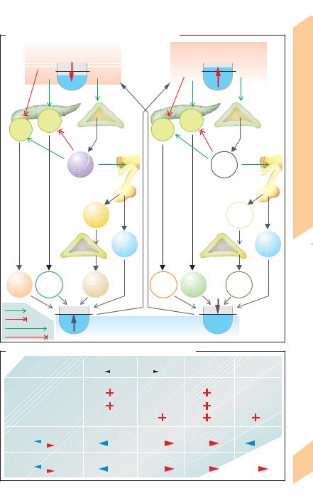
Secretion.

Insulin

is

secreted in pulsatile


1. Glycolysis generally refers to

the anaerobic


bursts, mainly in response to increases in the


conversion of glucose to lactate (!p. 72). This oc-


blood levels of glucose (!B right), as fol-


curs in the red blood cells, renal medulla, and skeletal


muscles (!p. 72). Aerobic oxidation of glucose oc-

lows: plasma glucose "!glucose in B cells

and

curs in the CNS, heart, skeletal muscle and in most

"!glucose oxidation "!cytosolic ATP

other organs.


"!closure

of

ATP-gated K+

channels

Hormones

2. Glycogenesis, i.e., the synthesis of glycogen

!depolarization !opening of voltage-gated

muscle can only be used by that muscle.

and (b) re-opening of K+ channels (deactivated


from glucose (in liver and muscle), facilitates the

Ca2+

channels

!cytosolic Ca2+". The rising


storage of glucose and helps to maintain a constant

Ca2+ in B cells leads to (a) exocytosis of insulin


plasma glucose concentration. Glycogen stored in a


11

3. Glycogenolysis is the breakdown of glycogen

by

feedback

control).

Stimulation.

Insulin

to glucose, i.e., the opposite of glycogenesis.

secretion is stimulated mainly during food


4. Gluconeogenesis is the production of glucose

digestion via acetylcholine (vagus nerve),


(in liver and renal cortex) from non-sugar molecules

gastrin, secretin,

GIP

(!p. 234)

and

GLP-1


such as amino acids (e.g., glutamine), lactate (pro-

(glucagon-like

peptide

= enteroglucagon), a


duced by anaerobic glycolysis in muscles and red


peptide that dissociates from intestinal pro-


cells), and glycerol (from lipolysis).


glucagon. Certain amino acids (especially ar-


5. Lipolysis is the breakdown of triacylglycerols


ginine and leucine), free fatty acids, many


into glycerol and free fatty acids.


6. Lipogenesis is the synthesis of triacylglycerols

pituitary hormones and some steroid hor-


(for storage in fat depots).


mones also increase insulin secretion. Inhibi-


Islets of Langerhans in the pancreas play a pri-

tion. Epinephrine

and

norepinephrine (α2-


adrenoceptors; !A, B), SIH (!p. 273 B) and


mary role in carbohydrate metabolism. Three


the neuropeptide galanin inhibit insulin secre-


cell types (A, B, D) have been identified so far


tion. When hypoglycemia occurs due, e.g., to


(!p. 273 B). 25% of all islet cells are type A (α)


fasting or prolonged physical exercise, the low


cells that produce glucagon, 60% are B (") cells


blood glucose concentration is sensed by cen-


that synthesize insulin, and 10% are D (δ) cells


tral chemosensors for glucose, leading to reflex


that secrete

somatostatin (SIH). These hor-


activation of the sympathetic nervous system.


mones mutually influence the synthesis and


secretion of each other (!p. 273 B). Islet cells

The insulin receptor is a heterotetramer (α2"2) con-


in the pancreas head synthesize pancreatic

sisting of two extracellular α subunits and two trans-


polypeptide,

the physiological

function of

membranous " subunits. The α subunits bind the


which is not yet clear. High concentrations of

hormone. Once the " subunits are autophosphory-


lated, they

act

as

receptor tyrosine

kinases that


these hormones reach the liver by way of the


phosphorylate insulin receptor substrate-1 (IRS-1). In-


portal venous circulation.


tracellular proteins with SH2 domains are phospho-


Function. Pancreatic hormones (1) ensure


rylated by IRS-1 and pass on the signal (!p. 277 C3).


that ingested food is stored as glycogen and fat

Action of insulin (!A, B, C). Insulin has ana-


(insulin); (2) mobilize energy reserves in re-


bolic and lipogenic effects, and promotes the


sponse to food deprivation, physical activity or


storage of glucose, especially in the liver, where

282

stress (glucagon and the non-pancreatic hor-

it activates enzymes that promote glycolysis

mone epinephrine); (3) maintain the plasma


!

Despopoulos, Color Atlas of Physiology © 2003 Thieme

All rights reserved. Usage subject to terms and conditions of license.

A. Glucose metabolism (simplified overview)

Food


g/L

mmol/L

1.8

10

Blood glucose


0.8

4.5

Red cell

Glucose

in urine Anaerobic glycolysis

liver

by liver


Glucose released from

Glucose taken up

Lactate


Lactate


Glucose


Muscle


Glycogen


Glucose


E


taken up


E

by cell


Ι


Ι


Glucose


CNS


Ι


Ι


Oxidation


Anaerobic glycolysis

Oxidation (aerobic)


(aerobic)in CNS

Energy


Energy


Lactate


CO2 + H2O

CO2 + H2O


Proteins


To liver


G


C


Gluconeogenesis


G


Amino

Ι


acids

Glycogenolysis

Ι C

Glycerol


Lipolysis


Ι

C


Glycogenesis

G

E

G


Lipogenesis


E


Triacyl-


Free


Glycogen

Fat tissue

glycerol

Liver

fatty acids


(fat)


Stimulated by:


Ketogenesis


Ι

Insulin


(after meals)

C

Cortisol


Epinephrine


Glucagon


E (exercise, etc.)

G

(hunger)

Despopoulos, Color Atlas of Physiology © 2003 Thieme

All rights reserved. Usage subject to terms and conditions of license.

Plate 11.9 Carbohydrate Metabolism I

283


!


and glycogenesis and suppresses those involved


in gluconeogenesis. Insulin also increases the


number of GLUT-4 uniporters in skeletal myo-


cytes. All these actions serve to lower the


plasma glucose concentration (which in-


creases after food ingestion). About two-thirds


of the glucose absorbed by the intestines after


a meal (postprandial) is temporarily stored


and kept ready for mobilization (via glucagon)

Reproduction

during the interdigestive phase. This provides

a relatively constant supply of glucose for the


glucose-dependent CNS and vital organs in ab-


sence of food ingestion. Insulin increases the


storage of amino acids (AA) in the form of pro-


teins, especially in the skeletal muscles (ana-

and

bolism). In addition, it promotes growth, inhib-

its extrahepatic lipolysis (!p. 257, D) and af-

Hormones

(35 mg/dL) produce glucose deficiencies in the brain,


fects K+ distribution (!p. 180).


Hypoglycemia develops when the insulin concentra-


tion is too high. Glucose levels of !2 mmol/L

11

which can lead to coma and hypoglycemic shock.

The excessive intake of carbohydrates can over-


load glycogen stores. The liver therefore starts to


convert glucose into fatty acids, which are trans-


ported to and stored in fatty tissues in the form of tri-


acylglycerols (!p. 257 D).


Diabetes mellitus (DM). One type of DM is in-


sulin-dependent diabetes mellitus (IDDM), or type 1


DM, which is caused by an insulin deficiency. Another


type is non-insulin-dependent DM (NIDDM), or type 2


DM, which is caused by the decreased efficacy of in-


sulin and sometimes occurs even in conjunction with


increased insulin concentrations. DM is characterized


by an abnormally high plasma glucose concentration


(hyperglycemia), which leads to glucosuria (!p. 158).


Large quantities of fatty acids are liberated since


lipolysis is no longer inhibited (!p. 257 D). The fatty


acids can be used to produce energy via acetyl-


coenzyme A (acetyl-CoA); however, this leads to the


formation of acetoacetic acid, acetone (ketosis), and


!-oxybutyric acid (metabolic acidosis, !p. 142). Be-


cause hepatic fat synthesis is insulin-independent


and since so many fatty acids are available, the liver


begins to store triacylglycerols, resulting in the


development of fatty liver.


Glucagon, Somatostatin and Somatotropin


Glucagon released from A cells is a peptide


hormone (29 AA) derived from proglucagon


(glicentin). The granules in which glucagon is


stored are secreted by exocytosis. Secretion is

284stimulated by AA from digested proteins (especially alanine and arginine) as well as by hy-

poglycemia (e.g., due to fasting, prolonged physical exercise; !B) and sympathetic impulses (via !2 adrenoceptors; !A). Glucagon secretion is inhibited by glucose and SIH (!p. 273, B) as well as by high plasma concentrations of free fatty acids.

The actions of glucagon (!A, B, C) (via cAMP; !p. 274) mainly antagonize those of insulin. Glucagon maintains a normal blood glucose level between meals and during phases of increased glucose consumption to ensure a constant energy supply. It does this (a) by increasing glycogenolysis (in liver not muscle) and (b) by stimulating gluconeogenesis from lactate, AA (protein degradation = catabolism) and glycerol (from lipolysis).

Increased plasma concentrations of amino acids (AA) stimulate insulin secretion which would lead to hypoglycemia without the simultaneous ingestion of glucose. Hypoglycemia normally does not occur, however, since AA also stimulate the release of glucagon, which increases the blood glucose concentration. Glucagon also stimulates gluconeogenesis from AA, so some of the AA are used for energy production. In order to increase protein levels in patients, glucose must therefore be administered simultaneously with therapeutic doses of AA to prevent their metabolic degradation.

Somatostatin (SIH). Like insulin, SIH stored in D cells (SIH 14 has 14 AA) is released in response to increased plasma concentrations of glucose and arginine (i.e., after a meal). Through paracrine pathways (via Gi-linked receptors), SIH inhibits the release of insulin (!p. 273, B). Therefore, SIH inhibits not only the release of gastrin, which promotes digestion (!p. 243, B3), but also interrupts the in- sulin-related storage of nutrients. SIH also inhibits glucagon secretion (!p. 273 B). This effect does not occur in the presence of a glucose deficiency because of the release of catecholamines that decrease SIH secretion.

Somatotropin (STH) = growth hormone (GH). The short-term effects of GH are similar to those of insulin; its action is mediated by somatomedins (!p. 280). In the long-term, GH increases the blood glucose concentration and promotes growth.

The effects of glucocorticoids on carbohydrate metabolism are illustrated on plate C and explained on p. 296.

Despopoulos, Color Atlas of Physiology © 2003 Thieme

All rights reserved. Usage subject to terms and conditions of license.

B. Hormonal control of blood glucose concentration


Blood glucose drops


Blood glucose rises


below normal


above normal level


5mmol/L


5mmol/L


II


Pancreas


Metabolism


B


B


A

cells


Adrenal


A

cells


cells


cells


rec.


Carbohydrate


medulla


rec.


2


2

α


Hypothalamus


β

Epi-


Epi-


nephrine


nephrine


11.10


ACTH


ACTH

Plate


STH


STH


Adrenal cortex

long-term


short-term

Glucagon

Insulin


Cortisol

Glucagon

Insulin

Cortisol


Stimulates


Inhibits


No stimulation


Blood glucose returns to normal


No inhibition


C. Hormonal effects on carbohydrate and fat metabolism


Hormone

Insulin


Glucagon

Epinephrine

Cortisol


Glucose

Function

Satiated


Buffer


Hungry

Stress, exercise


Supply


Muscle,


Muscle,


Uptake by cell


fat


Muscle


fat


Glycolysis


Gluconeogenesis (liver)


Glycogen


Liver,


Liver


Liver,


Liver


Synthesis


Lysis


muscle


muscle


Fat


Liver,


Fat


Fat


Fat

285

Synthesis


Lysis


fat


Despopoulos, Color Atlas of Physiology © 2003 Thieme

All rights reserved. Usage subject to terms and conditions of license.


Thyroid Hormones

thyrocolloid. The phenol ring of one DIT (or


MIT) molecule links with another DIT


The thyroid gland contains spherical follicles

molecule (ether bridges). The resulting thyro-


(50–500 µm in diameter). Follicle cells synthe-

globulin chain now contains tetraiodothy-


size the two iodine-containing thyroid hor-

ronine residues and (less) triiodothyronine resi-


mones thyroxine (T4, tetraiodothyronine) and

dues (!C). These are the storage form of T4 and


triiodothyronine (T3). T3 and T4 are bound to

T3.


the glycoprotein thyroglobulin (!B2) and

TSH also stimulates T3 and T4 secretion. The


stored in the colloid of the follicles (!A1, B1).

iodinated thyroglobulin in thyrocolloid are re-

Reproduction

The synthesis and release of T3/T4 is controlled

absorbed by the cells via endocytosis (!B3,

by the thyroliberin (= thyrotropin-releasing

C). The endosomes fuse with primary lyso-


hormone, TRH)—thyrotropin (TSH) axis (!A,

somes to form phagolysosomes in which thy-


and p. 270ff.). T3 and T4 influence physical

roglobulin is hydrolyzed by proteases. This


growth, maturation and metabolism. The par-

leads to the release of T3 and T4 (ca. 0.2 and


afollicular cells (C cells) of the thyroid gland

1–3 mol per mol of thyroglobulin, respec-

and

synthesize calcitonin (!p. 292).

tively). T3 and T4 are then secreted into the

Thyroglobulin, a dimeric glycoprotein (660 kDa) is

bloodstream (!B3). With the aid of deiodase,

Hormones

Imeanwhile is split from concomitantly re-

synthesized in the thyroid cells. TSH stimulates the


leased MIT and DIT and becomes reavailable


transcription of the thyroglobulin gene. Thyro-


for synthesis.


globulin is stored in vesicles and released into the col-


loid by exocytosis (!B1 and p. 30).

Control of T3/T4 secretion. TSH secretion by

11

Iodine uptake. The iodine needed for hormone

the anterior pituitary is stimulated by TRH, a

hypothalamic tripeptide (!p. 280) and in-


synthesis is taken up from the bloodstream as


hibited by somatostatin (SIH) (!A and p. 270).


iodide (I). It enters thyroid cells through sec-


The effect of TRH is modified by T4 in the


ondary active transport by the Na+-I symport


plasma. As observed with other target cells,


carrier (NIS) and is concentrated in the cell ca.


the T4 taken up by the thyrotropic cells of the


25 times as compared to the plasma (!B2).


anterior pituitary is converted to T3 by 5!-deio-


Via cAMP, TSH increases the transport capacity


dase. T3 reduces the density of TRH receptors in


of basolateral Iuptake up to 250 times. Other


the pituitary gland and inhibits TRH secretion


anions competitively inhibit Iuptake; e.g.,


by the hypothalamus. The secretion of TSH and


ClO4, SCNand NO2.


consequently of T3 and T4 therefore decreases


Hormone synthesis. Iions are continuously


(negative feedback circuit). In neonates, cold


transported from the intracellular Ipool to the


seems to stimulate the release of TRH via neu-


apical (colloidal) side of the cell by a I/Cl anti-


ronal pathways (thermoregulation, !p. 224).


porter, called pendrin, which is stimulated by


TSH is a heterodimer (26 kDa) consisting of an


TSH. With the aid of thyroid peroxidase (TPO)


α subunit (identical to that of LH and FSH) and


and an H2O2 generator, they are oxidized to el-


a " subunit. TSH controls all thyroid gland func-


ementary I20 along the microvilli on the colloid


tions, including the uptake of I, the synthesis


side of the cell membrane. With the help of


and secretion of T3 and T4 (!A-C), the blood


TPO, the I0 reacts with about 20 of the 144 ty-


flow and growth of the thyroid gland.


rosyl residues of thyroglobulin (!C). The


phenol ring of the tyrosyl residue is thereby

Goiter (struma) is characterized by diffuse or nodu-


iodinated at the 3 and/or 5 position, yielding a

lar enlargement of the thyroid gland. Diffuse goiter


protein chain containing either diiodotyrosine

can occur due to an iodine deficiency, resulting in


(DIT) residues and/or monoiodotyrosine (MIT)

T3/T4 deficits that ultimately lead to increased secre-


tion of TSH. Chronic elevation of TSH leads to the


residues. These steps of synthesis are stimu-


proliferation of follicle cells, resulting in goiter


lated by TSH (via IP3) and inhibited by


development (hyperplastic goiter). This prompts an


thiouracil, thiocyanate, glutathione, and other


increase in T3/T4 synthesis, which sometimes nor-


reducing substances. The structure of the thy-

malizes the plasma concentrations of T3/T4 (euthyroid

286

roglobulin molecule allows the iodinated ty-

goiter). This type of goiter often persists even after


rosyl residues to react with each other in the

the iodide deficiency is rectified.


!

Despopoulos, Color Atlas of Physiology © 2003 Thieme

All rights reserved. Usage subject to terms and conditions of license.

A. Thyroid hormones (overview)


SIH

TRH


Thyrocytes

T4

Thyroxin


HVL


(tetraiodothyronine)


Capillaries


5’-Deiodase


TSH


Thyroid gland

Colloid


T3 Triiodothyronine


Iodine uptake,


hormone synthesis


and secretion

Metabolism, growth, maturation, etc.


(See B.)

B. Hormone synthesis and secretion

1 Thyroglobulin synthesis


Thyroglobulin vesicle


Amino


Carbohydrates


acids


Thyroglobulin


Peptides


Exocytosis


Ribosomes


Endoplasmic


Golgi apparatus


Capillary

reticulum

Thyroid cell (thyrocyte)

Colloid


2 Iodine uptake, hormone synthesis and storage


SCN


Thyroid


CIO4


peroxidase


NO2


Ipool

T3/T4 synthesis

T4 T3


I


(see C.)


Na+ symport


NIS Na+


TSH


TSH


T3/T4 storage


(bound to thyroglobulin)

3 Hormone secretion

Primary lysosomes

TSH

with proteases


Endocytosis

T4

T3


Fusion


Thyroglobulin

I


cleavage

Deiodase


MIT, DIT

Plate 11.11 Thyroid Hormones I

287

Despopoulos, Color Atlas of Physiology © 2003 Thieme

All rights reserved. Usage subject to terms and conditions of license.

11 Hormones and Reproduction

288

!

Hypothyroidism occurs when TSH-driven thyroid enlargement is no longer able to compensate for the T3/T4 deficiency (hypothyroid goiter). This type of goiter can also occur due to a congenital disturbance of T3/T4 synthesis (see below) or thyroid inflammation. Hyperthyroidism occurs when a thyroid tumor (hot node) or diffuse struma (e.g., in Grave’s disease) results in the overproduction of T3/T4, independent of TSH. In the latter case, an autoantibody against the TSH receptor binds to the TSH receptor. Its effects mimic those of TSH, i.e., it stimulates T3/T4 synthesis and secretion.

T3/T4 transport. T3 and T4 occur at a ratio of 1 : 40 in the plasma, where !99% of them (mainly T4) are bound to plasma proteins: thy- roxine-binding globulin (TBG), thyroxine-bind- ing prealbumin (TBPA), and serum albumin. TBG transports two-thirds of the T4 in the blood, while TBPA and serum albumin transport the rest. Less than 0.3% of the total T3/T4 in blood occurs in an unbound (free) form, although only the unbound molecules have an effect on the target cells. Certain drugs split T3 and T4 from protein bonds, resulting in increased plasma concentrations of the free hormones.

Potency of T3/T4. T3 is 3–8 times more potent than T4 and acts more rapidly (half-life of T3 is 1 day, that of T4 7 days). Only ca. 20% of all circulating T3 originate from the thyroid; the other 80% are produced by the liver, kidneys, and other target cells that cleave iodide from T4. The conversion of T4 to T3 is catalyzed by microsomal 5!-deiodase, which removes iodine from the 5! position on the outer ring (!D). T3 is therefore the more potent hormone, while T4 is mainly ascribed a storage function in plasma.

The inactive form of T3 called reverse T3 (rT3) is produced from T4 when the iodine is split from the inner ring with the aid of a 5- (not 5!-)deiodase. Approximately equal amounts of T3 and rT3 are normally produced in the periphery (ca. 25 µg/day). When a person fasts, the resulting inhibition of 5!-deiodase decreases T3 synthesis (to save energy, see below) while rT3 synthesis increases. Pituitary 5!-deiodase is not inhibited, so TSH secretion (unwanted in this case) is suppressed by the negative feedback.

T3/T4 receptors are hormone-sensitive transcription factors located in the cell nuclei. Hor- mone–receptor complexes bind to regulator

proteins of certain genes in the nuclei and influence their transcription.

The actions of T3/T4 are numerous and mainly involve the intermediate metabolism. The thyroid hormones increase the number of mitochondria and its cristae, increase Na+-K+- ATPase activity and modulate the cholesterol metabolism. This results in an increase in energy turnover and a corresponding rise in O2 consumption and heat production. T3 also specifically stimulates heat production by increasing the expression of the uncoupling protein thermogenin in brown fat (!p. 222). T3 also influences the efficacy of other hormones. Insulin, glucagon, GH and epinephrine lose their energy turnover-increasing effect in hypothyroidism, whereas the sensitivity to epinephrine increases (heart rate increases, etc.) in hyperthyroidism. T3 is thought to increase the density of !-adrenoceptors. T3 also stimulates growth and maturation, especially of the brain and bones.

Cretinism occurs due to neonatal T3/T4 deficiencies and is marked by growth and maturation disorders (dwarfism, delayed sexual development, etc.) and central nervous disorders (intelligence deficits, seizures, etc.). The administration of thyroid hormones in the first six months of life can prevent or reduce some of these abnormalities.

Iodine metabolism (!D). Iodine circulates in the blood as either (1) inorganic I(2–10 µg/L),

(2)organic non-hormonal iodine (traces) and

(3)protein-bound iodine (PBI) within T3 and T4 (35–80 µg iodine/L). The average daily requirement of iodine is ca. 150 µg; larger quantities are required in fever and hyperthyroidism (ca. 250–500 µg/day). Iodine excreted from the body must be replaced by the diet (!D). Sea salt, seafood, and cereals grown in iodine-rich soil are rich in iodine. Iodized salt is often used to supplement iodine deficiencies in the diet. Since iodine passes into the breast milk, nursing mothers have a higher daily requirement of iodine (ca. 200 µg/day).

Despopoulos, Color Atlas of Physiology © 2003 Thieme

All rights reserved. Usage subject to terms and conditions of license.

Соседние файлы в предмете [НЕСОРТИРОВАННОЕ]