Добавил:
Upload Опубликованный материал нарушает ваши авторские права? Сообщите нам.
Вуз: Предмет: Файл:

Color Atlas of Physiology 2003 thieme

.pdf
Скачиваний:
110
Добавлен:
15.02.2016
Размер:
36.12 Mб
Скачать

stimuli (!p. 170), whereas the release of melatonin is subject to afferent neuron control (!p. 334).

Some of these hormones (e.g., angiotensin II) and tissue hormones or mediators exert paracrine effects within endocrine and exocrine glands, the stomach wall, other organs, and on inflammatory processes. Bradykinin (!pp. 214 and 236), histamine (!pp. 100 and 242), serotonin (5-hydroxytryptamine, !p. 102) and eicosanoids are members of this group.

Eicosanoids. Prostaglandins (PG), thromboxane (TX), leukotrienes and epoxyeicosatrienoates are eicosanoids (Greek ει!%σι = twenty [C atoms]) derived in humans from the fatty acid arachidonic acid (AA). (Prostaglandins derived from AA have the index number 2). AA occurs as an ester in the phospholipid layer of the cell membranes and is obtained from dietary sources (meat), synthesized from linoleic acid, an essential fatty acid, and released by phospholipase A2 (!p. 252).

Pathways of eicosanoid synthesis from arachidonic acid (AA):

1.Cyclooxygenase pathway: Cyclooxygenase (COX)-1

and COX-2 convert AA into PGG2, which gives rise to PGH2, the primary substance of the biologically active compounds PGE2, PGD2, PGF, PGI2 (prostacyclin) and TXA2. COX-1 and 2 are inhibited by nonsteroidal anti-inflammatory drugs (e.g., Aspirin!).

2.Lipoxygenase pathway: Leukotriene A4 is synthesized from AA (via the intermediate 5-HPETE = 5-hy- droperoxyeicosatetraenoate) by way of 5-lipoxy- genase (especially in neutrophilic granulocytes).

Leukotriene A4 is the parent substance of the leukotrienes C4, D4 and E4. The significance of 12lipoxygenase (especially in thrombocytes) is not yet clear, but 15-lipoxygenase is known to produce vasoactive lipoxins (LXA4, LXB4).

3.Cytochrome P450-epoxygenase produces epoxyeicosatrienoates (EpETrE = EE).

Typical effects of eicosanoids:

PGE2 dilates the bronchial and vascular musculature (and keeps the lumen of the fetal ductus arteriosus and foramen ovale open; !p. 220), stimulates intestinal and uterine contractions, protects the gastric mucosa (!p. 242), inhibits lipolysis, increases the glomerular filtration rate (GFR), plays a role in fever development (!p. 224), sensitizes nociceptive nerve endings (pain) and increases the

permeability of blood vessels (inflammation). PGD2 induces bronchoconstriction. PGI2 (prostacyclin) synthesized in the endothelium, is vasodilatory and inhibits platelet aggregation. TXA2, on the other hand, occurs in platelets, promotes platelet aggregation and acts as a vasoconstrictor (!p. 102). 11,12-EpETrE has a vasodilatory effect (= EDHF, !p. 214).

Hormones (h.) of the hypothalamus and pituitary

Name* Abbreviation/synonyme

Hypothalamus

The suffix “-liberin” denotes releasing

h. (RH) or factor (RF); “-statin” is used for releaseinhibiting h. (IH) or factors (IF)

Corticoliberin

Corticotropin RH, CRH, CRF


Gonadoliberin

Gonadotropin RH, Gn-RH; ICSH


Prolactostatin

Prolactin IH, PIH, PIF, dopamine

Hormones

Somatoliberin

Somatotropin RH, SRH, SRF,


GHRH, GRH


Somatostatin**

Somatotropin (growth h.) IH, SIH


Thyroliberin

Thyrotropin RH, TRH, TRF


Anterior lobe of the pituitary


Corticotropin

Adrenocorticotropic h. (ACTH)


Follitropin

Follicle-stimulating h. (FSH)


Lutropin

Luteinizing h. (LH), interstitial


cell-stimulating h. (ICSH)


Melanotropin

α-Melanocyte-stimulating h.


(α-MSH), α-melanocortin


Somatotropin

Somatotropic h. (STH), growth h.


(GH)


Thyrotropin

Thyroid stimulating h. (TSH)


Prolactin

PRL, lactogenic (mammotropic)


h.


Posterior lobe of the pituitary


Oxytocin


Adiuretin

Anti-diuretic h. ADH,


(arginine-) vasopressin (AVP)


** Names generally recommended by IUPAC-IUB Committee on Biochemical Nomenclature.

** Also synthesized in gastrointestinal organs, etc.

269

Despopoulos, Color Atlas of Physiology © 2003 Thieme

All rights reserved. Usage subject to terms and conditions of license.

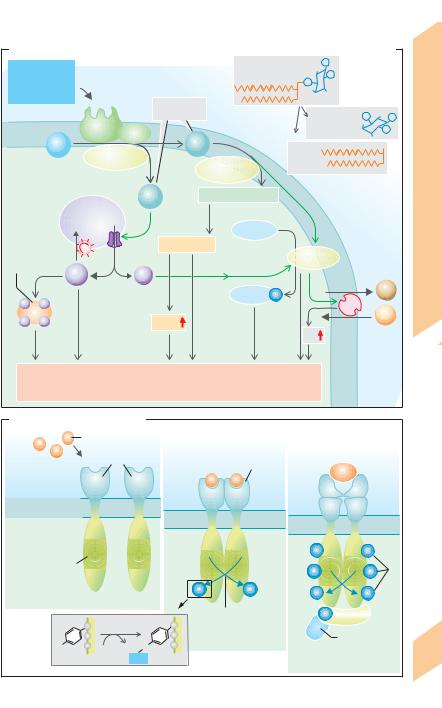
A. The hormones (simplified overview excluding tissue hormones)


Hypothalamus

Anterior pituitary


Peripheral sites of


hormone synthesis


FSH


Testes


Gn-RH


Ovarian


LH (ICSH)


follicles


Corpus


Reproductionand


luteum


control


PIH (= dopamine)


Prolactin (PRL)


Liver


Thyroid gland


TRH


TSH


(follicle cells)


Hormones11

systemnervouscentralUnder


Somatostatin (SIH)


GH-RH (=SRH)


STH (=GH)


cortexAdrenal


Reticular zone


Angiotensinogen


(liver)


Glomerular zone


CRH


ACTH


Fascicular zone


Posterior


pituitary


Axoplasmic transport

ADH


Axoplasmic transport

Oxytocin


Adrenal medulla


Stimulates release


control


Kidneys


D cells


Paracrine:


Pancreas


Peptides


humoral


A cells


Inhibits release


B cells


Glyco-


proteins


Affects


Under


Parathyroid gland


Steroids, etc.


Secretes


Thyroid, etc.:


C cells


Tyrosine


Effect


derivatives


270

Despopoulos, Color Atlas of Physiology © 2003 Thieme

All rights reserved. Usage subject to terms and conditions of license.

End-hormone

Functions (simplified)

Testosterone

Estrogens

Gestagens (progesterone)

Thyroxin (T4)

Deiodinization

Triiodothyronine (T3)

Somatomedins (IGF)

Angiotensin II

Mineralocorticoids

Per-

Glucocorticoids mis- sive

Androgens


Epinephrine


(norepinephrine)


and others


Erythropoietin


Calcitriol


Somatostatin (SIH)


Glucagon


Antagonistic


Insulin


bloodCirculation,


Parathyroid hormone


andWater balancemineral


Maturation

Reproduction


Metabolism


Antagonistic


Calcitonin (CT)


Plate 11.2 u. 11.3 Hormones

271

Despopoulos, Color Atlas of Physiology © 2003 Thieme

All rights reserved. Usage subject to terms and conditions of license.

11 Hormones and Reproduction

272

Humoral Signals: Control and Effects

Hormones and other humoral signals function to provide feedback control, a mechanism in which the response to a signal feeds back on the signal generator (e.g., endocrine gland). The speed at which control measures are implemented depends on the rate at which the signal substance is broken down—the quicker the degradation process, the faster and more flexible the control.

In negative feedback control, the response to a feedback signal opposes the original signal. In the example shown in A1, a rise in plasma cortisol in response to the release of corticoliberin (corticotropin-releasing hormone, CRH) from the hypothalamus leads to down-regulation of the signal cascade “CRH ! ACTH ! adrenal cortex,” resulting in a decrease in cortisol secretion. In shorter feedback loops, ACTH can also negatively feed back on the hypothalamus ( !A2), and cortisol, the end-hormone, can negatively feed back on the anterior pituitary (!A3). In some cases, the metabolic parameter regulated by a hormone (e.g., plasma glucose concentration) rather then the hormone itself represents the feedback signal. In the example (!B), glucagon increases blood glucose levels (while insulin decreases them), which in turn inhibits the secretion of glucagon (and stimulates that of insulin). Neuronal signals can also serve as feedback (neuroendocrine feedback) used, for example, to regulate plasma osmolality (!p. 170).

In positive feedback control, the response to the feedback amplifies the original signal and heightens the overall response (e.g., in autocrine regulation; see below).

The higher hormone not only controls the synthesis and excretion of the end-hormone, but also controls the growth of peripheral endocrine gland. If, for example, the end-hor- mone concentration in the blood is too low despite maximum synthesis and secretion of the existing endocrine cells, the gland will enlarge to increase end-hormone production. This type of compensatory hypertrophy is observed for instance in goiter development (!p. 288) and can also occur after surgical excision of part of the gland.

Therapeutic administration of a hormone (e.g., cortisone, a cortisol substitute) have the same effect on higher hormone secretion (ACTH and CRH in the example) as that of the end-hormone (cortisol in the example) normally secreted by the peripheral gland (adrenal cortex in this case). Long-term administration of an end-hormone would therefore lead to inhibition and atrophy of the endocrine gland or cells that normally produce that hormone. This is known as compensatory atrophy.

A rebound effect can occur if secretion of the higher hormone (e.g., ACTH) is temporarily elevated after discontinuation of end-hor- mone administration.

The principal functions of endocrine hormones, paracrine hormones and other humoral transmitter substances are to control and regulate:

enzyme activity by altering the conformation (allosterism) or inhibiting/stimulating the synthesis of the enzyme (induction);

transport processes, e.g., by changing the rate of insertion and synthesis of ion channels/ carriers or by changing their opening probability or affinity;

growth (see above), i.e., increasing the rate of mitosis (proliferation), “programmed cell death” (apoptosis) or through cell differentiation or dedifferentiation;

secretion of other hormones. Regulation can occur via endocrine pathways (e.g., ACTH-me- diated cortisol secretion; !A5), a short portal vein-like circuit within the organ (e.g., effect of CRH on ACTH secretion, !A4), or the effect of cortisol from the adrenal cortex on the synthesis of epinephrine in the adrenal medulla, (!A6), or via paracrine pathways (e.g., the effect of somatostatin, SIH, on the secretion of insulin and glucagon; !B).

Cells that have receptors for their own humoral signals transmit autocrine signals that function to

exert negative feedback control on a target cell, e.g., to discontinue secretion of a transmitter (e.g., norepinephrine; !p. 84);

coordinate cells of the same type (e.g., in growth);

exert positive feedback control on the secreting cell or to cells of the same type. These mechanisms serve to amplify weak signals as is observed in the eicosanoid secretion or in T cell clonal expansion (!p. 96ff.).

Despopoulos, Color Atlas of Physiology © 2003 Thieme

All rights reserved. Usage subject to terms and conditions of license.

A. Regulation of cortisol and epinephrine concentrations in plasma


Limbic system


Effects


Medial/rostral


Lateral

Autonomic


hypothalamus

hypothalamus

centers

Signal generator

and


1


Hypothalamic


Control


2


neurosecretion


ADH


CRH


Signals:


Sympathetic


feedback


Humoral

Anterior


nervous system


pituitary

4


3


Negative


Plate 11.4


ACTH


5


Adrenal


Adrenal


medulla


cortex


6


Cortisol

Epinephrine


Response to signal


(hormone release)


(norepinephrine)


B. Control of blood glucose


Insulin


Glucose supply

Glucagon


Liver


SIH


Pancreas

D

B


A


cells


cells

Ingested

cells


glucose


Glucose


Blood glucose


Blood


273


Despopoulos, Color Atlas of Physiology © 2003 Thieme

All rights reserved. Usage subject to terms and conditions of license.

Cellular Transmission of Signals from Extracellular Messengers


Hormones,

neurotransmitters

(!p. 55 and


p. 82), cytokines and chemokines (!p. 94ff.)


act as messenger substances (first messengers)


that are transported to their respective target


cells by extracellular pathways. The target cell


has a high-affinity binding site (receptor) for

Reproduction

its specific messenger substance.


Glycoprotein and peptide messengers as


well as catecholamines bind to cell surface re-


ceptors on the target cell. Binding of the mes-


senger to its receptor usually triggers certain


protein-protein interactions (and sometimes

and

protein-phospholipid interactions). This leads

to the release of secondary messenger sub-

Hormones

stances (second messengers) that forward the


signal within the cell. Cyclic adenosine mono-


phosphate (cAMP), cyclic guanosine mono-


phosphate (cGMP), inositol 1,4,5-tris-

11

phosphate (IP3), 1,2-diacylglycerol (DAG) and

Ca2+ are such second messengers. Since the


molecular structure of the receptor ensures


that the effect of the first messenger will be


specific, multiple first messengers can use the


same second messenger. Moreover, the intra-


cellular concentration of the second mes-


senger can be raised by one messenger and


lowered by another. In many cases, different


types of receptors exist for a single first mes-


senger.


cAMP as a Second Messenger


For a cAMP-mediated response to occur, the


cell membrane must contain stimulatory (Gs)


or inhibitory (Gi) G proteins (guanyl nu-


cleotide-binding proteins) (!A1). These G


proteins consist of three subunits—alpha (αS or


αi), beta (") and gamma (γ)—and are therefore


heterotrimers. Guanosine diphosphate (GDP) is


bound to

the α-subunit of

an inactive


G protein. Once the first messenger (M) binds


to the receptor (Rec.), the M–Rec. complex


conjugates with the Gs-GDP (or Gi-GDP)


molecule (!A2). GDP is then replaced by cyto-


solic GTP, and the -subunit and the M–Rec.


complex dissociate from the α-subunit if Mg2+


is present (!A3). αs-GTP or αi-GTP remain as

274

the final products. Adenylate cyclase on the in-

side of the cell membrane is activated by αs-


GTP (cytosolic cAMP concentration rises) and inhibited by αi-GTP (cAMP concentration falls;

!A3).

Gs-activating messengers. ACTH, adenosine (A2A and A2B rec.), antidiuretic hormone = vasopressin (V2 rec.), epinephrine and norepinephrine ("1, "2, "3 adrenoceptors), calcitonin, CGRP, CRH, dopamine (D1 and D5 rec.), FSH, glucagon, histamine (H2 rec.), oxytocin (V2 rec., see above), many prostaglandins (DP, IP, EP2 and EP4 rec.), serotonin = 5-hydroxytrypt- amine (5-HT4 and 5-HT7 rec), secretin and VIP activate Gs proteins, thereby raising cAMP levels. TRH and TSH induce partial activation.

Gi-activating messengers. Some of the above messenger substances also activate Gi proteins (thereby lowering cAMP levels) using a different binding receptor. Acetylcholine (M2 and M4 rec.), adenosine (Al and A3 rec.), epinephrine and norepinephrine (α2 adrenoceptors), angiotensin II, chemokines, dopamine (D2, D3 and D4 rec.), GABA (GABAB rec.), glutamate (mGLU2–4 and mGLU6–8 rec.), melatonin, neuropeptide Y, opioids, serotonin = 5-hydroxytryptamine (5-HTl rec.), somatostatin and various other substances activate Gi proteins.

Effects of cAMP. cAMP activates type A protein kinases (PKA = protein kinase A) which then activate other proteins (usually enzymes and membrane proteins, but sometimes the receptor itself) by phosphorylation (!A4). The specific response of the cell depends on the type of protein phosphorylated, which is determined by the type of protein kinases present in the target cell. Phosphorylation converts the proteins from an inactive to an active form or vice versa.

Hepatic glycogenolysis, for instance, is dually increased by cAMP and PKA. Glycogen synthase catalyzing glycogen synthesis is inactivated by phosphorylation whereas glycogen phosphorylase stimulating glycogenolysis is activated by cAMP-mediated phosphorylation.

Signal transduction comprises the entire signaling pathway from the time the first messenger binds to the cell to the occurrence of cellular effect, during which time the signal can be (a) modified by other signals and

(b) amplified by many powers of ten. A single adenylate cyclase molecule can produce numerous cAMP and PKA molecules, which in turn can phosphorylate an enormous number of enzyme molecules. The interposition of more kinases can lead to the formation of long

Despopoulos, Color Atlas of Physiology © 2003 Thieme

!

All rights reserved. Usage subject to terms and conditions of license.


A. cAMP as second messenger


Extracellular space


First messenger


Deactivation:


Ms level


Stimulatory

Ms


Mi

Inhibitory


Messenger (M)


decreases


messenger


messenger


binds to


Bi

level


e.g., epinephrine


e.g., epinephrine


receptor (R)


I

(β adrenoceptors)


2 adrenoceptor)


decreases

Transmission


Rs


Ri


cAMP


Cell


β


γ


cAMP


γ

β


membrane

GDP


GDP


αs


αi


β


γ

β

γ


1

Gs protein


Gi protein

GDP


GDP

Signal


Adenylate cyclase


αs


αi


Gs

Gi


Intracellular space


Cellular


Ms


M-R complex


Mi


Rs


binds to


11.5


G protein


Ri


β


β

γ


β

γ


β

γ

Plate


γ


GDP


GDP


αs


αi


2


Adenylate cyclase


Return


to inactive


state


Ms


α-GTP


Mi


influences


Rs


adenylate cyclase


Ri


Mg2+


5


Mg2+

GTP


GTP


GTP


αs


αi


α


GDP


GDP


GTPase


3

Stimulates


Inhibits


splits to


form GDP


Adenylate cyclase


Second


Phospho-


ATP


cAMP

diesterase


messenger


5’-AMP


Protein kinase A


Activates


Degradation of


Phosphatases


cAMP


6


Proteins


PO4

Proteins


Protein


Phosphorylation


4


ATP


ADP


Cell response


275


Despopoulos, Color Atlas of Physiology © 2003 Thieme

All rights reserved. Usage subject to terms and conditions of license.


!


kinase cascades that additionally amplify the


original signal while receiving further regula-


tory signals.


Deactivation of the signaling cascade (!A,


right panel) is induced by the α-subunit in that


its GTP molecule splits off GDP and Pi after re-


acting with its GTPase (!A5), and the subunit


subsequently binds to a subunit to again


form the trimeric G protein. Phosphodiesterase

Reproduction

also converts cAMP into inactive 5!-AMP

(!A4, A6), and phosphatases dephosphorylate


the protein previously phosphorylated by pro-


tein kinase A (!A4). Another way to inactivate


a receptor in the presence of high messenger


concentrations is to make the receptor insensi-

and

tive by phosphorylating it (desensitization).

Cholera toxin inhibits the GTPase, thereby blocking

Hormones

its deactivating effect on adenylate cyclase (!A5).


This results in extremely high levels of intracellular


cAMP. When occurring in intestinal cells, this can lead


to severe diarrhea (!p. 262). Pertussis (whooping

11

cough) toxin and forskolin also lead to an increase in

the cytosolic cAMP concentration. Pertussis toxin


does this by inhibiting Gi protein and thereby block-


ing its inhibitory effect on adenylate cyclase, while


forskolin directly activates adenylate cyclase.


Theophylline and caffeine inhibit the conversion of


cAMP to 5!-AMP, which extends the life span of cAMP


and prolongs the effect of the messenger.


Certain ion channels are regulated by Gs, Gi and


other G proteins (Go) with or without the aid of


adenylate cyclase. Some Ca2+ channels are acti-


vated by Gs proteins and inactivated by Go pro-


teins, whereas some K+ channels are activated


by Go proteins and (the subunits of) Gi pro-


teins (!p. 83 B). Golf in olfactory receptors,


transducin in retinal rods (!p. 348ff.), and α-


gustducin in gustatory sensors are also mem-


bers of the G protein family (!p. 338).


IP3 and DAG as Second Messengers


As in the case of Gs proteins, once the first mes-


senger using this transduction pathway binds


to its receptor outside the cell, the αq subunit


dissociates from the heterotrimeric Gq protein


and activates phospholipase C-" (PLC-") on the


inside of the cell membrane (!B1). PLC-" con-


verts phosphatidylinositol 4,5-bisphosphate


(PIP2), to inositol 1,4,5-trisphosphate (IP3) and


diacylglycerol (DAG). IP3 and DAG function as

276

parallel second messengers with different ac-

tions that are exerted either independently or jointly (!B1).

IP3 is a hydrophilic molecule carried via the cytosol to Ca2+ stores within the cell (mainly in the endoplasmic reticulum; !p. 36). IP3 binds there to Ca2+ channels to open them (!B2), leading to an efflux of Ca2+ from the intracellular stores into the cytosol. In the cytosol, Ca2+ acts as a third messenger that regulates various cell functions, e.g., by interacting with the cAMP signaling chain. Many Ca2+-related activities are mediated by calmodulin, a calciumbinding protein (!pp. 36 and 70).

DAG is a lipophilic molecule that remains in the cell membrane and has two main functions:

DAG is broken down by phospholipase A2 (PLA-2) to yield arachidonic acid, a precursor of eicosanoids (!B3 and p. 269).

DAG activates protein kinase C (PKC). PKC is Ca2+-dependent (hence the “C”) because the Ca2+ released by IP3 (see above) is needed to transfer PKC from the cytosol to the intracellular side of the cell membrane (!B4). Thus activated PKC phosphorylates the serine or threonine residues of many proteins.

PKC triggers a series of other phosphorylation reactions (high signal amplification) that ultimately lead to the phosphorylation of MAP kinase (mitogen-ac- tivated protein kinase). It enters the cell nucleus and activates Elk-1, a gene-regulating protein. NF- B, another gene-regulating protein, is also released in response to PKC phosphorylation. In addition, PKC activates Na+/H+ antiporters, thereby raising the cellular pH—a stimulus that triggers many other cellular reactions.

IP3 and DAG activating messengers include acetylcholine (M1 and M3 cholinoceptors), antidiuretic hormone = vasopressin (V1 rec.), epinephrine and norepinephrine (α1 adrenoceptor), bradykinin, CCK, endothelin, gastrin, glutamate (mGLU1 and mGLU5 rec.), GRP, histamine (H1 rec.), leukotrienes, neurotensin, oxytocin and various prostaglandins (FP, TP, and Ep1 rec.), serotonin = 5-hydroxytryp- tamine (5-HT2 rec.), tachykinin, thromboxane A2. TRH and TSH induce partial activation.

Deactivation of the signaling cascade can also be achieved through self-inactivation of the G proteins involved (GTP cleavage) and phosphatase (see above) as well as by degradation of IP3.

!

Despopoulos, Color Atlas of Physiology © 2003 Thieme

All rights reserved. Usage subject to terms and conditions of license.

B. Diacylglycerol (DAG) and inositol 1,4,5-trisphosphate (IP3) as second messengers

e.g.,


PIP2 (phosphatidylino-

5

P


epinephrine (α1),

Messenger binds


sitol 4,5-bisphosphate)


P


histamine (H1),

to specific receptors


P 1


4


CCK, etc.


Second


ECF


messengers


IP3

P

1

5 P


Cell


Gq


(inositol 1,4,5-

4

P


trisphosphate)

membrane


DAG


PIP2

Phospho-


DAG


(diacyl-


lipase C-β


Phospho-


Cytosol

1


glycerol)


lipase A2


IP3

Arachidonic acid


Ca2+ stores

2

3


4


Protein


Eicosanoids

Protein


Calmodulin


kinase C


Ca2+


Ca2+


Protein-

P


H+


cGMP


Na+


pH


Cell response


Neurons, exocrine and endocrine pancreas,


platelets, liver, adrenal cortex, leukocytes, oocytes, etc.


Plate 11.6 Cellular Signal Transmission II

C. Tyrosine kinase receptors


Messenger


Messenger-


Receptors


receptor


e.g.,


ECF


for messenger

EGF, etc.


binding


Extracellular


Insulin


Cell membrane

domains


α

α


Trans-


membrane


β

β


alpha helix


Cytosol

Cytosolic


residues


domains


P

P


Tyrosine


P

P

Phosphotyrosinyl


kinase


P

P


P

P


1a Inactive monomers


Tyr


Autophospho-

P

IRS-1


rylation


O

1b Active dimer


SH2 domain


OH

ATP

ADP


2 Target proteins bind to

277


PO32–


SH2 domains


Despopoulos, Color Atlas of Physiology © 2003 Thieme

All rights reserved. Usage subject to terms and conditions of license.

!

Enzyme-Linked Cell Surface Receptors For Messenger Substances


These (G protein-independent) receptors, to-


gether with their cytosolic domains, act as


enzymes that are activated when a messenger


binds to the receptor’s extracellular domain.


There are five classes of these receptors:


1. Receptor guanylyl cyclases convert GTP


into the second messenger cGMP, which acti-

Reproduction

vates protein kinase G (PKG; see below). The

atriopeptin receptor belongs to this class.


2. Receptor tyrosine kinases (!C),


phosphorylate proteins (of same or different


type) at the OH group of their tyrosyl residues.


The receptors for insulin and various growth

and

factors (GF) such as e.g., E[epidermal]GF, PDGF,

N[nerve]GF, F[fibroblast]GF, H[hepatocyte]GF,

Hormones

and I[insulin-like]GF-1 belong to this class of

and PDGF) are often transferred inside the cell via


receptors.


Signals regarding first messenger binding (e.g., EGF

11

binding of two receptors (dimerization; C1a ! C1b)

and subsequent mutual phosphorylation of their cy-


tosolic domain (autophosphorylation, !C1b). The


receptor for certain hormones, like insulin and IGF-1,


is from the beginning a heterotetramer (α2"2) that


undergoes autophosphorylation before phosphory-


lating another protein (insulin receptor substrate-1,


IRS-1) that in turn activates intracellular target pro-


teins containing SH2 domains (!C2).

3. Receptor serine/threonine kinases, which like the TGF-! receptor, function similar to kinases in Group 2, the only difference being that they phosphorylate serine or threonine residues of the target protein instead of tyrosine residues (as with PKC; see above).

4.Tyrosine kinase-associated receptors are those where the receptor works in combination with non-receptor tyrosine kinases (chiefly proteins of the Src family) that phosphorylate the target protein. The receptors for STH, prolactin, erythropoietin and numerous cytokines belong to this group.

5.Receptor tyrosine phosphatases remove phosphate groups from tyrosine residues. The CD45 receptor involved in T cell activation belongs to this group.

Hormones with Intracellular Receptors

Steroid hormones (!p. 270ff., yellow areas), 278 calcitriol and thyroid hormones are like other

hormones in that they induce a specific cell response with the difference being that they activate a different type of signaling cascade in the cell. They are lipid-soluble substances that freely penetrate the cell membrane.

Steroid hormones bind to their respective cytoplasmic receptor protein in the target cell (!D). This binding leads to the dissociation of inhibitory proteins (e.g., heat shock protein, HSP) from the receptors. The hormone–recep- tor protein complex (H–R complex) then migrates to the cell nucleus (translocation), where it activates (induces) or inhibits the transcription of certain genes. The resulting increase or decrease in synthesis of the respective protein (e.g., AIPs; !p. 182) is responsible for the actual cell response (!D).

Triiodothyronine (T3; !p. 286ff.) and calcitriol (!p. 292) bind to their respective receptor proteins in the cell nucleus (nuclear receptors). These receptors are hormone-acti- vated transcription factors. Those of calcitriol can induce the transcription of calcium-bind- ing protein, which plays an important role in interstitial Ca2+ absorption (!p. 262).

Recent research indicates that steroid hormones and calcitriol also regulate cell function by non-genomic control mechanisms.

Nitric Oxide as a Transmitter Substance

In nitrogenergic neurons and endothelial tissues, nitric (mon)oxide (NO) is released by Ca2+/calmodulin-mediated activation of neuronal or endothelial nitric oxide synthase (NOS) (!E). Although NO has a half-life of only a few seconds, it diffuses into neighboring cells (e.g., from endothelium to vascular myocytes) so quickly that it activates cytoplasmic guanylyl cyclase, which converts GTP into cGMP (!E). Acting as a second messenger, cGMP activates protein kinase G (PKG), which in turn decreases the cytosolic Ca2+ concentration [Ca2+]i by still unexplained mechanisms. This ultimately leads to vasodilatation (e.g., in coronary arteries).

Penile erections are produced by cGMP-mediated vasodilatation of the deep arteries of the penis (!p. 308). The erection can be prolonged by drugs that inhibit cGMP-specific phosphodiesterase type 5, thereby delaying the degradation of cGMP (e.g., sildenafil citrate = Viagra!).

Despopoulos, Color Atlas of Physiology © 2003 Thieme

All rights reserved. Usage subject to terms and conditions of license.

Соседние файлы в предмете [НЕСОРТИРОВАННОЕ]