

ECHO 2013 / Acute Coronary Syndromes Echo in the Assessment of Complications
.pdfState-of-the-Art Echo 2013
Acute Coronary Syndromes II.
Echo in the Assessment of
Complications
Rebecca T. Hahn, MD
Columbia University
No Disclosures
Complications of MI Detected by  Echo
Echo
Hemodynamic States
–Globally reduced LV contractility
–Hypovolema
–Right ventricular infarction
–Ischemic MR
Mechanical Complications
– Papillary muscle rupture
–Ventricular septal rupture
–Free wall rupture and tamponade
Other
–Left ventricular aneurysm
–Mural thrombus
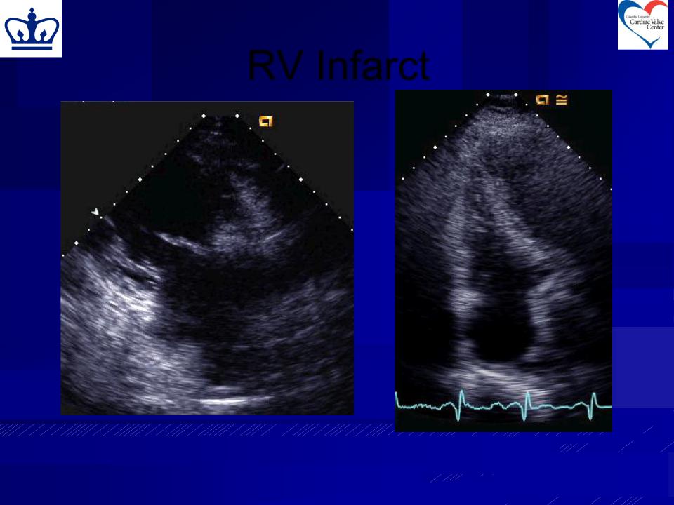
Echo Signs of RV Infarct
Primary
–RV dilatation
–Segmental wall motion abnormalities
–Decreased descent of RV base
Secondary
–Paradoxical septal motion
–Tricuspid regurgitation
–Tricuspid papillary muscle rupture
–Dilated IVC/Plethora of IVC
–Right-to-left IAS bowing
–Right-to-left PFO shunt
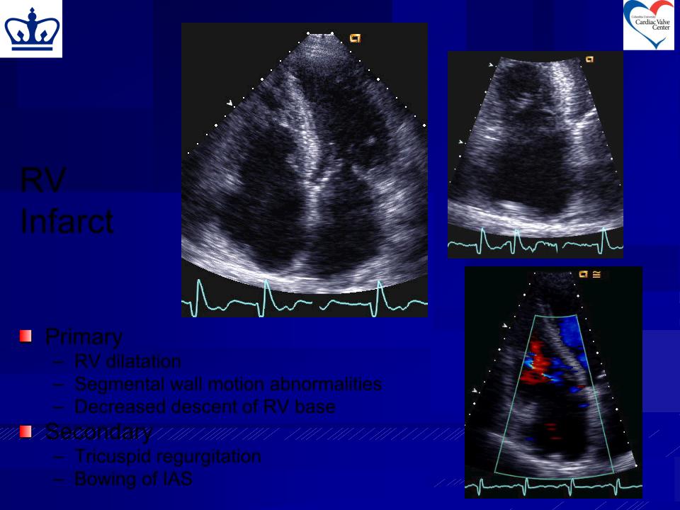
RV
Infarct
Primary
–RV dilatation
–Segmental wall motion abnormalities
–Decreased descent of RV base
Secondary
–Tricuspid regurgitation
–Bowing of IAS
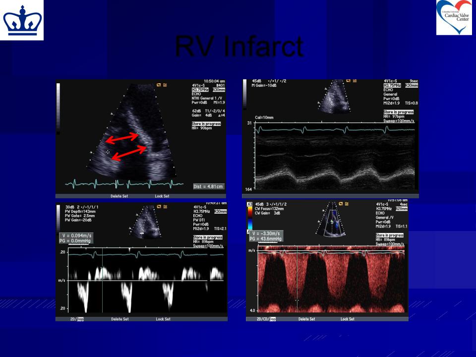
RV Infarct
RVD = 4.8 cm |
TAPSE = |
|
|
|
12mm |
TRV = 3.3 m/s
S’ = 9 cm
RV Infarct
Other Important Views: RV Inflow and Apical 2Chamber
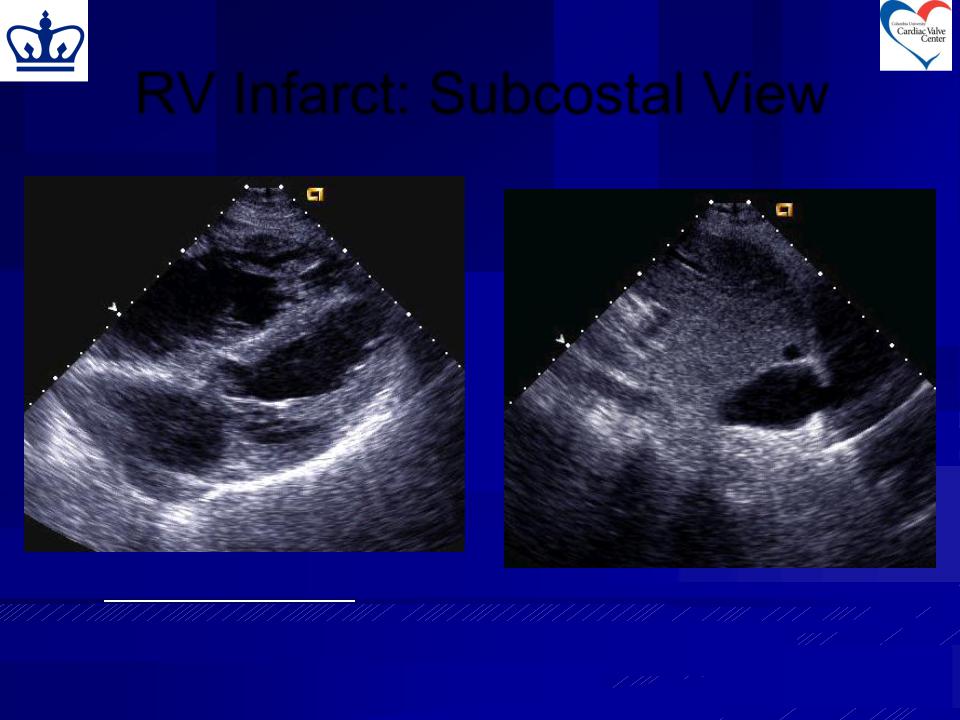
RV Infarct: Subcostal View
Secondary Signs: |
Dilated IVC/Plethora of IVC |
Dilated IVC/Plethora of IVC |
|
Right-to-left IAS bowing |
|
Right-to-left PFO shunt |
|
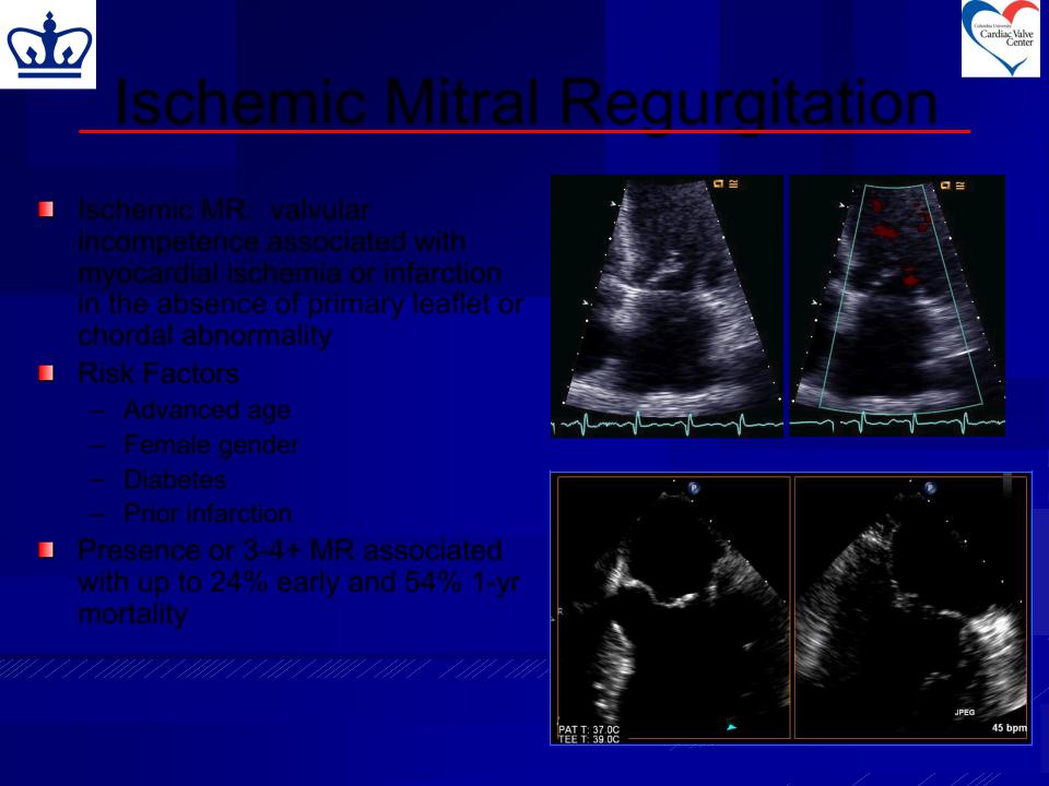
Ischemic Mitral Regurgitation
Ischemic MR: valvular incompetence associated with myocardial ischemia or infarction in the absence of primary leaflet or chordal abnormality
Risk Factors
–Advanced age
–Female gender
– Diabetes
– Prior infarction
Presence or 3-4+ MR associated with up to 24% early and 54% 1-yr mortality
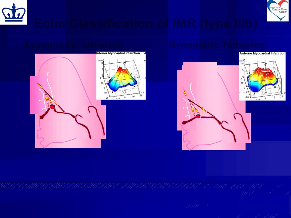
Echo Classification of IMR (type IIIb)
Asymmetric tethering |
Symmetric Tethering |
||
|
|
|
|
|
|
|
|
|
|
|
|
Posterior MI |
Anterior MI |
||
1. |
“Asymmetric” displacement of the |
||
|
|||
posteromedial papillary muscle |
1. Global LV remodeling and |
||
|
|||
2. |
Deficit in coaptation is due to |
apical displacement of both |
|
|
|||
tethering of a portion of P2 and P3 |
papillary muscles. |
|
3. Malcoaptation of the lateral
scallops
Agricola E et al. Eur J Echocardiography 2004 5, 326-334
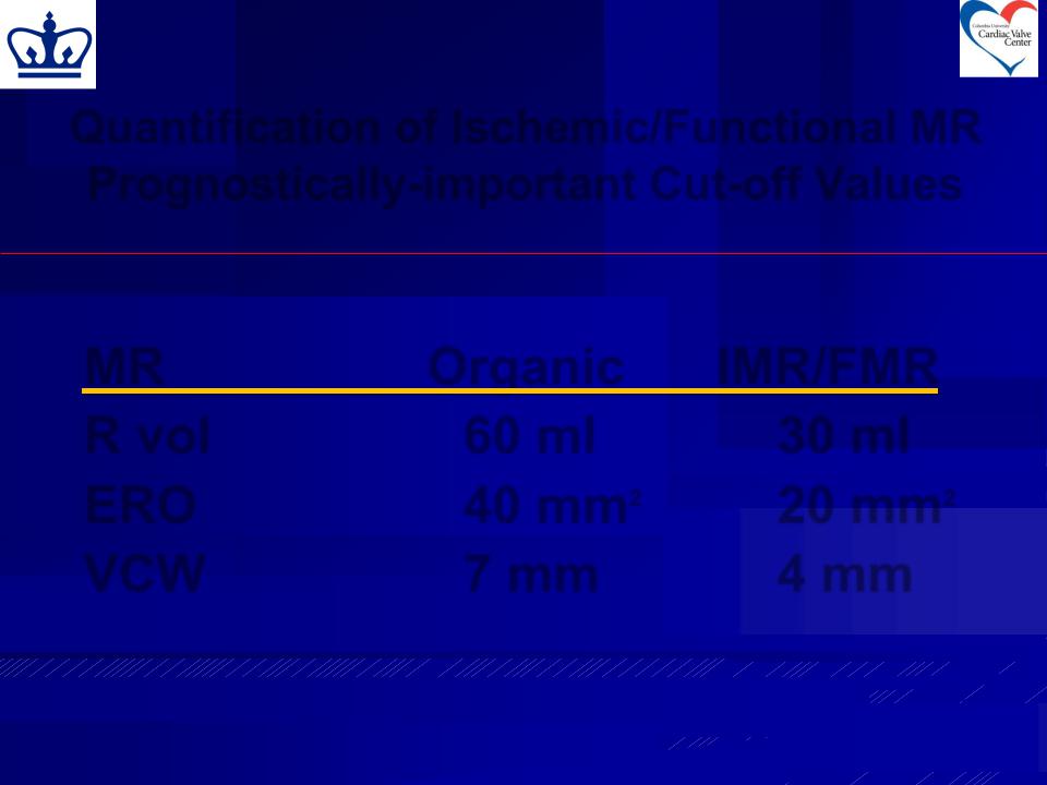
Quantification of Ischemic/Functional MR
Prognostically-important Cut-off Values
MR |
Organic |
IMR/FMR |
R vol |
60 ml |
30 ml |
ERO |
40 mm2 |
20 mm2 |
VCW |
7 mm |
4 mm |